Wir verwenden Cookies ausschließlich zu dem Zweck, technisch notwendige Funktionen wie das Login oder einen Warenkorb zu ermöglichen, oder Ihre Bestätigung zu speichern. Mehr Informationen zur Datenerhebung und -verarbeitung finden Sie in unserer Datenschutzerklärung.
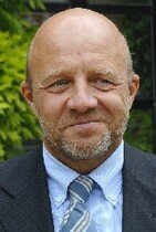
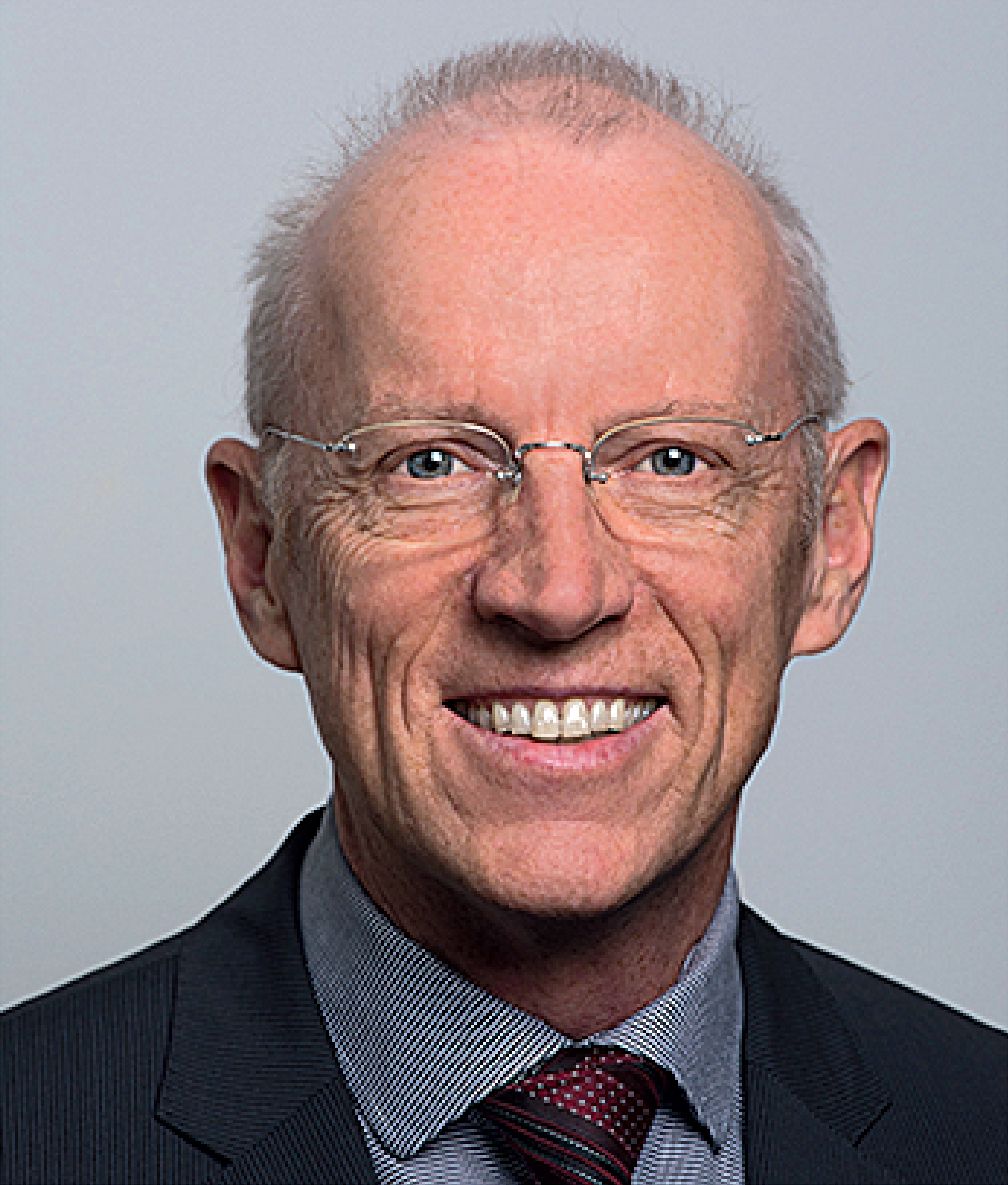
Prof Marc Quirynen graduated as a dentist in 1980 from the Catholic University of Leuven, Belgium, where he went on to complete training in periodontology and obtain his PhD. In 1990, he was appointed as a professor at Catholic University and is currently Chairman of the Department of Periodontology. His research focuses on simplification and optimization of periodontal therapy, and over the past decade, he has explored platelet concentrates in soft and hard tissue regeneration. Prof Quirynen has published over 450 papers in international peer-reviewed journals and serves on several editorial boards, including for the Journal of Clinical Periodontology and Clinical Oral implants Research. His most inspiring lectures deal with the clinical benefits of L-PRF and the prevention of peri-implantitis.
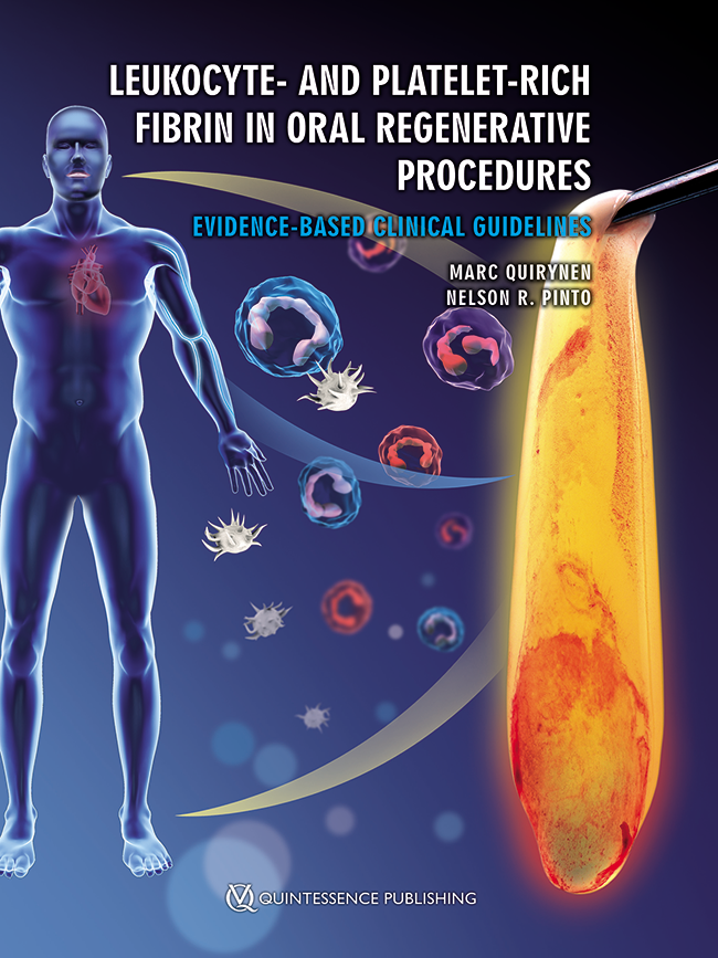
1. Auflage 2022 Buch Hardcover; 21 x 28 cm; incl. 18 Videos via App, 304 Seiten, 830 Abbildungen Sprache: Englisch Kategorien: Parodontologie, Implantologie, Mund-Kiefer-Gesichtschirurgie Artikelnr.: 7732 ISBN 978-1-78698-105-9 QP Deutschland
£155.00
Veranstaltungen
2nd European Congress Ceramic Implant Dentistry
European Society for Ceramic Implantology20. Okt. 2022 — 22. Okt. 2022Seminarhotel Bocken, Horgen, Schweiz
Referenten: Owen Addison, Bilal Al-Nawas, Rodrigo Beltrão, Michael M. Bornstein, André Chen, Jérôme Chevalier, Jospeh Choukroun, Alessandro Devigus, Michael Gahlert, Shahram Ghanaati, Elisabeth Jacobi-Gresser, Ronald Jung, Ralf J. Kohal, Sebastian Kühl, Frank-Michael Maier, Jochen Mellinghoff, Michael Payer, Mattias Pettersson, Marc Quirynen, Stefan Röhling, Jens Tartsch
European Society for Ceramic Implantology - ESCI
EuroPerio10
15. Juni 2022 — 18. Juni 2022Bella Center Copenhagen, Copenhagen, Dänemark
Referenten: Mario Aimetti, Zvi Artzi, Serhat Aslan, Georgios Belibasakis, Florian Beuer, Juan Blanco Carrión, Michael M. Bornstein, Nagihan Bostanci, Philippe Bouchard, Darko Božić, Olivier Carcuac, Maria Clotilde Carra, Nelson Carranza, Iain L. C. Chapple, Pierpaolo Cortellini, Jan Cosyn, Mike Curtis, Francesco D'Aiuto, Bettina Dannewitz, Luca De Stavola, Jan Derks, Nikolaos Donos, Peter Eickholz, Bahar Eren Kuru, Ricardo Faria Almeida, Roberto Farina, Magda Feres, Elena Figuero, Dagmar Fosså Bunæs, Rok Gašperšič, William Giannobile, Cecilie Gjerde Gjengedal, Moshe Goldstein, Marjolaine Gosset, Klaus Gotfredsen, Filippo Graziani, Adrian Guerrero, George Hajishengallis, Hady Haririan, Lisa J. A. Heitz-Mayfield, Palle Holmstrup, Marc Hürzeler, Mark Ide, Søren Jepsen, Ronald Jung, Sérgio Kahn, Anhgela R. Kamer, Alpdogan Kantarci, Moritz Kebschull, Björn Klinge, Thomas Kocher, Odd Carsten Koldsland, Kenneth Kornman, Marja Laine, Markus Laky, Isabelle Laleman, Evanthia Lalla, France Lambert, Luca Landi, Niklaus P. Lang, Antonio Liñares, Tomas Linkevičius, Bruno Loos, Rodrigo Lopez, Eli Machtei, Aslan Mammadov, Mauro Merli, Andrea Mombelli, Eduardo Montero, Niki Moutsopoulos, Jose Nart, Gustavo G. Nascimento, Ian Needleman, Tiernan O'Brien, William Papaioannou, Panos N. Papapanou, Michael A. Pikos, Pawel Plakwicz, Constanza Pontarolo, Philip M. Preshaw, Marc Quirynen, Mia Rakic, Christoph Andreas Ramseier, Hélène Rangé, Papageorgiou Spyridon, Maurizio S. Tonetti, Leonardo Trombelli, Istvan Urban, Fridus van der Weijden, Fabio Vignoletti, Charalambos Vlachopoulos, Nicola West, Asaf Wilensky, Ion Zabalegui, Egija Zaura, Nicola Zitzmann, Giovanni Zucchelli, Otto Zuhr, Fardal Øystein
European Federation of Periodontology (EFP)
Abschiedssymposium Christoph Hämmerle
Rekonstruktive Zahnmedizin: Wo stehen wir, wo gehen wir hin?22. Jan. 2022, 9:00 — 17:30 Uhr (CET)Universität Zürich Campus Irchel, Zürich, Schweiz
Referenten: Thomas Attin, Beatrice Beck Schimmer, Tord Berglundh, Rino Burkhardt, Lyndon F. Cooper, Christer Dahlin, Stefano Gracis, Christoph Hämmerle, Lisa J. A. Heitz-Mayfield, Marc Hürzeler, Ronald Jung, Nadja Nänni, Mutlu Özcan, Marc Quirynen, Irena Sailer, Christian S. Stohler, Daniel Thoma
Implantology: Beyond your expectations12. Okt. 2021 — 14. Okt. 2021online
Referenten: Enrico Agliardi, Alessandro Agnini, Andrea Mastrorosa Agnini, Mauricio Araujo, Goran Benic, Juan Blanco Carrión, Daniel Buser, Raffaele Cavalcanti, Tali Chackartchi, Luca Cordaro, Jan Cosyn, Holger Essig, Vincent Fehmer, Stefan Fickl, Alberto Fonzar, Helena Francisco, German O. Gallucci, Ramin Gomez-Meda, Oscar Gonzalez-Martin, Robert Haas, Arndt Happe, Alexis Ioannidis, Ronald Jung, Niklaus P. Lang, Tomas Linkevičius, Iva Milinkovic, Sven Mühlemann, Katja Nelson, Sergio Piano, Michael A. Pikos, Bjarni E. Pjetursson, Marc Quirynen, Franck Renouard, Isabella Rocchietta, Dennis Rohner, Irena Sailer, Henning Schliephake, Shakeel Shahdad, Massimo Simion, Ali Tahmaseb, Hendrik Terheyden, Jochen Tunkel, Stefan Vandeweghe, Piero Venezia, Stijn Vervaeke, Martin Wanendeya, Georg Watzek, Giovanni Zucchelli
European Association for Osseintegration (EAO)
Zeitschriftenbeiträge dieses Autors
International Journal of Periodontics & Restorative Dentistry, Pre-Print
DOI: 10.11607/prd.6989, PubMed-ID: 387174383. Mai 2024,Seiten: 1-22, Sprache: EnglischDhondt, Rutger A.L. / Lahoud, Pierre / Siawasch, Manoetjer / Castro, Ana B. / Quirynen, Marc / Temmerman, Andy
Objective: This study aims to collect data on implant survival, bone volume maintenance, and
complications associated with the socket shield technique. Background data: The socket shield
technique was introduced in 2010. Since then, several systematic reviews have been published,
showing good clinical outcomes. The behaviour of the buccal bone plate is so far not completely
understood. Methods: The study involved the placement of 23 implants using the socket shield
technique in 20 patients. AstraTech EV implants were used, and no bone substitutes or connective
tissue grafts were applied. Patients were monitored for 18 months, recording implant survival,
volumetric bone analysis on CBCT scans, interproximal bone levels, bone sounding, pink esthetic
scores, and complications. Prosthetic procedures were also described, including temporary and final
restorations. Results: A 95.7% cumulative 18-month implant survival rate was obtained using the
socket shield technique, with a significant but limited reduction in buccal bone thickness (BBT) after
implant placement. One implant did not integrate and two shields were partially exposed. The mean
pink esthetic score, 1 year after loading was 12.93 ± 1.22. Conclusion: The study suggests that the
socket shield technique can result in limited reduction of the buccal bone volume, with a high implant
survival rate. Re-entry studies are recommended to investigate the causes of bone resorption.
Schlagwörter: bone preservation, immediate implant, implant survival, oral implant, partial extraction, socket shield
Purpose: To clinically evaluate oral implant restorations placed by undergraduate students in the dental clinical curriculum at KU Leuven (Belgium) in terms of function and esthetics.
Materials and methods: A retrospective observational cohort study was designed. The esthetic and functional evaluations of implant-supported restorations placed in the framework of the undergraduate implant dentistry clinical training program using White/Pink Esthetic Score (WES/PES) and visual analog scale (VAS) scoring was performed. Furthermore, complications were registered based retrospectively on the patient's medical file. The following research questions were stipulated: (1) How well do implant-supported restorations placed by undergraduate students perform esthetically? and (2) Which complications occurred and how were these managed?
Results: Between August 2008 and July 2014 (6 academic terms), 251 implants (Brånemark System Mk III, Nobel Biocare) were placed in 113 patients by 155 students (> 40% of all students enrolled in the training program). Of these implants, 228 were restored in 101 patients by 118 students with varying restoration types. Esthetic scoring of the restorations in 83 of these patients revealed a satisfying mean WES of 8.14 ± 2.09 (out of 10) and PES of 9.56 ± 3.14 (out of 14). Complications were registered in 18.9% of the cases.
Discussion: Clinical training in implant dentistry for undergraduates contributes to the development of advanced skills in the dental student's Master education. Overall, patients were satisfied with their implant-supported restorations. Implant and restoration success rates and complication incidence were confirmed by long-term data in the oral implant literature.
The International Journal of Oral & Maxillofacial Implants, 3/2023
DOI: 10.11607/jomi.9773, PubMed-ID: 37279221Seiten: 503-515, Sprache: EnglischQuirynen, Marc / Van der Veken, Dominique / Lahoud, Pierre / Neven, Jan / Politis, Constantinus / Jacobs, Reinhilde
Purpose: To propose diffuse osteomyelitis as risk indicator for peri-implantitis following the loss of several dental implants in patients that present with highly sclerotic bone areas.
Materials and Methods: A total of six “nightmare cases”—three of which were treated at the Department of Periodontology of the University Hospitals of the Catholic University Leuven and three of which were referred there for a second opinion—were retrospectively analyzed using radiographs obtained via contact with referring clinicians in order to fully reconstruct the treatment pathway and dental history for each of these patients.
Results: All patients suffered from early implant failures and/or severe peri-implantitis with bone loss and crater formation up to the apical level, as well as the loss of all or nearly all implants. Re-examination of their preand postoperative CBCTs, in combination with several bone biopsies, confirmed the diagnosis of a diffuse sclerosing osteomyelitis in the treated area. Osteomyelitis could be linked to a longstanding history of chronic and/or therapyresistant periodontal/endodontic pathology.
Conclusion: The current retrospective case series seems to suggest that diffuse osteomyelitis should be considered as a risk indicator for severe peri-implantitis.
Schlagwörter: bone quality, bone density, dental implants, implant failure, peri-implantitis
The aim of the present study was to compare leukocyte- and platelet-rich fibrin (L-PRF) membranes with a connective tissue graft (CTG) in combination with a coronally advanced flap (CAF) in the treatment of Miller Class I or II localized gingival recessions. A randomized controlled clinical trial with 17 recessions in each group was initiated; the control group received treatment with CAF+CTG, and the test group received CAF+L-PRF. The following variables were measured before treatment and after 1, 3, and 6 months: gingival recession depth (RD), gingival recession width (RW), gingival thickness (GT), probing depth (PD), clinical attachment level (CAL), and keratinized tissue height (KTH). Also, the root coverage percentage (RC), the pain score, postoperative complications, and the root coverage esthetic score (RES) were recorded after surgery. Both treatments presented significant improvements in the RD, RW, and CAL at 1, 3, and 6 months. CTG achieved a significantly higher RC at 1, 3, and 6 months and a significantly higher RES score at 6 months. L-PRF presented a significantly lower pain score and less postoperative complications. Both strategies were effective for the treatment of localized gingival recessions. The CTG obtained higher RC and esthetic results, and L-PRF had less pain and postsurgical complications.
Purpose: To clinically evaluate oral implant restorations placed by undergraduate students in the dental clinical curriculum at KU Leuven (Belgium) in terms of function and esthetics.
Materials and methods: A retrospective observational cohort study was designed. The esthetic and functional evaluations of implant-supported restorations placed in the framework of the undergraduate implant dentistry clinical training program using White/Pink Esthetic Score (WES/PES) and visual analog scale (VAS) scoring was performed. Furthermore, complications were registered based retrospectively on the patient's medical file. The following research questions were stipulated: (1) How well do implant-supported restorations placed by undergraduate students perform esthetically? and (2) Which complications occurred and how were these managed?
Results: Between August 2008 and July 2014 (6 academic terms), 251 implants (Brånemark System Mk III, Nobel Biocare) were placed in 113 patients by 155 students (> 40% of all students enrolled in the training program). Of these implants, 228 were restored in 101 patients by 118 students with varying restoration types. Esthetic scoring of the restorations in 83 of these patients revealed a satisfying mean WES of 8.14 ± 2.09 (out of 10) and PES of 9.56 ± 3.14 (out of 14). Complications were registered in 18.9% of the cases.
Discussion: Clinical training in implant dentistry for undergraduates contributes to the development of advanced skills in the dental student's Master education. Overall, patients were satisfied with their implant-supported restorations. Implant and restoration success rates and complication incidence were confirmed by long-term data in the oral implant literature.
Purpose: To prospectively assess marginal bone loss and implant survival with Astra Tech (Dentsply Sirona, Charlotte, NC, USA) (group A) and Brånemark (Nobel Biocare, Zurich, Switzerland) (group B) implants in a split-mouth study conducted over a 20-year follow-up period.
Materials and methods: A total of 95 implants (n = 50, group A and n = 45, group B) were randomly placed in the left or right side of the maxilla or mandible in 18 patients. Clinical and radiographic examinations were performed, and results were reported at 5, 10, 15 and 20 years after insertion of the prosthesis.
Results: Ten patients were followed up for 20 years (n = 26 implants, group A and n = 25 implants, group B). No implant loss or prosthetic failures were observed. After 20 years of follow-up, no significant differences in marginal bone loss were found between both implant groups (P = 0.25). The proportion of marginal bone loss ≥ 0.5 mm was not significantly different between implant types (P > 0.05), and no statistically significant relationships were found between marginal bone loss and time (P ≥ 0.05). More specifically, there was no significant difference in marginal bone level between year 20 and baseline in group A (P = 0.70), whereas a difference of 0.5 to 1.0 mm was found in group B (P = 0.15).
Conclusions: After 20 years of follow-up, marginal bone loss around screw-shaped titanium implants was clinically insignificant. Furthermore, no significant differences in survival and marginal bone loss were found between group A and B implants over the follow-up period.
Schlagwörter: bone remodelling, implant system, marginal bone level, osseointegration, split-mouth design
Conflict-of-interest statement: The authors declare there are no conflicts of interest relating to this study.
Purpose: Sinus floor elevation (SFE) and simultaneous implant placement is predictable and reproducible.
However, the graft material for the antral cavity remains a topic of debate. Considering the high osteogenic
potential of the sinus membrane, most graft materials are generally accepted. This study aimed to assess
the outcome of simultaneous SFE and implant placement, using leukocyte- and platelet-rich fibrin (L-PRF)
as a sole graft material.
Materials and Methods: This study was designed as a single cohort prospective
study. Clinical and radiographic measurements (cone beam computed tomography [CBCT]) were performed
immediately after implant placement and at abutment connection (6 months later). The amount of newly
formed bone was linearly recorded on cross-sectional images. Four measurements (mesial, distal, buccal,
palatal) were registered with the axis of the implant as reference.
Results: Six lateral and 22 transalveolar
SFEs were performed in 26 patients with simultaneous implant placement. Six months after surgery, 27/29
implants were clinically integrated. The mean vertical bone gain was 3.4 ± 1.2 mm and 5.4 ± 1.5 mm
for transalveolar SFE and lateral SFE, respectively. The level of the new sinus floor was in all cases in
continuation with the apex of the implant, and the peri-implant crestal bone height was stable.
Conclusion:
L-PRF as a sole graft material during simultaneous SFE and implant placement proved to be a practical, safe,
and economical subsinus graft material, resulting in natural bone formation.
Purpose: The leukocyte- and platelet-rich fibrin block (L-PRF block) is a composite graft that combines a xenograft that is acting as a scaffold with L-PRF membranes that serve as a bioactive nodule with osteoinductive capacity. This study evaluated the properties of the L-PRF block and its components in terms of release of growth factors, cellular content, and structure.
Materials and Methods: The concentration of transforming growth factor-β1 (TGF-β1), vascular endothelial growth factor (VEGF), platelet-derived growth factor-AB (PDGF-AB), and bone morphogenetic protein-1 (BMP-1) released by an L-PRF membrane and an L-PRF block were examined with ELISA for five time intervals (0 to 4 hours, 4 hours to 1 day, 1 to 3 days, 3 to 7 days, 7 to 14 days). Those levels in L-PRF exudate and liquid fibrinogen were also evaluated. The cellular content of the liquid fibrinogen, L-PRF membrane, and exudate was calculated. The L-PRF block was also analyzed by means of a microcomputed tomography (micro-CT) scan and scanning electron microscopy (SEM).
Results: TGF-β1 was the most released growth factor after 14 days, followed by PDGF-AB, VEGF, and BMP-1. All L-PRF blocks constantly released the four growth factors up to 14 days. L-PRF membrane and liquid fibrinogen presented high concentrations of leukocytes and platelets. The micro-CT and SEM images revealed the bone substitute particles surrounded by platelets and leukocytes, embedded in a dense fibrin network.
Conclusion: The L-PRF block consists of deproteinized bovine bone mineral particles surrounded by platelets and leukocytes, embedded in a fibrin network that releases growth factors up to 14 days.
Schlagwörter: blood platelets, cell count, fibrinogen, growth factors, scanning electron microscopy, tissue engineering
Purpose: Osteotome sinus floor elevation (OSFE) is a technique aimed at simplifying implant placement in the posterior atrophic maxilla. The necessity of bone grafting under the elevated sinus membrane has been widely debated. The aim was to compare the evolution over 5 years of implant stability in sites grafted or left ungrafted.
Materials and methods: A total of 12 patients (9 female and 3 male) presenting ≤ 4 mm initial bone height (IBH) in the posterior maxillary sites were recruited. Implants (n = 37) were placed using OSFE. According to the randomisation, the sinuses received either bone graft (n = 20, control group) or no graft (n = 17, test group). Patients received both these treatments when both sinuses fulfilled the inclusion criteria. Control assessments were performed 1 week, 10 weeks, 12 weeks, and then 1 year, 3 years and 5 years after the implant placement. Periapical radiographs were taken and the implant stability quotient (ISQ) was measured at different time points.
Results: At implant surgery, the mean ISQ was 58.9 ± 11.2 for the test group and 53.8 ± 10.2 for the control group; it plummeted 10 weeks after the implant placement and rose thereafter. Five years after the implant placement, the mean ISQ reached 80.8 ± 4.2 for the control group, and 79.7 ± 4.3 for the test group. The difference between the groups was not significant. The IBH significantly affected implant stability at implant insertion and 5 years after the implant insertion, but not at the other time points.
Conclusions: The implants performed using OSFE in ungrafted sites were as stable as the implants placed in grafted sites.
Schlagwörter: bone graft, implant stability quotient (ISQ), osteotome sinus floor elevation (OSFE), posterior maxilla, resonance frequency analysis (RFA)
Conflict of interest statement: All authors report no conflict of interest.
Aim: The objective of this systematic review was to evaluate the diagnostic value of CBCT compared with 2D imaging and clinical gold standard techniques in peri-implant bone defect detection and measurement.
Materials and methods: Literature search was performed using MEDLINE, Embase and Web of Science databases up to July 2017. Clinical, ex vivo, in vitro and animal studies that assessed and measured peri-implant bone defects using different imaging modalities were included in this review. Two reviewers performed data extraction and qualitative analysis. The methodological quality of each study was reviewed using the QUADAS-2 tool.
Results: The initial search revealed 2849 unique papers. Full-text analysis was performed on 60 articles. For the present review, nine studies were considered eligible to be included for qualitative analysis. CBCT performed similar to intraoral radiography in mesiodistal defect detection and measurements. Additional buccolingual visualisation and volumetric and morphological assessment of peri-implant bone defects are major advantages of 3D visualisation with CBCT. Nevertheless, one must be aware of metal artefacts masking osseointegration, shallow bony defects and other peri-implant radiolucencies, thus impeding early diagnosis of intrabony lesions.
Conclusions: The present review did not provide evidence to support the use of CBCT as standard postoperative procedure to evaluate peri-implant bone. Up to date, we are clinically forced to remain with intraoral radiography, notwithstanding the inherent limitations related to restricted field of view and two-dimensional overlap. A 3D imaging approach for postoperative implant evaluation is crucial, making further development of an optimised and artefact-free CBCT protocol indispensable.
Schlagwörter: bone defects, CBCT, imaging, implant dentistry, peri-implantitis