Wir verwenden Cookies ausschließlich zu dem Zweck, technisch notwendige Funktionen wie das Login oder einen Warenkorb zu ermöglichen, oder Ihre Bestätigung zu speichern. Mehr Informationen zur Datenerhebung und -verarbeitung finden Sie in unserer Datenschutzerklärung.
2003 Fachzahnarzt für Parodontologie, ZÄKWL. 2003 Spezialist der Deutschen Gesellschaft für Parodontologie. 2006 Master of Oral Medicine in Implantology (MOM). 2007 Fachzahnarzt für Oralchirurgie, ZÄKWL. 2007 Hospitations- und Supervisionsreferent DGParo/APW. 2008 1. Vorsitzender des Referats „Chirurgische Parodontologie“ im Berufsverband Deutscher Oralchirurgen. 2011 Hospitations-und Supervisionsreferent DGI/APW. 2018 ITI Fellow. 2004 - 2015 Lehrbeauftragter und Gastdozent an der Poliklinik für Parodontologie des Universitätsklinikums Münster. 2019 ITI Speaker. 2020 Gründung der Social Media Learning Platform „Regenerative Bioversity“. Seit 2007 Niederlassung in spezialisierter Praxis für Parodontologie, Implantologie & Oralchirurgie zusammen mit Dr. Carolin Tunkel, Fachzahnärztin für Kieferorthopädie in Bad Oeynhausen, Ostwestfalen.
Veranstaltungen
International Esthetic Days
Digital excellence across disciplines19. Sept. 2024 — 21. Sept. 2024Palau de Congressos de Palma, Palma de Mallorca, Spanien
Referenten: Eirik Aasland Salvesen, Wael Att, Andrea Bazzucchi, Andre Chen, Maja Chmielewska, Karim Dada, Mona Eide-Gast, Vincent Fehmer, Roshi Frafjord, Fernando Franch, Ophir Fromovich, Nuno Gil, Oscar Gonzalez-Martin, Sanaa Kader, France Lambert, Adriaens Laurence , Diego Lops, Mark Lowe, James Mah, Patrice Margossian, Alberto Monje, Léon Parienté, Lucrezia Paternò Holtzman, Alessandro Perucchi, Sonia Presencia Pascual, Algirdas Puišys, Cristian Scognamiglio, Prav Solanki, Gabor Tepper, Jasper Thoolen, Jochen Tunkel, Kay Vietor, Eglė Vindašiūtė-Narbutė, Sarah Weston
Straumann GmbH
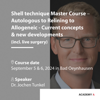
Shell technique Master Course – Autologous to Relining to Allogeneic – Current concepts & new developments (including live surgery)
5. Sept. 2024 — 6. Sept. 2024Tunkel & Tunkel Fachzahnarztpraxis, Bad Oeynhausen, Deutschland
Referenten: Jochen Tunkel
Stoma Dentalsysteme GmbH & Co KG
EAO Digital Days
Implantology: Beyond your expectations12. Okt. 2021 — 14. Okt. 2021online
Referenten: Enrico Agliardi, Alessandro Agnini, Andrea Mastrorosa Agnini, Mauricio Araujo, Goran Benic, Juan Blanco Carrión, Daniel Buser, Raffaele Cavalcanti, Tali Chackartchi, Luca Cordaro, Jan Cosyn, Holger Essig, Vincent Fehmer, Stefan Fickl, Alberto Fonzar, Helena Francisco, German O. Gallucci, Ramin Gomez-Meda, Oscar Gonzalez-Martin, Robert Haas, Arndt Happe, Alexis Ioannidis, Ronald Jung, Niklaus P. Lang, Tomas Linkevičius, Iva Milinkovic, Sven Mühlemann, Katja Nelson, Sergio Piano, Michael A. Pikos, Bjarni E. Pjetursson, Marc Quirynen, Franck Renouard, Isabella Rocchietta, Dennis Rohner, Irena Sailer, Henning Schliephake, Shakeel Shahdad, Massimo Simion, Ali Tahmaseb, Hendrik Terheyden, Jochen Tunkel, Stefan Vandeweghe, Piero Venezia, Stijn Vervaeke, Martin Wanendeya, Georg Watzek, Giovanni Zucchelli
European Association for Osseintegration (EAO)
Zeitschriftenbeiträge dieses Autors
Parodontologie, 4/2022
Seiten: 443-451, Sprache: DeutschTunkel, Jochen / Hofmann, Frederik / de Stavola, Luca
In den letzten Jahrzehnten wurden unterschiedliche Techniken zur Deckung parodontaler Rezessionen beschrieben. Ein Großteil davon ist nicht zur Deckung multipler tiefer benachbarter Cairo-Klasse-I-Rezessionen, sondern eher zur Korrektur multipler flacher bzw. einer tiefen mit mehreren benachbarten flachen Rezessionen geeignet. Der vorliegende Fallbericht beschreibt eine neue modifizierte Operationsmethode: den mehrfach gestielten koronal verschobenen Lappen, kombiniert mit Schmelz-Matrix-Proteinen und Bindegewebetransplantaten. Mithilfe dieser operativen Technik ist es möglich, selbst weit über die mukogingivale Grenze hinausgehende benachbarte Rezessionen mit einer hohen Wahrscheinlichkeit zu decken. Ein 25-jähriger Patient wurde eigenen Angaben zufolge wegen progredienter tiefer Cairo-Klasse-I-Rezessionen zur Therapie überwiesen. Die Wurzeldeckung erfolgte durch einen modifizierten mehrfach gestielten koronal verschobenen Lappen in Kombination mit einem aus dem Gaumen entnommenen Bindegewebetransplantat und einem Schmelzmatrixderivat. Die Evaluation des Ergebnisses erfolgte nach 6 Monaten. Hierbei zeigte sich eine Abdeckung von über 80 % der zuvor freiliegenden Wurzeloberflächen. Der mehrfach gestielte koronal verschobene Lappen ist eine nützliche Methode zur Deckung tiefer benachbarter parodontaler Rezessionen, die den Erhalt vorhandener respektive die Verbreiterung keratiniserter Gingiva ermöglicht und die Bildung von sichtbarem Narbengewebe auf ein geringes Ausmaß reduziert.
Manuskripteingang: 22.03.2022, Annahme: 26.09.2022
Schlagwörter: Schmelzmatrixproteine, Gingivarezession, koronal verschobener Lappen, lateral verschobener Lappen, Zahnwurzel, Wurzeldeckung, Bindegewebetransplantat
During bone augmentation procedures, primary wound healing determines the bone augmentation result. After a crestal incision in the maxilla, the palatal flap might not be an adequate length to correctly couple to the vestibular flap and to seal the wound with horizontal mattress and single sutures. Due to the histologic structure made of dense connective tissue, the palatal flap eversion is impossible, negatively impacting the wound seal and primary healing. This case report describes the effectiveness and efficacy of an incision design to improve palatal flap management during bone augmentation procedures in the maxilla. Indeed, palatal flap verticalization is achieved. The incision line is proportionally shifted on the vestibular side, based on the defect anatomy, to obtain a palatal flap length extending at least 4 mm coronal to the bone graft level prior to wound closure. The described approach simplifies the optimal adaptation of the inner faces of the palatal and vestibular flaps, reducing the risk of nonprimary wound healing.
Tooth loss generally leads to a corresponding loss of supporting bone structures, jeopardizing correct implant placement. Bone augmentation procedures facilitate reconstruction of the alveolar contours but lengthen treatment time by about 4 to 9 months. The aim of this case series report is to describe the short-term results of the combination of three-dimensional bone augmentation using the shell technique in conjunction with simultaneous implantation. A total of 10 patients who underwent autologous bone augmentation using the shell technique with simultaneous implantation were retrospectively examined. The shell technique is an augmentation procedure using thin cortical bone plates adapted to the buccal and oral walls of the defect to rebuild the contours of the alveolar ridge. The remaining spaces are filled with bone chips. Healing time before second stage surgery was 4 months. The vertical bone defect at the beginning (VD), the height of the vertical bone graft, resorption at the time of second-stage surgery (BR1) and 1 year after prosthetic rehabilitation (BR2), the total resorption between augmentation and 1 year (BRtot), and the vertical bone loss of the implant (VBL) were measured. VD was 3.1 mm. Values for BR1 and BR2 were 0.4 and 0.45 mm, respectively, resulting in a total bone loss of 0.85 mm of bone loss (BRtot). VBL was 0.45 mm 1 year after prosthetic rehabilitation. The simultaneous approach of vertical bone augmentation in the shell technique and implantation shows excellent results in bone reconstruction and stability up to 1 year after prosthetic reconstruction and can shorten treatment time by 4 to 9 months.
Traumatische parodontale Rezessionen lassen sich heute mittels einer Vielzahl von Behandlungsmethoden erfolgreich therapieren. Hierbei wird den Verschiebelappen in Kombination mit Bindegewebstransplantaten die beste Erfolgschance beigemessen. Es konnte jedoch gezeigt werden, dass die Applikation von Schmelzmatrixproteinen zu einem signifikant höheren Gewinn an parodontalem Attachment führt. Dies hat im Vergleich mit Verschiebelappen allein eine verbesserte Wurzeldeckung zur Folge, welche sich dagegen beim Einsatz von Verschiebelappen in Kombination mit Bindegewebstransplantaten nicht einstellt. Durch die Kombination von Schmelzmatrixproteinen und Bindegewebstransplantat scheint es zu einer Verbesserung des Ergebnisses zu kommen. Allerdings ist die Evidenz hierzu eingeschränkt, und weitere Studien müssen den Langzeiterfolg dieser Kombination unter Beweis stellen.
Schlagwörter: Rezession, Koronalverschiebung, Lateralverschiebung, Tunneltechnik, Bindegewebstransplantat, Schmelzmatrixproteine, Wurzeldeckung
Purpose: To define the role played by a suspended external-internal (SEI) suture in reducing marginal flap tension after bone augmentation in the maxilla and in enhancing primary wound healing.
Materials and Methods: Twenty partially edentulous patients requiring bone augmentation (either guided bone regeneration or autogenous bone block placement) before or simultaneous with implant insertion in the maxilla were enrolled in this clinical prospective cohort study. Flap tension was measured by a dynamometer, which was accurate to within 1 g. The force recorded was that needed to enable the vestibular extensible flap to reach the edge of the palatal nonextensible flap. Flap tension was recorded after the periosteum-releasing incision was made (before application of any suture; T1), and after the SEI suture was applied (T2). Final marginal flap adaptation was accomplished via horizontal mattress sutures and simple stitches. Wound healing was monitored at 1, 2, 4, and 16 weeks and classified as "obtained primary closure" or "compromised" as a result of dehiscence or marginal flap necrosis.
Results: The mean flap tension measured at T1 was 32.9 ± 7.7 g. After the SEI suture was applied, the mean marginal flap tension decreased to 4.1 ± 1.5 g. The marginal flap tension was reduced by 87.6% compared to the initial strain. All patients healed uneventfully, and no complications such as dehiscences or marginal flap necrosis were recorded.
Conclusion: The application of the SEI suture reduced the tension on the margins of the flaps and played a decisive role in obtaining primary wound healing. In case of passive wound closure (strain 5 g), the type of augmentation procedure (guided bone regeneration or autogenous block) was revealed to have no impact on the quality of wound healing.
Schlagwörter: bone augmentation, internal suture, primary wound closure, wound dehiscence
Zahnverlust aufgrund parodontaler oder endodontischer Probleme führt in der Regel zu einem Verlust knöcherner Strukturen, der eine Implantation in prothetisch korrekter Position einschränken kann. Die Möglichkeiten der autologen Knochenblockaugmentation oder gesteuerter Geweberegeneration lassen zwar eine vorhersagbare Wiederherstellung des knöchernen Lagers zu, verlängern aber die Dauer der implantologischen Behandlung um 4 bis 9 Monate. Die Kombination aus Knochenblockaugmentation in Schalentechnik und simultaner Implantation kann diese Therapiedauer erheblich verkürzen und somit die Compliance und Zustimmung des Patienten zu solchen Eingriffen erheblich verbessern.
Schlagwörter: Knochenblockaugmentation, Schalentechnik, Implantat, Therapiedauer
The aim of this study was to report the outcome of the management of alveolar crest vertical defects using the tunnel technique approach associated with autogenous bone blocks prior to implant placement in 10 partially dentate consecutively treated patients. Four clinical linear measurements were taken: maximal extension of the vertical defect (VD) at the time of the augmentation procedure (time 0), vertical bone graft (VBG) recorded at time 0, bone resorption at implant placement (time 1), and bone resorption during implant healing at the time of abutment connection (time 2). All patients healed uneventfully, and no complications were recorded. Both mean VD and VBG at time 0 were 6.50 ± 1.43 mm. Mean bone resorption at time 1 was 0.30 ± 0.48 mm and mean bone resorption at time 2 was 0.25 ± 0.26 mm, yielding an overall vertical bone remodeling of 0.55 ± 0.49 mm (8.4%) after 8 months. This study supports the capability of a minimally invasive approach to regenerate bone in vertical defects prior to implant placement.
Purpose: The aim of this case series is to propose an approach to help maintain autogenous bone grafts. This is done by applying a collagen membrane (CM) and anorganic bovine bone (ABBM) at the time of implant surgery, rather than at the time of ridge augmentation, to avoid volume loss after implants are inserted.
Materials and Methods: Ten patients with severe horizontal bone atrophy were consecutively enrolled in this study. A staged approach was chosen for implant placement following horizontal ridge augmentation. A block graft was harvested from the retromolar area and secured to the recipient site with fixation screws; contour overbuilding was avoided. The width of the ridge was measured before and after horizontal augmentation. After 4 months of healing, implants were inserted, the augmented site was relined with ABBM, and CM was applied to prevent bone volume loss. Another 4 months later, at the time of abutment placement, cone beam computed tomography was performed to quantify the end result.
Results: The mean horizontal ridge width prior to treatment was 2.1 ± 0.5 mm. Mean postsurgical crest width was 6.9 ± 0.5 mm. After the 4-month healing period, the mean alveolar crest width was 6.6 ± 0.6 mm. At the time of abutment connection, the mean width of the regenerated ridge, as measured on three-dimensional cone beam images, was 7.7 ± 0.8 mm.
Conclusions: Minimal bone loss occurred in unprotected autogenous bone grafts with respect to alveolar bone contour (0.25 ± 0.29 mm). After the implants were inserted, no further remodeling/resorption occurred with sites treated by ABBM and CM relining; moreover, an additional increase in alveolar crest width was evident. The nonresorbable ABBM osseointegrated clinically and radiologically, preventing bone loss prior to implant loading. This layer appeared to maintain the regenerated crest volume.
Schlagwörter: autogenous bone graft, bone augmentation, bone resorption, volume maintenance
The aim of this case series report is to compare the results of the increase in keratinized mucosa using three different techniques of stage-two surgery. Thirty-two patients with one to eight dental implants who received prosthetic rehabilitation of the maxilla were included. Patients were divided into three groups based on preoperative anatomical considerations. Stage-two surgery was performed using either the apically repositioned flap (ARF; n = 14), the roll flap (RF; n = 10), or an apically repositioned flap combined with a connective tissue graft (ARFCT; n = 8). The height of the keratinized mucosa and relative tissue thickness were measured preoperatively and postoperatively at 2 weeks and 3, 6, and 12 months after surgery. The mean gains of keratinized mucosa and tissue thickness were calculated from these measurements. After 1 year, the mean gains in tissue thickness and keratinized tissue were 1.37 and 4.63 mm in the ARF group, 2.41 and 1.35 mm in the RF group, and 3.10 and 4.10 mm in the ARFCT group, respectively. There was no significant statistical difference between the 12-month and postoperative measurements (P > .05). In patients with deficient tissue thickness, a roll flap or an apically repositioned flap should be performed, while a lack of keratinized mucosa indicates the use of an apically repositioned flap with or without a connective tissue graft. When an increase in both keratinized mucosa and tissue thickness is necessary, an apically repositioned flap combined with a free connective tissue graft can be recommended. After a 12-month healing period, the obtained results showed excellent stability.
Vor Einführung der Implantologie hatte die präprothetische Chirurgie zur Verbesserung des Prothesenlagers eine große Bedeutung in der Zahnmedizin. Doch auch im Rahmen implantatgetragener Versorgungen kann mit Hilfe einer Vestibulumplastik z. B. nach Kazanjian die Breite der fixierten Gingiva vergrößert und somit die Pflege wie auch die prothetische Versorgung von Implantaten erleichtert werden. Nach Präparation eines Mukosalappens und anschließendem Ablösen des Muskels vom Periost können unter Darstellung des Knochens und ggf. des Nervus mentalis die Implantate inseriert werden. Dann wird das Periost zunächst mit dem lingualen Mukoperiostanteil und anschließend der Mukosaanteil am bukkalen Periost fixiert. Die Vestibulumplastik nach Kazanjian zeigt zwar eventuell eine höhere Rezidivtendenz als alternative Techniken, aber die geringere Resorption der bukkalen Knochenwand erweist sich in der Implantologie als Vorteil gegenüber anderen Vestibulumplastiken wie etwa der nach Edlan und Mejchar.
Schlagwörter: Vestibulumplastik, Kazanjian-Plastik, Edlan-Mejchar-Plastik, Freilegung, fixierte Mukosa