Wir verwenden Cookies ausschließlich zu dem Zweck, technisch notwendige Funktionen wie das Login oder einen Warenkorb zu ermöglichen, oder Ihre Bestätigung zu speichern. Mehr Informationen zur Datenerhebung und -verarbeitung finden Sie in unserer Datenschutzerklärung.
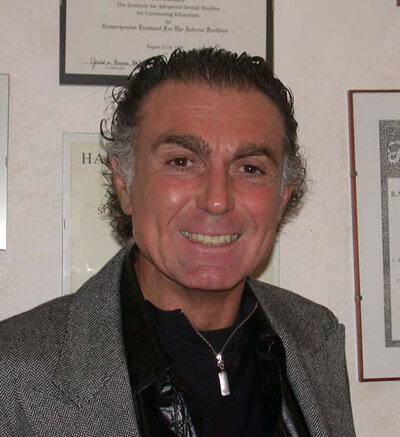
Stefano Parma-Benfenati, MD, DDS, MScD, is a visiting professor in postgraduate periodontology at the University of Turin and a professor in postgraduate oral surgery and pathology at the University of Parma. Dr Parma-Benfenati has been an active member of the Italian Society of Periodontology since 1987 and maintains a private practice limited to periodontology and implantology in Ferrara, Italy.
Veranstaltungen
SEPA 24 Bilbao
Clinical evidence based on scientific evidence29. Mai 2024 — 1. Juni 2024Bilbao, Spanien
Referenten: Eduardo Anitua, Sofia Aroca, Serhat Aslan, Gustavo Avila-Ortiz, Juan Blanco Carrión, Gonzalo Blasi, Nagihan Bostanci, Iain L. C. Chapple, Jan Cosyn, Glécio Vaz de Campos, Luca De Stavola, Jan Derks, Vincent Fehmer, Elena Figuero, Sergio García, Alfonso L. Gil, Oscar Gonzalez-Martin, Adrian Guerrero, Sérgio Kahn, Alejandro Lanis, Antonio Liñares, Ferrán Llansana, Francesco Mangano, Dino Calzavara mantovani, Mauro Merli, Juan Mesquida, Alberto Monje, Eduardo Montero, Stefano Parma-Benfenati, Bjarni E. Pjetursson, Pablo Ramírez, Mariano Sanz, Ignacio Sanz Sànchez, Beatriz Solano Mendoza, Jacobo Somoza, Martina Stefanini, Maurizio S. Tonetti, Leonardo Trombelli, Ion Zabalegui
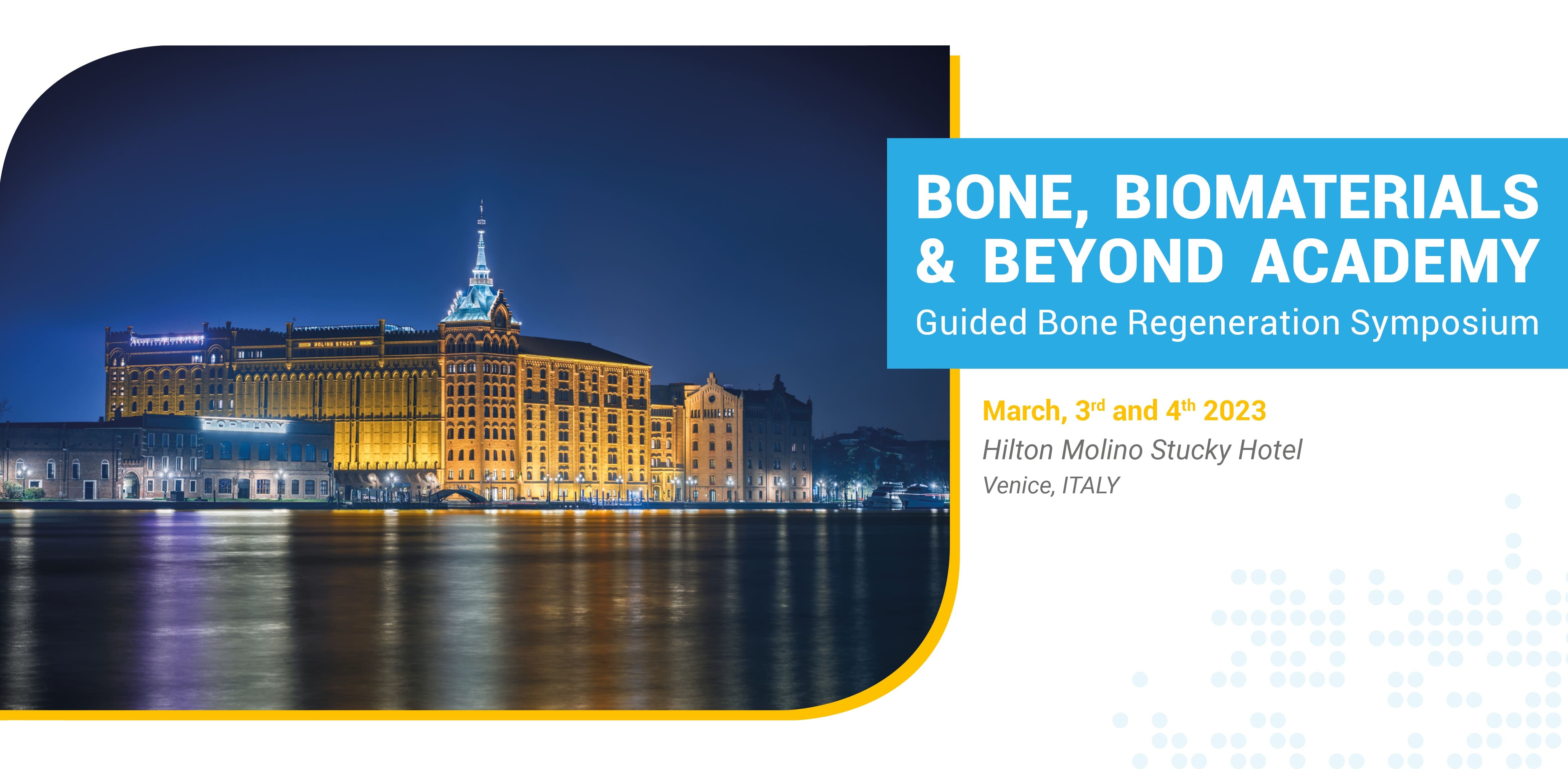
Bone, Biomaterials & Beyond Academy
Guided Bone Regeneration Symposium3. März 2023 — 4. März 2023Hilton Molino Stucky Venice, Venice, Italien
Referenten: Luca Comuzzi, Massimo del Fabbro, Abdelsalam Elaskary, Kai Fischer, Fernando Guerra, Arndt Happe, Göran Kjeller, Gerd Körner, Diego Lops, Christian Maischberger, Magda Mensi, Ulf Nannmark, Patrick Palacci, Stefano Parma-Benfenati, Adriano Piattelli, Claudio Stacchi, Tiziano Testori, Hannes Wachtel, Michael Weinländer
Bone, Biomaterials & Beyond Academy
The 14th International Symposium on Periodontics and Restorative Dentistry (ISPRD)
9. Juni 2022 — 12. Juni 2022Boston Marriott Copley Place, Boston, MA, Vereinigte Staaten von Amerika
Referenten: Tara Aghaloo, Edward P. Allen, Evanthia Anadioti, Wael Att, Vinay Bhide, Markus B. Blatz, Scotty Bolding, Lorenzo Breschi, Jeff Brucia, Daniel Buser, Luigi Canullo, Daniele Cardaropoli, Stephen J. Chu, Donald Clem, Christian Coachman, Lyndon F. Cooper, Daniel Cullum, Lee Culp, José Carlos Martins da Rosa, Sergio De Paoli, Marco Degidi, Nicholas Dello Russo, Serge Dibart, Joseph P. Fiorellini, Mauro Fradeani, Stuart J. Froum, David Garber, Maria L. Geisinger, William Giannobile, Luca Gobbato, Ueli Grunder, Galip Gürel, Chad Gwaltney, Christoph Hämmerle, Robert A. Horowitz, Marc Hürzeler, David Kim, Gregg Kinzer, Christopher Köttgen, Ina Köttgen, Purnima S. Kumar, Burton Langer, Lydia Legg, Pascal Magne, Kenneth A. Malament, Jay Malmquist, George Mandelaris, Pamela K. McClain, Michael K. McGuire, Mauro Merli, Konrad H. Meyenberg, Craig M. Misch, Julie A. Mitchell, Marc L. Nevins, Myron Nevins, Michael G. Newman, Miguel A. Ortiz, Jacinthe M. Paquette, Stefano Parma-Benfenati, Michael A. Pikos, Giulio Rasperini, Pamela S. Ray, Christopher R. Richardson, Isabella Rocchietta, Marisa Roncati, Marco Ronda, Paul S. Rosen, Maria Emanuel Ryan, Irena Sailer, Maurice Salama, David M. Sarver, Takeshi Sasaki, Todd Scheyer, Massimo Simion, Michael Sonick, Sergio Spinato, Dennis P. Tarnow, Lorenzo Tavelli, Douglas A. Terry, Tiziano Testori, Carlo Tinti, Istvan Urban, Hom-Lay Wang, Robert Winter, Giovanni Zucchelli
Quintessence Publishing Co., Inc. USA
Zeitschriftenbeiträge dieses Autors
International Journal of Periodontics & Restorative Dentistry, 3/2023
A 12-month prospective consecutive case series study was conducted to evaluate the effectiveness of a novel porcine collagen matrix to correct moderate to severe buccogingival recession defects. A total of 10 healthy patients (8 women and 2 men; age range: 30 to 68 years) with 26 maxillary and mandibular recession gingival defects > 4 mm deep were included. Healthy maturation of gingival tissues with natural color and texture matching the adjacent soft tissue areas were observed at all reevaluation visits. Complete root coverage was not achieved in all cases, possibly due to severe buccal bone loss in most of the selected cases, which negatively impacted the results. Nevertheless, when a novel porcine collagen matrix was used, a mean root coverage of 63.15% was achieved, and gains in clinical attachment level and keratinized tissue height were seen. Future randomized clinical trials are needed to further explore the potential of porcine collagen matrix for treatment of localized gingival recession defects.
This study aimed to describe the step-by-step procedure of the polydioxanone dome technique associated with guided bone regeneration (GBR) and to report the results up to 72 months after implant loading. Patients with maxillary horizontal bone defects (< 5 mm residual width, confirmed by CBCT scan) were treated with the proposed intervention. During the GBR procedure, four bone perforations were strategically prepared in a roughly square configuration. Segments of polydioxanone suture material were introduced in the perforations, forming a dome-shaped structure. Six months after bone augmentation, a new CBCT was performed. After implant restoration, periapical radiographs were taken, which were then repeated annually. The following outcomes were analyzed: implant survival, horizontal bone gain, marginal bone level, and complications. Twenty implants were placed in 11 patients with a survival rate of 100% in a mean follow-up of 38.18 ± 19.65 months after loading. Mean horizontal bone gain was 3.82 ± 1.67 mm and mean marginal bone level was –0.12 ± 1.17 mm. Only minor complications were observed. The present results suggest that the polydioxanone dome technique may represent a promising approach during horizontal GBR, alone or in combination with implant placement.
This study evaluated bone behavior during dynamic osseointegration. A total of 12 implants were placed in sheep tibia and analyzed at 15, 30, 60, and 90 days. Quantitative and qualitative bone behaviors were evaluated with histologic, histomorphometric, Alizarin Red S, and SEM-EDX (scanning electron microscopy with energy-dispersive x-ray spectroscopy) analysis. Twenty microanalyses were performed in chambers 1, 3, and 5 (a chamber is the distinctive space/bone volume between two coils of the implant screw) in distinctive zones: the titanium-bone interface (zone A), the middle chamber-bone front (zone B), the bone-surgical threading interface (zone C), and native bone (zone D; used as a control). The dynamic osseointegration index (DOI) and bone quality index (BQI) with calcium/phosphorus (Ca/P) content were detected to evaluate the osseointegration quality, bone-to-implant contact (BIC), and bone density around implants. At 15 days, initial bone formation with osteoid matrix deposition and different color intensities were observed (means: BIC = 23.3% ± 3.9%; DOI = 1.55). SEM-EDX analysis showed low mineralized bone/bone marrow with a very low Ca/P mean value. At 30 days, high new bone deposition with higher color intensity in the crestal portion was recorded (BIC = 77.3% ± 0.4%; DOI = 2.58). At 90 days, tight BIC to the middle and apical implant portions were detected, as well as several osteon structures in the crestal portion (BIC = 86.4% ± 0.6%; DOI = 0.96). During all observed time periods, the BQI showed 25% more Ca/P in zone A. Greater maturation degree and lower BQI were seen at zone A compared to the other zones. After 15 and 30 days, zones B and C (except for P in zone B) showed BQIs slightly over 50% and around 75%, respectively, confirming a progressively higher degree of bone maturation that proceeds with the osseointegration process. After 90 days, the BQI values of zones B and C (greater than 70% in zone B and around 90% in zone C) confirmed the bone mineralization and maturation process and an acceleration of implant osseointegration, while a lower BQI value (25%) was recorded in zone A. This study shows osseointegration as a variable dynamic process with a higher bone deposition in contact with the implant surface during the early phase, while in the active and later osseointegration times, the bone quality maturation showed higher values only "at distance" (growth of native bone to the implant surface, observed later in the osseointegration process). After 3 months (before loading), the BQI evaluation was lower (25%) in zone A, confirming that the healing and maturation process of the bone cannot be considered complete.
The aim of this classification is to diagnose and grade four different types of soft tissue deficiency around loaded, osseointegrated implants according to increasing severity. The suggested soft tissue augmentation to increase the width of the peri-implant keratinized mucosa will improve the long-term stability of peri-implant tissues.
MAPA-cision, named after those who first introduced the method, is a novel simplified regenerative technique for periodontal-orthodontic cases that can be used in all circumstances where bone thickening is required. It is an innovative, minimally invasive piezoelectric surgical procedure designed to facilitate orthodontic tooth movement while simultaneously increasing bone thickness with guided bone regeneration principles. A new regenerative device consisting of a resorbable collagen membrane with filling materials (a “bone bundle” or “small sausage”) is inserted through a tunneling procedure to increase the bone envelope width by allowing the teeth to move within an enhanced periodontal support.
The aim of this retrospective study was to evaluate long-term clinical and radiologic outcomes of submerged and nonsubmerged guided bone regenerative treatments for peri-implantitis lesions. Strict methods of implant-surface decontamination and detoxification were performed. Data on clinical probing depth, soft tissue measures, and marginal bone level that were documented by comparative radiographs were obtained from 45 patients, for a total of 57 implants prior to treatment and at the latest follow-up. The average followup period was 6.9 years (range: 2 to 21 years). Analysis of implant-based data revealed a success rate of 70.2% for a total of 40 implants. Recurrence of periimplantitis was observed on 9 implants, and 8 implants were removed. The regenerative procedures, under a strict periodontal control, were effective in the treatment of moderate to advanced peri-implantitis lesions.
This investigation was designed to evaluate the healing response of 9.3-μm CO2 laser–assisted periodontal therapy. Five patients presenting with moderate to severe periodontitis, with an initial pocket depth (PD) ≥ 5 mm and with teeth predetermined to be surgically extracted, were enrolled and consented to treatment with full-mouth CO2 laser–assisted therapy. The laser treatment was carried out in the Ultraguide Mode at a setting of 0.25-mm spot size, with an average power of 0.65 to 1.15 watts and 20% mist. The laser tip was passed from the gingival margin and down apically to the base of the pocket with a sweeping L motion. The teeth were intensely scaled with piezoultrasonic instrumentation afterwards. A second pass of the laser tip was performed for the study teeth. At 9 months, all patients were clinically reevaluated. For sites with an initial pocket depth of ≥ 7 mm, a mean PD reduction of 3.97 ± 1.36 mm and a mean clinical attachment level gain of 3.54 ± 1.54 mm were achieved, resulting in a mean PD of 3.91 ± 0.77 mm. En bloc biopsy samples of four teeth were obtained and analyzed; two demonstrated histologic evidence of new bone formation while the other two healed with a long junctional epithelium with minimal inflammatory infiltrate. Further long-term clinical studies are needed to investigate the treatment stability obtained with a 9.3-μm CO2 laser compared to conventional surgical therapy. Nevertheless, the encouraging clinical results indicated that adjunctive use of the 9.3-μm CO2 laser–assisted periodontal therapy can be beneficial for treatment of periodontally compromised patients.
Thirty-eight patients (aged 25 to 74) requesting extraction-socket and sinus augmentation procedures (27 and 11 patients, respectively) prior to implant placement volunteered to participate in this case series protocol. Surgical sites were grafted with either biphasic calcium phosphate (BCP) + collagen (for extractionsocket augmentation) or BCP with a collagen barrier membrane (for maxillary sinus augmentation). All patients completed the 1-year postloading follow-up, which consisted of clinical and radiographic evaluations. No implants were lost, and both healthy soft tissue support and good radiographic evidence of supporting bone were found around implants. The result of this short-term evaluation of implants placed in areas grafted with alloplasts seemed to be favorable and promising.
The goal of this investigation was to evaluate the bone-to-implant contact (BIC) of dental implants placed into fresh extraction sockets without pre-existing periapical pathology. When the extraction sites exhibited a gap distance of > 2 mm, autogenous bone harvested from surrounding surgical sites was grafted to fill that gap with no barrier membranes. All implants were clinically stable and successful at 6 months postoperative. The histologic examination demonstrated an average of 66.2% BIC for all five immediately placed dental implants. The results of this study provided sufficient histologic and histomorphometric knowledge to support immediate dental implant placement in carefully selected clinical scenarios.
The goal of this investigation was to evaluate the efficacy of dental implants with a surface that was sandblasted with large grit and acid etched in a human model. Eight patients volunteered to allow the biopsy of a small implant in exchange for complete dental rehabilitation at no cost. All biopsy sites received soft and hard tissue reconstruction, and this report provides observation of successful bone-to-implant contact and successful prosthesis construction for the patient. The patients enthusiastically reported improved quality of life as a result of participation in this study. The surgeons' confidence in this implant was reflected by the clinical and histologic result of the study.