Wir verwenden Cookies ausschließlich zu dem Zweck, technisch notwendige Funktionen wie das Login oder einen Warenkorb zu ermöglichen, oder Ihre Bestätigung zu speichern. Mehr Informationen zur Datenerhebung und -verarbeitung finden Sie in unserer Datenschutzerklärung.
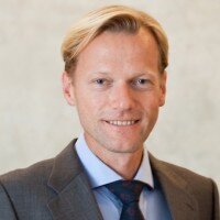
Seit 2016 stellv. Leiter der klinischen Abteilung für Orale Chirurgie und Kieferorthopädie der Univ. Klinik für Zahnmedizin und Mundgesundheit der Medizinischen Universität Graz. Seit 2014 assoziierte Professur an der klinischen Abteilung für Orale Chirurgie und Kieferorthopädie der Univ. Klinik für Zahnmedizin und Mundgesundheit der Medizinischen Universität Graz. 2013: Forschungsaufenthalt an der Klinik für Kronen- und Brückenprothetik und Materialkunde der Universität Zürich, Schweiz. 2012: Gast-Professor am Prince Phillip Dental Hospital, Dept. Implant Dentistry & Oral Rehabilitation, University of Hong Kong
2011: Forschungsaufenthalt an der Klinik für Orale Chirurgie der Heinrich Heine Universität, Düsseldorf. Seit 04/2010: Privatdozent an der klinischen Abteilung für Orale Chirurgie und Kieferorthopädie der Univ. Klinik für Zahnmedizin und Mundgesundheit der Medizinischen Universität Graz. 2009: DGI-APW Implantologie Curriculum (Germany). 2005-2010: Assistent an der klinischen Abteilung für Orale Chirurgie und Kieferorthopädie der Univ. Klinik für Zahnmedizin und Mundgesundheit der medzinischen Universität Graz. 2003-2006: Doktoratstudium der medizinischen Wissenschaften MedUni-Graz. 2004-2005: Postdoctoral research fellow am Department für Zell-Biologie, Histologie und Embryologie, Zentrum für Molekulare Medizin, MedUni Graz. Seit 2004: Privatpraxis in Graz. 1998-2004: Diplomstudium Zahnmedizin, Medizinische Universität Graz. 2000: Forschungsaufenthalt am Department of Maxillofacial Surgery, University of Pittsburgh (UPMC), USA. 1994-2000: Diplomstudium Humanmedizin, Karl-Franzens Universität, Graz.
Veranstaltungen
31st EAO Annual Scientific Meeting
Details make perfection24. Okt. 2024 — 26. Okt. 2024MiCo - Milano Convention Centre, Milano, Italien
Referenten: Bilal Al-Nawas, Gil Alcoforado, Federico Hernández Alfaro, Sofia Aroca, Wael Att, Gustavo Avila-Ortiz, Kathrin Becker, Anne Benhamou, Juan Blanco Carrión, Dieter Bosshardt, Daniel Buser, Francesco Cairo, Paolo Casentini, Raffaele Cavalcanti, Tali Chackartchi, Renato Cocconi, Luca Cordaro, Luca De Stavola, Nuno Sousa Dias, Egon Euwe, Vincent Fehmer, Alberto Fonzar, Helena Francisco, Lukas Fürhauser, German O. Gallucci, Oscar Gonzalez-Martin, Dominik Groß, Robert Haas, Alexis Ioannidis, Simon Storgård Jensen, Ronald Jung, France Lambert, Luca Landi, Georg Mailath-Pokorny jun., Silvia Masiero, Iva Milinkovic, Carlo Monaco, Jose Nart, José M. Navarro, Katja Nelson, Manuel Nienkemper, David Nisand, Michael Payer, Sergio Piano, Bjarni E. Pjetursson, Sven Reich, Isabella Rocchietta, Giuseppe Romeo, Irena Sailer, Mariano Sanz, Ignacio Sanz Martín, Frank Schwarz, Shakeel Shahdad, Massimo Simion, Ralf Smeets, Benedikt Spies, Bogna Stawarczyk, Martina Stefanini, Hendrik Terheyden, Tiziano Testori, Daniel Thoma, Ana Torres Moneu, Piero Venezia, Lukas Waltenberger, Hom-Lay Wang, Stefan Wolfart, Giovanni Zucchelli, Otto Zuhr
European Association for Osseintegration (EAO)
30th EAO Annual Scientific Meeting / 37th DGI Annual Congress
Berlin reloaded28. Sept. 2023 — 30. Sept. 2023CityCube Berlin, Berlin, Deutschland
Referenten: Samir Abou-Ayash, Bilal Al-Nawas, Thomas Bernhart, Florian Beuer, Stefan Bienz, Elena Calciolari, Najla Chebib, Andreas Dengel, Vincent Donker, Joke Duyck, Roberto Farina, Gary Finelle, Alberto Fonzar, Tobias Fretwurst, Rudolf Fürhauser, Oscar Gonzalez-Martin, Stefano Gracis, Knut A. Grötz, Christian Hammächer, Lisa J. A. Heitz-Mayfield, Detlef Hildebrand, Norbert Jakse, Jim Janakievski, Tim Joda, Daniel Jönsson, Gregg Kinzer, Vincent G. Kokich, Michael Krimmel, Cecilia Larsson Wexell, Martin Lorenzoni, Georg Mailath-Pokorny, Julia Mailath-Pokorny, Frank Georg Mathers, Gerry McKenna, Henny Meijer, Alberto Monje, Torsten Mundt, Nadja Nänni, David Nisand, Robert Nölken, Nicole Passia, Michael Payer, Christof Pertl, Aušra Ramanauskaitė, Eik Schiegnitz, Martin Schimmel, Ulrike Schulze-Späte, Frank Schwarz, Falk Schwendicke, Robert Stigler, Michael Stimmelmayr, Anette Strunz, Christian Ulm, Stefan Vandeweghe, Kay Vietor, Arjan Vissink, Asaf Wilensky, Stefan Wolfart, Werner Zechner, Anja Zembic, Nicola Zitzmann
European Association for Osseintegration (EAO)
2nd European Congress Ceramic Implant Dentistry
European Society for Ceramic Implantology20. Okt. 2022 — 22. Okt. 2022Seminarhotel Bocken, Horgen, Schweiz
Referenten: Owen Addison, Bilal Al-Nawas, Rodrigo Beltrão, Michael M. Bornstein, André Chen, Jérôme Chevalier, Jospeh Choukroun, Alessandro Devigus, Michael Gahlert, Shahram Ghanaati, Elisabeth Jacobi-Gresser, Ronald Jung, Ralf J. Kohal, Sebastian Kühl, Frank-Michael Maier, Jochen Mellinghoff, Michael Payer, Mattias Pettersson, Marc Quirynen, Stefan Röhling, Jens Tartsch
European Society for Ceramic Implantology - ESCI
Zeitschriftenbeiträge dieses Autors
The International Journal of Oral & Maxillofacial Implants, 6/2023
DOI: 10.11607/jomi.10284, PubMed-ID: 38085741Seiten: 1105-1114, Sprache: EnglischKarapataki, Sofia / Vegh, Daniel / Payer, Michael / Fahrenholz, Harald / Antonoglou, Georgios N.
Purpose: To assess the clinical performance of a two-piece zirconia implant system, with a focus on biologic complications. Materials and Methods: A total of 39 patients received 91 two-piece zirconia implants. The patients were recruited from two private clinics and were monitored for 5 to 12 years (median: 5.6 years). The primary outcomes were biologic complications, such as peri-implant infections (peri-implant mucositis and peri-implantitis), and the secondary outcome was radiographically evident marginal bone loss (MBL). Results: Three patients (7.7%) with 9 total implants (9.9%) presented with peri-implant mucositis. MBL that did not exceed the first thread was evident at 32 mesial sites (35%) and 25 distal sites (27.4%). MBL exceeding the first thread but not the third thread was evident at 6 mesial and 5 distal sites (thread pitch: 0.7 mm). Only one peri-implant pocket deepened (4 mm) and showed bleeding; however, the estimated MBL did not exceed 1.65 mm. No peri-implantitis occurred, and no implant was lost. Conclusions: This prospective study shows high survival rates and a seemingly low prevalence of biologic and prosthetic complications for this two-piece zirconia implant system over an observation period of up to 12 years.
Schlagwörter: bone resorption, dental implantation, osseointegration, peri-implantitis, two-piece zirconia implants
Titanium oral implants are still considered "state of the art" in implant dentistry, with well-documented survival rates. However, their grayish color and high prevalence of peri-implant infections have resulted in controversial discussion as to whether tooth-like-colored, metal-free zirconia ceramic implants provide sufficient potential to be considered equal regarding treatment outcomes. The present position paper has been composed upon invitation by the European Association of Osseointegration in order to provide an update on the current level of evidence regarding zirconia implants in clinical trials. To date, most available and scientifically documented zirconia implant systems are one-piece implants that require an experienced surgeon and prosthodontist due to the restricted flexibility in cases of compromised angulation or vertical positioning. Taking this limitation into account, there is evidence of a comparable outcome for one-piece zirconia implants compared to titanium implants for the fixed replacement of one to three missing teeth. In contrast, currently available clinical data evaluating two-piece zirconia implants with an adhesively bonded implant-abutment interface suggest an inferior outcome. Data evaluating the clinical applicability of screw-retained solutions, even if revealing sufficient fracture resistance in laboratory investigations, are still missing. High survival rates were reported for all-ceramic reconstructions supported by zirconia implants, but with increased technical complications; ie, fractures of the ceramic veneer in the case of bilayered restorations. Sufficient clinical evidence for recommending monolithic approaches is limited to single crowns.
Aims: While numerous materials are available for sinus floor elevation, plant-based alternatives still hold promise of overcoming concerns about allogeneic or xenogeneic materials. Thus, the present authors designed a randomised clinical trial to histologically compare an almost pure hydroxyapatite (HA) to a biphasic calcium phosphate comprising 80% β-tricalcium phosphate (β-TCP) and 20% hydroxyapatite (β-TCP/HA), all of phycogenic origin.
Materials and methods: Twenty patients scheduled for lateral window sinus floor elevation were randomised to either an HA or a β-TCP/HA group. Biopsy specimens were taken 3 months after sinus floor elevation and during implant surgery after 6 months. One ground section per biopsy specimen (N = 40) was stained, scanned and histomorphometrically analysed for new bone, old bone, soft tissue, graft, bone infiltration of graft, bone-to-graft contact and penetration depth.
Results: At 6 months, more new bone was seen in the β-TCP/HA group (P = 0.011), whereas more residual graft was present and in more extensive contact with new bone in the HA group. More pronounced alterations, and smaller particle sizes, of graft surrounded and infiltrated by bone were seen in the β-TCP/HA group. The less extensive bone-to-graft contact in the β-TCP/ HA group reflected a more advanced state of resorption, while infiltration of residual graft material by bone was also increased in this group.
Conclusions: Proper healing was seen in both groups, with the graft materials guiding the formation of new bone, which grew especially well through the particles of the highly osteoconductive and resorptive β-TCP/HA material. HA was very stable, without significant resorption, but was extensively in contact with new bone after 6 months.
Schlagwörter: biocompatible materials, bone substitutes, calcium phosphates, hydroxyapatites, sinus floor elevation
This study was partially supportedby Dentsply Sirona Implants(I-BI-14-077; Mannheim, Germany) and the materials investigated in the study were providedfree of charge for patients’ use;
Ziel: Die Herstellung individueller Zahnreplikate findet in der Zahnmedizin zwei Anwendungen: autogene Zahntransplantationen und wurzelanaloge Zahnimplantate. Diese Anwendungen erfordern ein besonders hohes Maß an Präzision. Ziel dieser Studie war es, eine Methode zur Herstellung individueller, 3-D-gedruckter Zahnreplikate zu etablieren und zu evaluieren.
Material und Methode: Zehn Patienten, die die Extraktion eines Weisheitszahnes und eine präoperative digitale Volumentomographie (DVT) benötigten, wurden eingeschlossen. Ausschlusskriterien waren eine intraoperative Zerteilung oder Fraktur des Zahnes. 3D Slicer 4.6.2 wurde für die Zahnsegmentierung und Modellgenerierung auf Basis von DVT-Daten verwendet. Die Zahnreplikate wurden durch selektives Laserschmelzen hergestellt. Die extrahierten Zähne und 3-D-gedruckten Replikate wurden 3-D-gescannt und in CloudCompare 2.8.1 auf Oberflächenabweichungen getestet.
Ergebnisse: Die mittlere absolute Oberflächenabweichung zwischen den 3-D-gedruckten Zähnen und den entsprechenden extrahierten Zähnen lag im Bereich von 0,13 bis 0,25 mm mit Standardabweichungen von 0,10 bis 0,21 mm; 95 % der gemessenen Oberflächenpunkte wichen weniger als 0,474 mm ab; Die Oberfläche wurde um -6,0 % und das Volumen um -3,4 % verringert. Der quadratische Mittelwert betrug 0,238 mm und die mittlere maximale absolute Oberflächenabweichung 0,927 mm. Die selektive Laserschmelztechnik zeigte eine hohe Präzision mit einer mittleren absoluten Abweichung von 0,045 mm und einer Standardabweichung von 0,04 mm.
Schlussfolgerung: Auf Basis von DVT-Daten konnten 3-D-gedruckte Zahnreplikate mit sehr hoher Genauigkeit hergestellt werden. Das beschriebene Verfahren eignet sich zur Herstellung von Zahnreplikaten zur Verwendung bei autogenen Zahntransplantationen oder zur Erzeugung individueller wurzelanaloger Implantate.
Schlagwörter: 3-D-Druck, Rapid Prototyping, selektives Laserschmelzen, Zahnreplikate, Digitale Volumentomographie, autologe Zahntransplantation, wurzelanaloge Zahnimplantate, Implantologie
Das Ziel dieser Studie war die Bestimmung des Zellwachstums humaner Osteoblasten auf Polyetheretherketon (PEEK)-Oberflächen mit definierter Rauigkeit und geometrischer Beschaffenheit im Vergleich zu Standardoberflächen aus Titan- und Zirkonoxid.
Methode: Für die Zellexperimente wurden Plättchen mit einem Durchmesser von 14 mm und einer Dicke von 1 mm aus den Materialien Reintitan (cp-Ti), Yttriumoxid-stabilisiertes Zirkoniumdioxid (Y-TZP) und PEEK mit jeweils glatter Oberfläche verwendet. Auf allen drei Oberflächen wurden humane fetale Osteoblasten (hFOB) gezüchtet, um nach einem, drei und sieben Tagen die Zellvitalität, die Laktatdehydrogenase-Freisetzung (LDH), PCNA, Ki67 mRNA-Expression und Vinculin-Expression zu bestimmen.
Ergebnisse: Zelladhäsion und Vitalität waren auf PEEK-Oberflächen im Vergleich zu den Kontrolloberflächen reduziert. Zwischen den Titan- und Zirkonoxid-Oberflächen konnte kein signifikanter Unterschied beobachtet werden.
Schlussfolgerung: Reintitan und Zirkonoxid sind gemäß der Ergebnisse der vorliegenden Untersuchung im Hinblick auf die zellulären Parameter als gleichwertig anzusehen. Hingegen erscheinen bei den untersuchten PEEK-Proben aufgrund der reduzierten Zelladhäsion und Vitalität, vor allem für zukünftige osseointegrative Anwendungsgebiete, noch weitere Untersuchungen mit modifizierten Oberflächen sinnvoll.
Schlagwörter: Biokompatibilität, Osteoblasten, Polyetheretherketon, PEEK, Titan, Zirkonoxid
The aim of the study was to test whether or not, for primary bone augmentation, the use of a xenogenic bone block loaded with rhBMP-2 results in similar bone quantity and quality compared to an autogenous bone block and to evaluate patient morbidity following the surgical procedure with the two treatment modalities. 24 patients requiring implant therapy for the reconstruction of 1 to 4 missing teeth and insufficient bone volume for implant placement were randomly assigned to receive one out of two treatment modalities. Both treatment modalities were successful in regenerating bone to allow for dental implant placement at 4 months. Histologically, a higher amount of mineralised tissue was observed for the control group at 4 months. The use of a second surgical site in the control group tended to a higher patient morbidity compared to the test group, but did not reach statistical significance.
Schlagwörter: dental implants, guided bone regeneration, growth factors, bone morphogenetic protein-2, autogenous bone blocks