We use cookies to enable the functions required for this website, such as login or a shopping cart. You can find more information in our privacy policy.
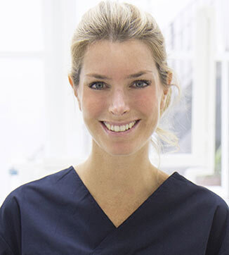
Isabella Rocchietta, DDS, MSc, is an honorary senior research associate to the Department of Periodontology and a lecturer for the Diploma in Implant Dentistry at the UCL Eastman Dental Institute in London. She is an elected board member of the European Academy for Osseointegration and a member of the Experts Council of the Osteology Foundation. She is the author of several national and international peer-reviewed publications and book chapters. Dr Rocchietta performs clinical work limited to periodontics and implant dentistry in London and Milan and lectures internationally.
Events
31st EAO Annual Scientific Meeting
Details make perfection24. Oct 2024 — 26. Oct 2024MiCo - Milano Convention Centre, Milano, Italy
Speakers: Bilal Al-Nawas, Gil Alcoforado, Federico Hernández Alfaro, Sofia Aroca, Wael Att, Gustavo Avila-Ortiz, Kathrin Becker, Anne Benhamou, Juan Blanco Carrión, Dieter Bosshardt, Daniel Buser, Francesco Cairo, Paolo Casentini, Raffaele Cavalcanti, Tali Chackartchi, Renato Cocconi, Luca Cordaro, Luca De Stavola, Nuno Sousa Dias, Egon Euwe, Vincent Fehmer, Alberto Fonzar, Helena Francisco, Lukas Fürhauser, German O. Gallucci, Oscar Gonzalez-Martin, Dominik Groß, Robert Haas, Alexis Ioannidis, Simon Storgård Jensen, Ronald Jung, France Lambert, Luca Landi, Georg Mailath-Pokorny jun., Silvia Masiero, Iva Milinkovic, Carlo Monaco, Jose Nart, José M. Navarro, Katja Nelson, Manuel Nienkemper, David Nisand, Michael Payer, Sergio Piano, Bjarni E. Pjetursson, Sven Reich, Isabella Rocchietta, Giuseppe Romeo, Irena Sailer, Mariano Sanz, Ignacio Sanz Martín, Frank Schwarz, Shakeel Shahdad, Massimo Simion, Ralf Smeets, Benedikt Spies, Bogna Stawarczyk, Martina Stefanini, Hendrik Terheyden, Tiziano Testori, Daniel Thoma, Ana Torres Moneu, Piero Venezia, Lukas Waltenberger, Hom-Lay Wang, Stefan Wolfart, Giovanni Zucchelli, Otto Zuhr
European Association for Osseintegration (EAO)
360 IMPLANTOLOGY by MIS – MIS Global Conference
12. Sep 2024 — 14. Sep 2024Palau de Congressos de Palma, Palma de Mallorca, Spain
Speakers: Tara Aghaloo, Serhat Aslan, Nitzan Bichacho, Darko Božić, Tali Chackartchi, Victor Clavijo, Gustavo Giordani, Moshe Goldstein, Galip Gürel, Stefen Koubi, Hilal Kuday, Alberto Monje, Stavros Pelekanos, Mario Alonso Puig, Ariel J. Raigrodski, Aušra Ramanauskaitė, Isabella Rocchietta, Mariano Sanz, Ignacio Sanz Sànchez, Venceslav Stankov
The 9th World Dental Meeting in Japan 2023
No Dentistry, No Wellness!29. Sep 2023 — 1. Oct 2023Pacifico Yokohama Conference Center, Yokohama, Japan
Speakers: Alessandro Agnini, Andrea Mastrorosa Agnini, Wael Att, Gustavo Avila-Ortiz, Markus B. Blatz, Victor Clavijo, Karim Dada, Glécio Vaz de Campos, Vincent Fehmer, Naoki Hayashi, Mario Imburgia, Guillaume Jouanny, Sérgio Kahn, Bertrand Khayat, Sandra Khong Tai, Christopher Köttgen, Stefen Koubi, Tomas Linkevičius, Nazariy Mykhaylyuk, Ravindra Nanda, Andreas Nolte, Léon Parienté, Jose Manuel Reuss , Domenico Ricucci, Isabella Rocchietta, Irena Sailer, Todd R. Schoenbaum, Werner Schupp, Istvan Urban, Eric Van Dooren, Débora R. Vilaboa, Otto Zuhr
Quintessence Publishing Co. Ltd. Japan
4th Buser & Sculean International Symposium
Regeneration and Esthetics in Periodontology and Implant Dentistry2. Dec 2022 — 3. Dec 2022Congress Center, Kursaal Bern, Bern, Switzerland
Speakers: Sofia Aroca, Daniel Buser, Stephen Chen, Nikolaos Donos, Marc Hürzeler, Sascha Jovanovic, Ronald Jung, Giulio Rasperini, Isabella Rocchietta, Frank Schwarz, Anton Sculean, Shakeel Shahdad, Andreas Stavropoulos, Martina Stefanini, Leonardo Trombelli, Istvan Urban, Giovanni Zucchelli
Buser & Sculean Academy
The 14th International Symposium on Periodontics and Restorative Dentistry (ISPRD)
9. Jun 2022 — 12. Jun 2022Boston Marriott Copley Place, Boston, MA, United States of America
Speakers: Tara Aghaloo, Edward P. Allen, Evanthia Anadioti, Wael Att, Vinay Bhide, Markus B. Blatz, Scotty Bolding, Lorenzo Breschi, Jeff Brucia, Daniel Buser, Luigi Canullo, Daniele Cardaropoli, Stephen J. Chu, Donald Clem, Christian Coachman, Lyndon F. Cooper, Daniel Cullum, Lee Culp, José Carlos Martins da Rosa, Sergio De Paoli, Marco Degidi, Nicholas Dello Russo, Serge Dibart, Joseph P. Fiorellini, Mauro Fradeani, Stuart J. Froum, David Garber, Maria L. Geisinger, William Giannobile, Luca Gobbato, Ueli Grunder, Galip Gürel, Chad Gwaltney, Christoph Hämmerle, Robert A. Horowitz, Marc Hürzeler, David Kim, Gregg Kinzer, Christopher Köttgen, Ina Köttgen, Purnima S. Kumar, Burton Langer, Lydia Legg, Pascal Magne, Kenneth A. Malament, Jay Malmquist, George Mandelaris, Pamela K. McClain, Michael K. McGuire, Mauro Merli, Konrad H. Meyenberg, Craig M. Misch, Julie A. Mitchell, Marc L. Nevins, Myron Nevins, Michael G. Newman, Miguel A. Ortiz, Jacinthe M. Paquette, Stefano Parma-Benfenati, Michael A. Pikos, Giulio Rasperini, Pamela S. Ray, Christopher R. Richardson, Isabella Rocchietta, Marisa Roncati, Marco Ronda, Paul S. Rosen, Maria Emanuel Ryan, Irena Sailer, Maurice Salama, David M. Sarver, Takeshi Sasaki, Todd Scheyer, Massimo Simion, Michael Sonick, Sergio Spinato, Dennis P. Tarnow, Lorenzo Tavelli, Douglas A. Terry, Tiziano Testori, Carlo Tinti, Istvan Urban, Hom-Lay Wang, Robert Winter, Giovanni Zucchelli
Quintessence Publishing Co., Inc. USA
EAO Digital Days
Implantology: Beyond your expectations12. Oct 2021 — 14. Oct 2021online
Speakers: Enrico Agliardi, Alessandro Agnini, Andrea Mastrorosa Agnini, Mauricio Araujo, Goran Benic, Juan Blanco Carrión, Daniel Buser, Raffaele Cavalcanti, Tali Chackartchi, Luca Cordaro, Jan Cosyn, Holger Essig, Vincent Fehmer, Stefan Fickl, Alberto Fonzar, Helena Francisco, German O. Gallucci, Ramin Gomez-Meda, Oscar Gonzalez-Martin, Robert Haas, Arndt Happe, Alexis Ioannidis, Ronald Jung, Niklaus P. Lang, Tomas Linkevičius, Iva Milinkovic, Sven Mühlemann, Katja Nelson, Sergio Piano, Michael A. Pikos, Bjarni E. Pjetursson, Marc Quirynen, Franck Renouard, Isabella Rocchietta, Dennis Rohner, Irena Sailer, Henning Schliephake, Shakeel Shahdad, Massimo Simion, Ali Tahmaseb, Hendrik Terheyden, Jochen Tunkel, Stefan Vandeweghe, Piero Venezia, Stijn Vervaeke, Martin Wanendeya, Georg Watzek, Giovanni Zucchelli
European Association for Osseintegration (EAO)
This author's journal articles
International Journal of Periodontics & Restorative Dentistry, 1/2021
Pages 99-104, Language: EnglishNevins, Myron / Chen, Chia-Yu / Kerr, Eric / Mendoza-Azpur, Gerardo / Isola, Gaetano / Soto, Claudio P. / Stacchi, Claudio / Lombardi, Teresa / Kim, David / Rocchietta, Isabella
The goal of this multicenter randomized controlled study was to evaluate the effectiveness of a newly developed ionic-sonic electric toothbrush in terms of plaque removal and reduction of gingival inflammation. A total of 78 subjects from three dental centers were invited to join the study. They were randomized to receive either a manual toothbrush (control group) or an ionic-sonic electric brush (test group). Full-mouth prophylaxis and oral hygiene instructions based on the stationary bristle technique were provided 1 week prior to the baseline visit. At baseline and at each follow-up appointment, Plaque Index (PI) and Gingival Index (GI) were recorded. In addition, probing depth (PD) and bleeding on probing were recorded at baseline and at the last appointment (week 5). At completion of the study, subjects in the test group were given a questionnaire regarding their satisfaction with the toothbrush. Sixty-four subjects completed the study (control: 28; test: 36). The mean age of the subjects was 36.90 ± 12.19 years. No significant difference between the baseline and 5-week PD was found. Plaque removal efficacy and reduction in gingival inflammation were more significant for the test group at week 2. Both the control and test groups showed statistically significant improvement in PI and GI from baseline to week 5. The ionic-sonic toothbrush was more effective than manual toothbrush after a 1-week application.
This investigation was designed to evaluate the reestablishment of bone-toimplant contact on infected dental implant surfaces following decontamination with an erbium, chromium:yttrium-scandium-gallium-garnet (Er,Cr:YSGG) laser and reconstructive therapy. Three patients presenting with at least one failing implant each were enrolled and consented to treatment with the Er,Cr:YSGG laser surface decontamination and reconstruction with a bone replacement allograft and a collagen membrane. The laser treatment was carried out at a setting of 1.5 W, air/water of 40%/50%, and pulse rate of 30 Hz. At 6 months, all three patients returned for the study. En bloc biopsy samples of four implants were obtained and analyzed. Two patients had excellent clinical outcomes, while one patient with two adjacent failing implants experienced an early implant exposure during the follow-up period. There was histologic evidence of new bone formation with two implant specimens and less bone gain with the others. Despite the small sample size, these were optimistic findings that suggested a positive role of Er,Cr:YSGG laser in debridement of a titanium implant surface to facilitate subsequent regenerative treatment. This investigation provides histologic evidence as well as encouraging clinical results that use of the Er,Cr:YSGG laser can be beneficial for treatment of peri-implantitis, but further long-term clinical studies are needed to investigate the treatment outcome obtained.
The objective of this proof-of-principle multicenter case series was to examine the bone regenerative potential of a newly introduced equine-derived bone mineral matrix (Equimatrix) to provide human sinus augmentation for the purpose of implant placement in the posterior maxilla. There were 10 patients requiring 12 maxillary sinus augmentations enrolled in this study. Histologic results at 6 months demonstrated abundant amounts of vital new bone in intimate contact with residual graft particles. Active bridging between residual graft particles with newly regenerated bone was routinely observed in intact core specimens. A mean value of 23.4% vital bone formation was observed at 6 months. This compared favorably with previous results using xenografts to produce bone in the maxillary sinus for the purpose of dental implant placement. Both the qualitative and quantitative results of this case series suggest comparable bone regenerative results at 6 months to bovine-derived xenografts.
Soft tissue augmentation around dental implants in the esthetic region remains a challenging and unpredictable procedure. The ideal surgical technique would include of an off-the-shelf product to minimize morbidity after autogenous grafting procedures. The aim of this study was to use a resorbable collagen matrix (Mucograft) to serve as a scaffold to recombinant human platelet-derived growth factor BB (rhPDGF-BB) to increase peri-implant soft tissue volume in anterior maxillary sites. A total of six patients who had previously undergone a bone regeneration procedure were included in this study. The collagen matrix was applied during stage-two surgery (expanded polytetrafluoroethylene membrane removal and implant placement). Measurements were performed through customized stents by means of endodontic files, and at abutment connection, a soft tissue biopsy specimen was harvested for histologic examination. The healing period was uneventful in all six patients. Measurements were taken apically, centrally, and occlusally for each site. The mean gains in volume from baseline to the 4-month measurement at the apical, central, and occlusal aspects were 0.87 ± 2.13 mm, 2.14 ± 3.27 mm, and 0.35 ± 3.20 mm, respectively. The results showed a moderate increase in the soft tissue volume in esthetic peri-implant sites when applying a collagen matrix infused with rhPDGF-BB. However, the measuring techniques available need to be further improved to record exact changes in the soft tissue volume.
Autogenous soft tissue augmentation procedures around natural teeth and dental implants are performed daily by clinicians. However, patient morbidity is often associated with the second surgical site; hence, research is moving toward an era where matrices may substitute autogenous grafts. The aim of this study was to assess the soft tissue response to a collagen matrix in an animal model. Nine pigs were included in this study. Each animal received four collagen matrices, two for each mandible. Three cohorts were included in the study: group A, where the matrix was applied as an onlay on a partial-thickness flap; group B, where the matrix was inserted under a partial-thickness flap; and group C, where the matrix was inserted in an inverted position under a full-thickness flap. Sacrifice occurred at 7, 15, and 30 days postoperatively for histologic assessment. The collagen matrix was seen in place for the first 2 weeks, and it was completely replaced by healthy connective tissue within 30 days in the inlay cohorts. No inflammatory adverse reactions were noticed in any specimen, resulting in optimal integration of the device. This study showed an optimal integration within 30 days postoperative of the placement of experimental collagen matrix in the soft tissues of an animal model. Its proven safety in this model provides an optimal starting point for further research projects considering its clinical applications.
International Journal of Periodontics & Restorative Dentistry, 3/2011
PubMed ID (PMID): 21556379Pages 227-235, Language: EnglishNevins, Myron / Camelo, Marcelo / De Angelis, Nicola / Hanratty, James J. / Khang, Wahn G. / Kwon, Jong-Jin / Rasperini, Giulio / Rocchietta, Isabella / Schupbach, Peter / Kim, David M.
The objective of this study was to investigate the potential of xenograft (cancellous bovine bone) granules to form vital bone in non-natural boneforming areas of maxillary sinuses. Fourteen sinus augmentations were performed in 14 patients. Surgical outcomes were uneventful, and sufficient radiopaque volume was present radiographically to place dental implants in all sites. Clinical reentry at 6 months revealed bone formation at the osteotomy site. Histologic evaluation of the obtained bone cores revealed that xenograft granules were integrated and surrounded by woven bone and lamellar bone that were in close contact with the particles. The average percentage of newly formed bone at 6 months was 27.5% ± 8.9%. Vital bone formation using the xenograft granules was supported by both clinical and histologic evidence.
The goal of classifying complications in guided bone regeneration procedures with nonresorbable membranes is to provide the clinician with an instrument for easier identification of both the problem and treatment modality. A standardized terminology represents a key point for proper communication among clinicians and provides guidelines for managing these drawbacks.
This preclinical study evaluated the efficacy of purified recombinant human platelet-derived growth factor (rhPDGF-BB), combined with a novel equine hydroxyapatite and collagen (eHAC) bone block, in providing vertical bone regeneration in critical-size defects simulating localized mandibular alveolar bone atrophy. In addition, the impact of barrier membrane placement in growth factor-mediated bone regeneration was also studied. Bilateral posterior mandibular defects simulating severe localized bony atrophy were created in 12 adult foxhounds following removal of all four mandibular premolars. Three months later, the defects were grafted as follows: group A: eHAC block alone; group B: eHAC block + collagen membrane; group C: eHAC block + rhPDGF-BB; group D: eHAC block + rhPDGF-BB + membrane. The animals were sacrificed after 5 months and the grafted areas were examined histologically, radiographically, and clinically. Groups A and B (controls) exhibited little to no vertical bone regeneration. Group C demonstrated significant vertical bone regeneration, with dense, well-vascularized bone, high bone-to-implant contact, and accelerated replacement of graft particles with newly formed bone. In group D, with the imposition of a barrier membrane, robust bone regeneration was less evident when compared to group C. As in the first study in this series, the importance of the periosteum as a source of osteoprogenitor cells in growth factor-mediated regenerative procedures is examined.
The present investigation was performed to compare the biocompatibility, safety, and manageability of a newly developed bone block and a deproteinized bovine bone block (Bio-Oss) for the treatment of localized bone defects in a dog model. Two male beagle dogs were used for this study. The mandibular premolars were extracted and two saddle-type defects were created bilaterally in the edentulous area. The defects were filled according to a randomized design with Bio-Oss bone block or with an equine hydroxyapatite plus collagen bone block (eHAC). Most control and test sites developed dehiscences during healing. After 4 weeks, the animals were euthanized and each hemimandible was prepared for histologic examination. No significant difference in terms of local tolerance was observed between test and control sites, and test and control sites showed similar histologic findings. However, a significant difference was noticed between the Bio-Oss block and the new bone block in terms of manageability.
Objectives: The aim of this cohort study was to evaluate the safety and the acceptability of an electric toothbrush used on the peri-implant mucosa of implants placed in the aesthetic area.
Methods: One hundred consecutive patients rehabilitated with implants positioned in the maxillary aesthetic area were recruited. Implants had to be restored at least 6 months prior to baseline. At baseline, subjects were provided with Oral-B Professional Care 7000 and received appropriate instructions to brush twice a day over a 12-month period. Papillary bleeding index, recession and probing depth were measured at baseline and at 3, 6, and 12 months.
Results: Ninety-eight (98) patients completed the study. There was an overall reduction of recession (mean 0.2 mm) of borderline statistical significance. All of the changes occurred at the first followup visit (P=0.09) and persisted thereafter. The statistical analyses regarding the probing depth found a highly significant decrease over time (mean 0.3 mm). The bleeding score showed a gradual decrease over time, with a reduction at 12 months by more than half (0.65) in comparison with the baseline (1.50) and was shown to be highly significant (Wilcoxon sign-rank test: P0.001). No patient showed adverse effects such as ulcerations or desquamation. A high score of satisfaction by the patients using the electric toothbrush was reported (94% would continue to use it).
Conclusion: The electric toothbrush Oral B Professional Care 7000 appears to be safe for patients with fixed prosthesis on implants in aesthetic areas. Successive randomised clinical trials are needed to compare this instrument with other therapeutic devices for mechanical plaque control.
Keywords: electric toothbrush, implant, maintenance, peri-implant mucosa, powered toothbrush