We use cookies to enable the functions required for this website, such as login or a shopping cart. You can find more information in our privacy policy.
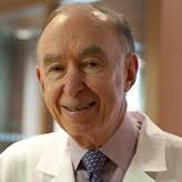
Burton Langer, DMD, MScD, was one of the first periodontists trained in osseointegration by Professor Per-Ingvar Brånemark. He has lectured extensively throughout the world and has written over 50 articles and chapters for ten textbooks. He is the recipient of the 1992 Isador Hirschfeld Award for Clinical Excellence, the 1997 American Academy of Periodontology Master Clinician Award, and the 2015 Robert Eskow New York University College of Dentistry/Implant Dentistry Award. Dr Langer was named an honorary member of the American College of Prosthodontists in 2016.
Events
The 14th International Symposium on Periodontics and Restorative Dentistry (ISPRD)
9. Jun 2022 — 12. Jun 2022Boston Marriott Copley Place, Boston, MA, United States of America
Speakers: Tara Aghaloo, Edward P. Allen, Evanthia Anadioti, Wael Att, Vinay Bhide, Markus B. Blatz, Scotty Bolding, Lorenzo Breschi, Jeff Brucia, Daniel Buser, Luigi Canullo, Daniele Cardaropoli, Stephen J. Chu, Donald Clem, Christian Coachman, Lyndon F. Cooper, Daniel Cullum, Lee Culp, José Carlos Martins da Rosa, Sergio De Paoli, Marco Degidi, Nicholas Dello Russo, Serge Dibart, Joseph P. Fiorellini, Mauro Fradeani, Stuart J. Froum, David Garber, Maria L. Geisinger, William Giannobile, Luca Gobbato, Ueli Grunder, Galip Gürel, Chad Gwaltney, Christoph Hämmerle, Robert A. Horowitz, Marc Hürzeler, David Kim, Gregg Kinzer, Christopher Köttgen, Ina Köttgen, Purnima S. Kumar, Burton Langer, Lydia Legg, Pascal Magne, Kenneth A. Malament, Jay Malmquist, George Mandelaris, Pamela K. McClain, Michael K. McGuire, Mauro Merli, Konrad H. Meyenberg, Craig M. Misch, Julie A. Mitchell, Marc L. Nevins, Myron Nevins, Michael G. Newman, Miguel A. Ortiz, Jacinthe M. Paquette, Stefano Parma-Benfenati, Michael A. Pikos, Giulio Rasperini, Pamela S. Ray, Christopher R. Richardson, Isabella Rocchietta, Marisa Roncati, Marco Ronda, Paul S. Rosen, Maria Emanuel Ryan, Irena Sailer, Maurice Salama, David M. Sarver, Takeshi Sasaki, Todd Scheyer, Massimo Simion, Michael Sonick, Sergio Spinato, Dennis P. Tarnow, Lorenzo Tavelli, Douglas A. Terry, Tiziano Testori, Carlo Tinti, Istvan Urban, Hom-Lay Wang, Robert Winter, Giovanni Zucchelli
Quintessence Publishing Co., Inc. USA
This author's journal articles
International Journal of Periodontics & Restorative Dentistry, 3/2015
There has been renewed interest in intentionally placing dental implants in proximity to or in contact with tooth root fragments. In clinical practice, human teeth are usually extracted due to nonrestorable caries, vertical or horizontal root fractures, periodontal disease, or endodontic failure, which is commonly accompanied by inflammation and bacterial contamination. The aim of this case series is to present the adverse effects in humans of clinically undetected root-to-implant contact (CURIC), where implants were unintentionally placed in proximity to undetected retained root fragments. The adverse effects of small (3 to 5 mm) root fragments were detectible 6 to 48 months post implant placement. Three out of seven implants in six patients were removed due to severe coronal bone loss. This differs from retrograde peri-implantitis, where only the apical area of the implant is affected and the coronal portion remains integrated. The detrimental effect of root fragment-to-implant contact is described along with its clinical management. Based on the review of currently relevant data, mixed results have been documented regarding the success of dental implants in proximity to tooth-root fragments. Careful evaluation of long-term, postloading results in humans where hopeless teeth have been extracted due to infection and significant bone loss are required before intentional root fragment retention is considered a safe and reliable clinical option for implant placement.
Since the introduction of implants into the armamentarium of the dental profession, periodontal disease is often overlooked or not treated appropriately. Patients with early or moderate disease that could be successfully treated with a positive long-term prognosis are allowed to smolder until they are virtually hopeless.
Most patients prefer to preserve their own dentition as opposed to extractions and implants even if it requires periodontal surgery. This is especially true when we consider the regenerative possibilities that periodontists have to offer. Do not lose sight that our primary objective is to achieve optimum periodontal health using the most conservative measures, which may mean proper oral hygiene, subgingival scaling, and pocket elimination where possible, so that ancillary personnel can maintain this health with routine maintenance visits.
The periodontal pocket is likely the most frequently encountered dental malady, but the treatment regimes run the course of paradox. Recently, an auditorium audience filled to the brim with clinicians expressing a keen interest in periodontology was asked if they would prefer to have significant pocket depth in their own dentitions. "Of course not," the incredulous audience proclaimed. Yet, how many practices have a double standard when contrasting their personal dental health with that of their patients?
If one accepts the bacteriologic roles in the etiology of periodontal disease and is aware of the investigations conducted by Socransky and associates as to the presence or absence of periodontal pathogens (the red complex) located in the pocket, it would appear that the conclusions would be self-evident.
The endpoint goal of all periodontal treatment should be the creation of an environment that the patient and hygienist can maintain. Dentitions exhibiting deep pocketing with compromised alveolar support are evidence of the patient's susceptibility to disease. Accepting the results of "soft tissue" therapies that do not result in a cleansable environment but provide pink, nonbleeding gingiva is only the first plateau of treatment. If the patient can't floss the depth of the probing and the hygienist cannot remove the accretions, the problem is not solved. All probing depths greater than 5 mm require more sophisticated analysis and treatment.
The naysayers proclaim that this is an impossible goal or not lucrative, but are health care goals really impossible or beyond the knowledge of our therapies? W. Somerset Maughan wrote: "It's a funny thing about life; if you refuse to accept anything but the best, you very often get it."
The obstacles to be encountered are predictable: the third parties will be aghast and patients wish to avoid surgery. Those who wait for statistical evidence will obviate the need to reach the previously stated endpoint goals. The manufacturers of products designed as alternatives to surgery would double their efforts to influence the clinical decisions of the less educated practitioners.
Dentistry has come a long way in the last two decades. Let us maintain the momentum for the continued benefit of our patients.
Thus, the litmus test is: What would I do if this were my own dentition?
This article reports on the efficacy of a technique involving expansion of edentulous ridges of less than 3 mm in width by means of a planned green stick fracture of the labial plate and simultaneous implant placement without the need for membranes. This retrospective case series includes long-term results from 21 patients with 36 sites and 37 implants with a mean follow-up of 4 years, 5 months from the date of restoration. Freeze-dried bone allograft was used in 22 sites (61%) to augment the ridge. Reentry at stage-two surgery confirmed the preservation of the displaced labial plate after implant integration. Three implants were removed prior to the planned uncovering because of incomplete healing of the overlying gingival tissue; therefore, the survival rate of the labial advancement was 92%. No implants failed after definitive prosthetic loading; therefore, the cumulative survival rate of loaded implants was 100%. No significant bone loss was detected at the final follow-up visit. Follow-up after loading ranged from 9 to 148 months. Advancement of the labial plate with simultaneous implant placement to gain horizontal ridge width dimension was shown to be a reliable and practical procedure for single-tooth sites where other grafting methods are often difficult.
This retrospective case series presents results from eight patients treated with demineralized freeze-dried bone allograft particles and barrier membranes using either miniscrews or implants to support the membrane in seven patients. In all patients, the amount of vertical bone regeneration enabled placement of one or more implants in the graft sites, followed by loading with definitive prostheses at least 5.5 months after implant placement. Marginal bone heights around the implants have remained stable throughout 4 to 13 years of follow-up.
The aim of this report was to describe the bone tissue response to Brånemark oral implants retrieved from patients. The material consisted of consecutively received Brånemark threaded oral implants and related patient data provided by clinicians. The implant samples were processed into undecalcified sections for evaluation under the light microscope. The analysis demonstrated a lower percentage of bone-to-implant contact for the unloaded implants as compared to the loaded implants. When the threads were divided into four different regions, the loaded implants had a lower percentage of bone-contacting length at the thread top as compared to the other three regions.
Thirty-nine patients with implant fractures treated by three of the authors have been analyzed as to probable causes. Thirty-five (90%) of the fractures occurred in the posterior region. Thirty (77%) of the prostheses were supported by one or two implants, which were exposed to a combination of cantilever load magnification and bruxism or heavy occlusal forces. It was concluded that prostheses supported by one or two implants and replacing missing posterior teeth are subjected to an increased risk of bending overload. The literature review indicates that the fracture frequency is low in these situations and this study demonstrates that with appropriate treatment planning, such overload situations can essentially be prevented.
Keywords: bending overload, bone resorption, implant fracture, mechanical problems, osseointegrated implant
This paper describes a new 5.0-mm-diameter self-tapping implant, a modification of the standard Brånemark fixture designed to increase the amount of bone-to-titanium surface contact and to capture dense bone at the lateral and crestal borders of the alveolus. Indications for its use include (1) areas of inadequate bone height, (2) areas of poor bone quality (type IV), and (3) immediate replacement of non-integrated or fractured implants. This preliminary report discusses the design alterations and surgical techniques available to treat these difficult bone situations.
Keywords: 5.0-mm fixture, osseointegration, rescue fixture, type IV bone, wide fixture
This long-term study has demonstrated the successful use of osseointegrated implants to replace posterior teeth in the partially dentate patient. A total of 1,203 Nobelpharma implants placed by two periodontists practicing in traditional office settings were included in the survey. Of the 551 implants placed in the mandible, 25 failed, for a success rate of 95.5%. Of the 247 mandibular prostheses fabricated for 200 mandibles, 8 failed, for a prosthesis stability rate exceeding 97%. Of the 652 implants placed in the maxilla, 31 failed, for a success rate of 95.2%. Of the 250 maxillary prostheses fabricated for 193 maxillae, 2 failed, for a success rate exceeding 99%. The results compare favorably to previous reports in terms of implant survival and stability.
Keywords: implant prosthesis stability, mandibular posterior implants, maxillary posterior implants, osseointegration, retrospective clinical study