We use cookies to enable the functions required for this website, such as login or a shopping cart. You can find more information in our privacy policy.
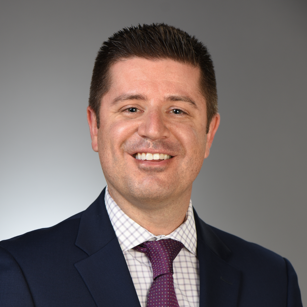
Dr. Christopher Barwacz is an Associate Professor with dual appointments in the Department of Family Dentistry and the Craniofacial Clinical Research Program (CCRP) at The University of Iowa College of Dentistry & Dental Clinics. Dr. Barwacz received his DDS degree from The University of Iowa College of Dentistry & Dental Clinics. He obtained his AEGD Certificate from Baylor College of Dentistry, Texas A&M Health Science Center, and subsequently completed an Implant Fellowship at The University of Iowa College of Dentistry & Dental Clinics. Dr. Barwacz currently serves as the Implant Program Director in The Department of Family Dentistry. He is a Fellow in The Academy of General Dentistry, a Fellow in The International College of Dentists, serves on the Academy of Osseointegration’s Fellowship & Certificate Steering Committee, and is a member of the International Association for Dental Research, the Academy of General Dentistry, the American Dental Association, and Omicron Kappa Upsilon (OKU). Dr. Barwacz has clinical translational research funding in oral implantology, specifically related to transmucosal factors that optimize implant prosthetic and mucosal outcomes. He maintains an interdisciplinary faculty intramural practice limited to restorative and implant dentistry at The University of Iowa College of Dentistry & Dental Clinics.
The International Journal of Oral & Maxillofacial Implants, 5/2021
DOI: 10.11607/jomi.8840Pages 966-976e, Language: EnglishBarwacz, Christopher A / Shah, Kumar C / Bittner, Nurit / Parker, William / Neumeier, Toni T / Thalji, Ghadeer N / De Kok, Ingeborg J
Purpose: To evaluate the clinical, radiographic, and prosthetic outcomes of a single custom CAD/CAM abutment system on implants from four manufacturers, with a mean clinical service of 4 years in a retrospective, multicenter case series study.
Materials and methods: Adult subjects (n = 142) previously restored with titanium or gold-shaded titanium nitride custom CAD/CAM abutments (Atlantis, Dentsply Sirona; n = 259) connected to implants from four different manufacturers (Dentsply Sirona Implants [AT], Biomet 3i [BM], Nobel Biocare [NB], Straumann [ST]), irrespective of implant-abutment interface, and replacing one or more teeth in any position were recalled for a single-visit examination by calibrated investigators at six university clinics. The primary outcomes evaluated included abutment success and survival; secondary outcomes included assessment of papillae fill, probing pocket depth (PD), bleeding on probing (BOP), marginal bone levels (MBLs), and patient-reported outcome measures (PROMs).
Results: Two-hundred fifty-five (98.5%) abutments supported cement-retained restorations, and four (1.5%) abutments supported screw-retained restorations. Forty-two patients had 64 AT implants (25%), 31 patients had 61 BM implants (24%), 26 patients had 50 NB implants (19%), and 43 patients had 84 ST implants (32%). The overall implant-CAD/CAM abutment success rate was 92.66% (95% CI: 88.78%, 95.53%) for all implants examined, and the survival rate was 98.84% (95% CI: 96.65%, 99.76%) for all implants evaluated. The Jemt papillae index demonstrated a minimum of at least 50% to complete fill of the interproximal papillae in 65.0% of sites. PDs had a median value of 3.0 mm, and BOP was present on 19% of abutment surfaces. Mean MBL changes from the time of placement to exam were -0.24 ± 0.99 mm (-4.9 to 2.3; P = .058) for mesial sites and -0.32 ± 0.96 mm (-4.9 to 2.1; P = .000) for distal sites. PROMs expressed 93% of patients reporting good to very good masticatory function, 97% of patients reporting being satisfied or very satisfied with their esthetic outcomes, and 94% of patients indicating that they were satisfied or very satisfied with their overall implant-restorative outcomes.
Conclusion: This retrospective, multicenter clinical study of FDA 510k-approved titanium CAD/CAM abutments of a single manufacturer on multiple implant systems demonstrated high levels of success and survival as well as stable peri-implant tissue outcomes, reflected by overwhelmingly positive PROMs.
Keywords: clinical trial, custom abutment, implant-abutment interface, patient outcomes
Purpose: Simplified and predictable immediate single-tooth replacement therapy can be optimised by leveraging recent advances in digital workflows that combine static surgical guidance and customised prosthesis design and fabrication. The present case report illustrates a novel approach to successfully executing immediate single-tooth replacement therapy via static computer-aided immediate implant placement and digital provisionalisation utilising angulated screw access, with a 1-year follow-up after delivery of the definitive restoration.
Materials and methods: A healthy 27-year-old woman with congenital agenesis of the mandibular second premolars and who required replacement of the mandibular left second premolar underwent immediate single-tooth replacement therapy via computer-aided, restrictive implant placement and provisionalisation using custom prosthetic components designed prior to the surgical intervention being performed.
Results: After a 6-month healing period, the provisional crown was replaced with the definitive restoration. After 1 year, the peri-implant keratinised mucosa demonstrated vertical gain in the mid-buccal portion and stability in the interproximal aspects, and no changes in marginal bone level were observed.
Conclusions: The present case report demonstrates the successful application of novel digital tools to facilitate both surgical and prosthetic aspects of immediate single-tooth replacement therapy. Digital workflows can optimise the clinical efficiency and predictability of tooth replacement therapy with dental implants by reducing surgical and restorative chair time and increasing patient satisfaction.
Conflict-of-interest statement: The authors declare there are no conflicts of interest relating to this study.
Keywords: CAD/CAM, computer-aided surgery, dental implant, digital dentistry, guided implant surgery, immediate loading, immediate placement, prosthetic procedure, prosthodontics
This study evaluated a panel of clinical, dimensional, volumetric, implant-related, histomorphometric, and patient-reported outcome measures (PROMs) following reconstruction of dehiscence defects in extraction sockets with a minimally invasive technique using particulate bone allograft and a nonresorbable dense polytetrafluoroethylene (dPTFE) membrane. Subjects (n = 17) presenting severe buccal dehiscence defects at the time of single-rooted tooth extraction participated in the study. The mean vertical dimension of the dehiscence defects at baseline was 5.76 ± 4.23 mm. Subjects were followed up at 1, 2, 5, and 20 weeks postoperatively. The dPTFE barrier was gently removed at 5 weeks. CBCT and intraoral scans were obtained at baseline and at 20 weeks. A bone core biopsy sample was harvested at 24 weeks (before implant placement). Linear radiographic measurements revealed a mean increase in buccal bone height from baseline to 20 weeks (5.66 ± 5.1 mm; P < .0001). A total alveolar bone volume gain of 9.12% was observed. Although approximately half of the sites required some degree of additional bone augmentation at the time of implant placement, all implants were placed in a favorable restorative position with adequate primary stability. Histomorphometric analyses revealed a mean mineralized tissue area of 31.04% ± 15.22%, and the proportions of remaining allograft material and nonmineralized tissue were 16.23% ± 10.63% and 52.71% ± 9.53%, respectively. All implants survived up to 12 months after placement. PROMs were compatible with minimal discomfort at different postoperative stages and a high level of overall satisfaction upon study completion. This study demonstrated that the reconstructive procedure employed was successful and predictable in treating large, postextraction alveolar ridge deformities to optimize tooth replacement therapy with implant-supported prostheses.
This study aimed to characterize extraction sockets based on indirect digital root analysis. The outcomes of interest were estimated socket volume and dimensions of the socket orifice. A total of 420 extracted teeth, constituting 15 complete sets of permanent teeth (except third molars), were selected. Teeth were scanned to obtain STL files of the root complex for digital analysis. After digitally sectioning each root 2.0 mm apical to the cementoenamel junction (CEJ), root volume was measured in mm3 and converted to cc. Subsequently, a horizontal section plane was drawn at the most zenithal level of the buccal CEJ, and the surface area (in mm2) and buccolingual and mesiodistal linear measurements of the socket orifice (in mm) were computed. Maxillary first molars exhibited the largest mean root volume (0.451 ± 0.096 cc) and mandibular central incisors the smallest (0.106 ± 0.02 cc). Surface area analysis demonstrated that mandibular first molars presented the largest socket orifice area (78.56 ± 10.44 mm2), with mandibular central incisors presenting the smallest area (17.45 ± 1.82 mm2). Maxillary first molars showed the largest mean socket orifice buccolingual dimension (11.08 ± 0.60 mm), and mandibular first molars showed the largest mean mesiodistal dimension (9.73 ± 0.84 mm). Mandibular central incisors exhibited the smallest mean buccolingual (5.87 ± 0.26 mm) and mesiodistal (3.52 ± 0.24 mm) linear dimensions. Findings from this study can be used by clinicians to efficiently plan extraction-site management procedures (such as alveolar ridge preservation via socket grafting and sealing) and implant provisionalization therapy, and by the industry to design products that facilitate site-specific execution of these interventions.
The International Journal of Oral & Maxillofacial Implants, 5/2018
DOI: 10.11607/jomi.6659, PubMed ID (PMID): 30231101Pages 1126-1135, Language: EnglishBarwacz, Christopher A. / Stanford, Clark M. / Diehl, Ursula A. / Cooper, Lyndon F. / Feine, Jocelyne / McGuire, Michael / Scheyer, E. Todd
Purpose: To evaluate the influence that three different implant-abutment interface designs had on periimplant mucosal outcomes as assessed by the pink esthetic score (PES) 3 years after delayed implant placement and immediate provisionalization.
Materials and Methods: Adult subjects (n = 141) requiring replacement of a bounded single tooth in the anterior maxilla as well as first premolar sites were randomized to receive one of three unique implant-abutment interface designs (conical interface [CI]; flat-to-flat interface [FI]; or platform-switch interface [PS]). Treatment included immediate provisionalization with prefabricated titanium abutments, followed by custom computer-aided design/computer-aided manufacturing (CAD/CAM) zirconia abutments and cement-retained, all-ceramic crowns delivered after 12 weeks. Bilateral (anterior sites) or unilateral (premolar sites) digital clinical photographs were made at 1, 3, 6, 12, 24, and 36 months post-implant placement. Five calibrated faculty evaluators who previously scored the 1-year PES image dataset scored the 24- and 36-month photographs using a digital, cloud-based tablet interface.
Results: Six hundred ten clinical photographs were evaluated, resulting in a total of 3,050 sum PES values and 21,350 individual PES values. Faculty evaluator intrarater and interrater reliability were found to be "substantial," with intraclass correlation coefficient (ICC) values of 0.76 and 0.77, respectively. All three implant-abutment interface groups demonstrated acceptable esthetics at 3 years (mean sum PES = 10.1 ± 1.9, 4.0 to 13.2), with no single group demonstrating significantly greater mean sum PES values than another at the 3-year follow-up or at any recall interval in between.
Conclusion: No significant differences were observed in mean sum PES scores for subjects randomized to one of three different implant-abutment interface geometries. Within the limitations of this study thus far, the first 6 months following definitive prosthesis delivery appear to still be the most significant with regard to improvement in PES outcomes for all three treatment groups.
Keywords: esthetics, immediate provisionalization, implant-abutment interface, peri-implant mucosa