We use cookies to enable the functions required for this website, such as login or a shopping cart. You can find more information in our privacy policy.
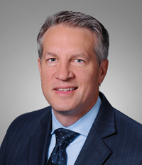
William Giannobile, DDS, MS, DMSc, is dean of the Harvard School of Dental Medicine and a professor in the Department of Oral Medicine, Infection, and Immunity. He has produced over 300 manuscripts, textbook chapters, and patents focused on periodontology, regenerative medicine, and oral health research and is coeditor of nine books focused on clinical, translational research, periodontology, and regenerative medicine. He is a consultant to the US Food and Drug Administration for dental devices and has received numerous awards, including the American Dental Association Norton Ross Award for Excellence in Clinical Research. In addition to his administrative and teaching responsibilities, he practices periodontics and implant dentistry at the Harvard Dental Center.
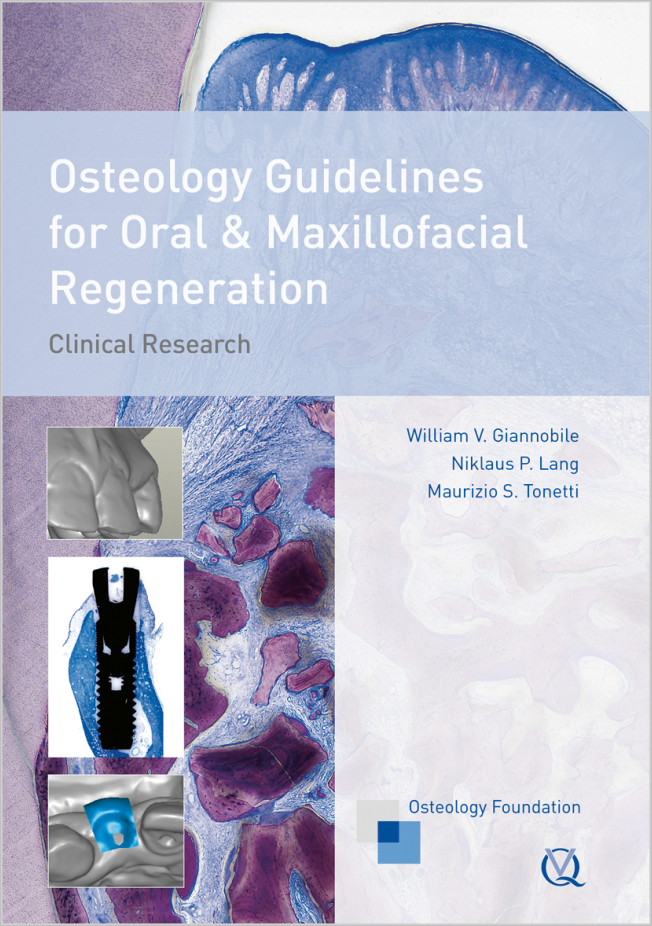
1st Edition 2011 Book Hardcover, 256 pages, 235 illus colored Language: English Categories: Oral/Maxillofacial Surgery, Oral Surgery ISBN 978-1-85097-211-2 QP Deutschland
15. Jun 2022 — 18. Jun 2022Bella Center Copenhagen, Copenhagen, Denmark
Speakers: Mario Aimetti, Zvi Artzi, Serhat Aslan, Georgios Belibasakis, Florian Beuer, Juan Blanco Carrión, Michael M. Bornstein, Nagihan Bostanci, Philippe Bouchard, Darko Božić, Olivier Carcuac, Maria Clotilde Carra, Nelson Carranza, Iain L. C. Chapple, Pierpaolo Cortellini, Jan Cosyn, Mike Curtis, Francesco D'Aiuto, Bettina Dannewitz, Luca De Stavola, Jan Derks, Nikolaos Donos, Peter Eickholz, Bahar Eren Kuru, Ricardo Faria Almeida, Roberto Farina, Magda Feres, Elena Figuero, Dagmar Fosså Bunæs, Rok Gašperšič, William Giannobile, Cecilie Gjerde Gjengedal, Moshe Goldstein, Marjolaine Gosset, Klaus Gotfredsen, Filippo Graziani, Adrian Guerrero, George Hajishengallis, Hady Haririan, Lisa J. A. Heitz-Mayfield, Palle Holmstrup, Marc Hürzeler, Mark Ide, Søren Jepsen, Ronald Jung, Sérgio Kahn, Anhgela R. Kamer, Alpdogan Kantarci, Moritz Kebschull, Björn Klinge, Thomas Kocher, Odd Carsten Koldsland, Kenneth Kornman, Marja Laine, Markus Laky, Isabelle Laleman, Evanthia Lalla, France Lambert, Luca Landi, Niklaus P. Lang, Antonio Liñares, Tomas Linkevičius, Bruno Loos, Rodrigo Lopez, Eli Machtei, Aslan Mammadov, Mauro Merli, Andrea Mombelli, Eduardo Montero, Niki Moutsopoulos, Jose Nart, Gustavo G. Nascimento, Ian Needleman, Tiernan O'Brien, William Papaioannou, Panos N. Papapanou, Michael A. Pikos, Pawel Plakwicz, Constanza Pontarolo, Philip M. Preshaw, Marc Quirynen, Mia Rakic, Christoph Andreas Ramseier, Hélène Rangé, Papageorgiou Spyridon, Maurizio S. Tonetti, Leonardo Trombelli, Istvan Urban, Fridus van der Weijden, Fabio Vignoletti, Charalambos Vlachopoulos, Nicola West, Asaf Wilensky, Ion Zabalegui, Egija Zaura, Nicola Zitzmann, Giovanni Zucchelli, Otto Zuhr, Fardal Øystein
European Federation of Periodontology (EFP)
The 14th International Symposium on Periodontics and Restorative Dentistry (ISPRD)
9. Jun 2022 — 12. Jun 2022Boston Marriott Copley Place, Boston, MA, United States of America
Speakers: Tara Aghaloo, Edward P. Allen, Evanthia Anadioti, Wael Att, Vinay Bhide, Markus Blatz, Scotty Bolding, Lorenzo Breschi, Jeff Brucia, Daniel Buser, Luigi Canullo, Daniele Cardaropoli, Stephen J. Chu, Donald Clem, Christian Coachman, Lyndon F. Cooper, Daniel Cullum, Lee Culp, José Carlos Martins da Rosa, Sergio De Paoli, Marco Degidi, Nicholas Dello Russo, Serge Dibart, Joseph P. Fiorellini, Mauro Fradeani, Stuart J. Froum, David Garber, Maria L. Geisinger, William Giannobile, Luca Gobbato, Ueli Grunder, Galip Gürel, Chad Gwaltney, Christoph Hämmerle, Robert A. Horowitz, Marc Hürzeler, David Kim, Gregg Kinzer, Christopher Köttgen, Ina Köttgen, Purnima S. Kumar, Burton Langer, Lydia Legg, Pascal Magne, Kenneth A. Malament, Jay Malmquist, George Mandelaris, Pamela K. McClain, Michael K. McGuire, Mauro Merli, Konrad H. Meyenberg, Craig M. Misch, Julie A. Mitchell, Marc L. Nevins, Myron Nevins, Michael G. Newman, Miguel A. Ortiz, Jacinthe M. Paquette, Stefano Parma-Benfenati, Michael A. Pikos, Giulio Rasperini, Pamela S. Ray, Christopher R. Richardson, Isabella Rocchietta, Marisa Roncati, Marco Ronda, Paul S. Rosen, Maria Emanuel Ryan, Irena Sailer, Maurice Salama, David M. Sarver, Takeshi Sasaki, Todd Scheyer, Massimo Simion, Michael Sonick, Sergio Spinato, Dennis P. Tarnow, Lorenzo Tavelli, Douglas A. Terry, Tiziano Testori, Carlo Tinti, Istvan Urban, Hom-Lay Wang, Robert Winter, Giovanni Zucchelli
Quintessence Publishing Co., Inc. USA
This author's journal articles
International Journal of Periodontics & Restorative Dentistry, 4/2018
DOI: 10.11607/prd.2018.4.e, PubMed ID (PMID): 29999503Pages 476-477, Language: EnglishMonje, Alberto / Asa'ad, Farah / Larsson, Lena / Giannobile, William V. / Wang, Hom-Lay
The International Journal of Oral & Maxillofacial Implants, 6/2017
DOI: 10.11607/jomi.6077, PubMed ID (PMID): 29140388Pages 1413-1420, Language: EnglishSuárez-López del Amo, Fernando / Rudek, Ivan / Wagner, Vivian Petersen / Martins, Manoela D. / O'Valle, Francisco / Galindo-Moreno, Pablo / Giannobile, William V. / Wang, Hom-Lay / Castilho, Rogerio M.
Purpose: To evaluate the effect of titanium (Ti) particles on oral epithelial cell homeostasis and the potential of dental implants to release Ti debris upon insertion.
Materials and Methods: Dental implants with varying surface treatments were employed to determine the feasibility of particle release during implant placement as well as the impact of free Ti debris on oral epithelial cells. Ti particles derived from implant surfaces were isolated and cultured in direct contact with normal oral epithelial cells for 48 hours. Further, cells were fixed and processed for immunofluorescence assay to detect the activation of the DNA damage response (DDR) using CHK2 and BRCA1 molecular markers. Positive cells demonstrating DNA damage were quantified and statistically analyzed.
Results: Ti particles derived from implants containing phosphate-enriched titanium oxide (PETO), fluoride-modified (FM), and grit-blasted (GB) surface treatments were able to activate CHK2 and trigger the recruitment of BRCA1 in oral epithelial cells. Also, implants with GB surfaces were able to release Ti particles upon implant placement.
Conclusion: The results indicate that Ti debris may be detached from the implant surface upon placement. Also, free Ti particles can trigger DDR signaling in oral epithelial cells. These findings suggest that Ti particles/debris released into a surgical wound may contribute to the disruption of epithelial homeostasis, and potentially compromise the oral epithelial barrier.
Keywords: dental implants, DNA damage, peri-implantitis, titanium
The International Journal of Oral & Maxillofacial Implants, 7/2016
Supplement Online OnlyPages 121-164, Language: EnglishAvila-Ortiz, Gustavo / Bartold, P. Mark / Giannobile, William / Katagiri, Wataru / Nares, Salvador / Rios, Hector / Spagnoli, Daniel / Wikesjö, Ulf M. E.
Purpose: The aim of this systematic review was to evaluate current and emerging regenerative approaches for implant site development in the edentulous atrophic maxilla using tissue engineering and regenerative medicine (TERM) principles and to identify priorities for future research.
Materials and Methods: Two independent examiners conducted a comprehensive search using specific keywords to identify original clinical studies using TERM for implant site development in the edentulous atrophic maxilla including indications for alveolar ridge preservation, horizontal alveolar augmentation, maxillary sinus augmentation, and augmentation of severe vertical or combined defects. Endpoints included clinical, radiographic, histologic, and patient-centered outcomes.
Results: The initial search identified 3,061 articles. The final selection included 89 articles, of which 12 evaluated alveolar ridge preservation, 6 horizontal defects, 61 maxillary sinus augmentation, and 11 management of severe vertical or combined defects. A summary of the main findings relative to the effect of TERM-based approaches applied for implant site development in the atrophic maxillary segments is presented. Marked heterogeneity among included studies prevented meaningful quantitative analysis. The following relevant effects of TERM-based therapies for site development in the edentulous atrophic maxilla were observed: (1) recombinant human bone morphogenetic protein-2 in an absorbable collagen sponge carrier increased bone augmentation; (2) recombinant human platelet-derived growth factor BB in combination with freeze-dried bone allograft or beta tricalcium phosphate accelerated bone formation through accelerated remodeling of carrier biomaterials; (3) autologous cell therapy enhanced clinical and radiographic outcomes; (4) autologous cell therapy in alveolar ridge preservation provided superior histomorphometric outcomes (vital bone formation) at 6 weeks; and (5) platelet-rich plasma formulations combined with autologous bone grafts for maxillary sinus augmentation increased radiographic density and accelerated bone mineralization at 6 months.
Conclusion: Clinical success has been demonstrated with the application of different TERM modalities for implant site development in the edentulous atrophic maxilla. However, indications are narrow and further study is needed. Clinical trials assessing meaningful outcomes, involving larger populations, and with longer follow-up are warranted to discern the effectiveness of the achieved results compared with a valid control.
Keywords: atrophic maxillae, biological agents, cell- and tissue-based therapy, dental implants, implant-supported dental prosthesis, tissue engineering
Until the turn of the century, oral health had been primarily assessed with objective indicators of caries such as the number of decayed, filled or missing teeth due to caries or with periodontal health indicators such as probing pocket depth or bone loss. Similarly, outcomes of dental treatment were primarily determined by considering clinical parameters. Following a powerful movement in medicine, clinicians and researchers in the oral health arena have begun to realize the value of considering patient-centered outcomes (PCO) and subjective indicators of oral health. The objectives of this general introduction to the relevance of PCOs in dental medicine are (a) to describe their importance for dental clinicians, researchers, and educators; (b) to specifically consider their role over the course of a patient's treatment from the time of diagnosis and treatment decision-making to the actual treatment phase and the post-treatment period; and (c) to discuss the relevance of PCOs to future research and clinical practice. PCOs such as patients' oral health-related quality of life and treatment satisfaction complement clinicians' understanding of patients' oral healthrelated experiences and treatment outcomes and thus can support their efforts to provide the best care possible. PCOs offer researchers an opportunity to comprehensively assess outcomes in basic science, translational, clinical, and behavioral research. Assuring that dental educators prepare future clinicians and researchers to understand the value of PCOs and to embrace their importance is therefore crucial. One ultimate benefit of PCOs is their value when communicating oral health-related concerns inter-professionally and to persons outside of the health professions.
Keywords: Patient-centered outcomes, patient-reported outcomes, oral health-related quality of life, treatment satisfaction
The International Journal of Oral & Maxillofacial Implants, 1/2013
DOI: 10.11607/jomi.2614, PubMed ID (PMID): 23377049Pages 68-76, Language: EnglishChang, Po-Chun / Seol, Yang-Jo / Goldstein, Steven A. / Giannobile, William V.
Purpose: It is currently a challenge to determine the biomechanical properties of the hard tissue-dental implant interface. Recent advances in intraoral imaging and tomographic methods, such as microcomputed tomography (micro-CT), provide three-dimensional details, offering significant potential to evaluate the boneimplant interface, but yield limited information regarding osseointegration because of physical scattering effects emanating from metallic implant surfaces. In the present study, it was hypothesized that functional apparent moduli (FAM), generated from functional incorporation of the peri-implant structure, would eliminate the radiographic artifact-affected layer and serve as a feasible means to evaluate the biomechanical dynamics of tissue-implant integration in vivo.
Materials and Methods: Cylindric titanium mini-implants were placed in osteotomies and osteotomies with defects in rodent maxillae. The layers affected by radiographic artifacts were identified, and the pattern of tissue-implant integration was evaluated from histology and micro-CT images over a 21-day observation period. Analyses of structural information, FAM, and the relationship between FAM and interfacial stiffness (IS) were done before and after eliminating artifacts.
Results: Physical artifacts were present within a zone of about 100 to 150 µm around the implant in both experimental defect situations (osteotomy alone and osteotomy + defect). All correlations were evaluated before and after eliminating the artifact-affected layers, most notably during the maturation period of osseointegration. A strong correlation existed between functional bone apparent modulus and IS within 300 µm at the osteotomy defects (r > 0.9) and functional composite tissue apparent modulus in the osteotomy defects (r > 0.75).
Conclusion: Micro-CT imaging and FAM were of value in measuring the temporal process of tissue-implant integration in vivo. This approach will be useful to complement imaging technologies for longitudinal monitoring of osseointegration.
Keywords: biomechanics, dental implants, finite element analysis, microcomputed tomography
Functional ankylosis of dental implants in alveolar bone is the current criterion to assess implant osseointegration from a biomechanical standpoint. In this literature review, the clinical significance and current available assessments of implant stability are discussed. However, these assessments demonstrate a variety of correlations to peri-implant structures and as such are difficult to translate to the clinical arena. Calculating the effective stiffness from homogenization of peri-implant tissues appears to be a more reliable approach to predict implant stability in preclinical studies, but the structure-biomechanical relationship remains a clinical challenge. Despite the limitations in functional assessments of dental implant stability and oral implant biomechanics, this review highlights some emerging approaches to adapt these measures to clinical situations.
International Journal of Periodontics & Restorative Dentistry, 5/2011
PubMed ID (PMID): 21845241Pages 457-469, Language: EnglishNeiva, Rodrigo / Pagni, Giorgio / Duarte, Frederico / Park, Chan Ho / Yi, Erica / Holman, Lindsay A. / Giannobile, William V.
The aims of this article were to perform a detailed evaluation of the healing of extraction sockets covered with a resorbable collagen membrane 12 weeks following exodontia and to determine if this device had ossifying properties. Ten consecutive subjects in need of extraction of maxillary premolars were recruited. Each subject had a hopeless maxillary premolar extracted with minimal trauma. Sockets were then covered with a collagen barrier membrane alone. At 12 weeks, reentry surgery was performed, clinical measurements were repeated, and bone core biopsies were obtained prior to dental implant placement for histologic and microcomputed tomography (micro-CT) analysis. Study sites showed mean bone regeneration horizontally of 7.7 mm (buccopalatally) and 4.6 mm (mesiodistally). Vertical bone repair showed a mean gain of 10.9 mm. Subtraction radiography showed a mean apical shift of the crestal bone at the center of the socket of 2.1 mm (range, 0.7 to 4.3 mm). Micro-CT and histology revealed formation of well-mineralized tissue at 12 weeks, with a mean percentage of vital bone of 45.87% ± 12.35%. No signs of membrane ossification were observed. A detailed analysis of tissue neogenesis in extraction sites protected by this barrier membrane has demonstrated that adequate bone formation for implant placement occurs as early as 12 weeks following exodontia, with minimal changes in alveolar ridge dimensions. No evidence of membrane ossification was observed.
The purpose of this investigation was to examine the clinical and microbiologic effects of apically repositioned flap surgery. Eighteen patients with chronic periodontitis received initial preparation (IP) including scaling and root planing, followed at 3 months by apically repositioned flap surgery at sites with pocket depth > 4 mm. Subjects were monitored clinically and microbiologically at baseline, 3 months after IP, and at 3, 6, 9, and 12 months postsurgery. Clinical assessments of plaque accumulation, gingival redness, suppuration, bleeding on probing, pocket depth, and attachment level were made at six sites per tooth. Subgingival plaque samples were taken from the mesial aspect of each tooth, and the presence and levels of 40 subgingival taxa were determined using checkerboard DNA-DNA hybridization. Significant reductions were seen in mean pocket depth and percentage of sites exhibiting gingival redness and bleeding on probing in both sites that received IP only and in sites receiving IP followed by surgery. Mean attachment level increased significantly for both sets of sites, but the increase was greater at the surgically treated sites. The total DNA probe counts were significantly reduced at sites in both treatment groups. At surgically treated sites, 19 of 40 taxa were significantly reduced posttherapy. At sites receiving IP only, 16 species were significantly reduced over time. While there were some reductions in mean counts after IP in this site group, the major reductions occurred after the surgical phase in these patients, even though these particular sites did not receive surgical therapy. The reduction in pocket depth by surgical means and the associated decrease in reservoirs of periodontal pathogens may be important in achieving sustained periodontal stability.
The purpose of this investigation was to examine the short-term effect of apically repositioned flap surgery on clinical and microbiologic parameters in patients with adult periodontitis. A total of 11 patients with moderate to advanced periodontitis received apically repositioned flap surgery. Subjects were monitored during a 3-month pretreatment phase, the baseline surgical phase, and for 3 months postsurgery. Clinical assessments including plaque accumulation, gingival redness, suppuration, bleeding on probing, pocket depth, and attachment level were made at 6 sites per tooth. Subgingival plaque samples were taken from the mesial aspect of each tooth and the presence and levels of 29 subgingival taxa were determined using whole genomic DNA probes and checkerboard DNA-DNA hybridization. The mean levels and percentage of sites colonized by each species (prevalence) were computed for each subject at each visit. After surgery, there was a significant decrease in mean pocket depth and percentage of sites exhibiting gingival redness. Significant decreases were seen in the percentage of sites that had attachment levels 4 mm, with a significant increase in the percentage of sites with attachment levels of 4 to 6 mm after therapy. The mean total DNA probe count for all bacterial species was significantly decreased by both scaling and root planing and surgical therapy. P gingivalis and B forsythus, 2 bacteria previously shown to be susceptible to mechanical therapy, exhibited statistically significant decreases in mean total DNA probe count. Because surgical therapy decreased levels of the suspected periodontal pathogens C rectus, P nigrescens, and C gracilis, it may be speculated that there was a potential added beneficial effect of surgery on the periodontal microbiota.