Quintessenz Zahnmedizin, 9/2021
Orale MedizinPages 1044-1050, Language: GermanEngelke, Wilfried / Kahn, Sandra / Saathoff, Frank / Claas, HolgerAtmung ist auf das Engste mit der athletischen Leistung (Leistungsperformance) verknüpft.
Ohne eine optimale Oxygenation aus der Atemluft ist sportliche Höchstleistung nicht denkbar.
Die Lungenfunktion ist daher seit jeher essenzieller Gegenstand der sportmedizinischen Forschung. Die oberen Luftwege, anatomisch im Wesentlichen aus Nase, Mund und Rachen bestehend und funktionell als luftleitende Hohlräume (Ventilationskompartimente) definiert, finden zunehmend mehr Beachtung. Hier ist neben anderen Disziplinen auch die Zahnmedizin beteiligt. Sie hat sich bisher in der Therapie obstruktiver Schlafapnoen mittels Protrusionsschienen etabliert. In der Sportzahnmedizin zeigen sich nun erste Interventionsansätze zur Optimierung der Ventilation der oberen Atemwege. Hierfür werden spezielle Apparaturen und Atmungsübungen entwickelt. Dieser Beitrag gewährt Einblicke in ein an Bedeutung gewinnendes Teilgebiet der Sportzahnmedizin und erklärt somit dessen Stellenwert für den Sportzahnarzt und seine fachliche Expertise.
Keywords: Sportzahnmedizin, Atmungsoptimierung, Performance, Sportmedizin, Schlafapnoe, biofunktionelles Modell, Sauerstoffmaximierung, Up-lock-Respiration
The International Journal of Oral & Maxillofacial Implants, 2/2018
DOI: 10.11607/jomi.5784, PubMed ID (PMID): 29534126Pages 383-388, Language: EnglishKauffmann, Philipp / Rau, Anna / Engelke, Wilfried / Troeltzsch, Markus / Brockmeyer, Phillip / Dagmar, Lauer-Saridakis / Cordesmeyer, RobertPurpose: Preoperative planning of the implant position as part of a coordinated prosthetic and surgical concept is becoming increasingly important regarding function and esthetics. The aim of this study was to investigate the transmission accuracy of template fixation during surgery in edentulous arches with hand fixation in comparison to intermediary screw fixation.
Materials and Methods: Preoperatively, 10 implant positions were planned using computed tomography (CT) with the system med3D for implant placement in four mandible models of the Goettingen study model, using a prosthetic diagnostic template. A total of 40 implant insertions were created. For every 20 insertions, the template was temporarily fixed with three screws and compared with the insertion using a hand-fixed template. The precision of the transmission was evaluated with and without screw fixation by re-evaluating the preimplant planning with additional CT scanning of the respective models.
Results: Compared with the hand-fixed procedure (HFG) in the model situation, there were no significant differences between the deviations of planned and final implant position in the screw-fixed group (FG). According to the study results, the fixed procedure leads to less depth deviation and lateral error of the implant base in relation to the HFG. Within both groups, there were significant differences between the radial deviation tendencies from the implant base to the implant apex (P = .033 for FG and P = .001 for HFG).
Conclusion: The use of CT-based implant planning succeeds in fixed and handfixed surgical procedures with high precision in the atrophic, edentulous mandible model. According to the results of this study, in cases demanding high depth precision, screw-fixation of the template can be helpful.
Keywords: dental implants, navigation-guided dental implant, template fixation
Kieferorthopädie, 4/2015
RepetitoriumPages 337-348, Language: GermanEngelke, Wilfried / Knösel, MichaelHintergrund und Zielsetzung: Das Kräftegleichgewicht zwischen Lippen und Zunge beeinflusst die Zahnstellung und kann Malokklusionen mitbedingen und verstärken. Die biologischen Funktionen des orofazialen Systems sind bisher nur unzureichend differenziert worden: Die Rolle des weichen Gaumens ebenso wie die Dynamik weiterer am Schluckvorgang beteiligter oraler Weichgewebe, wie Zunge und pharyngealer Sphinkter, sowie deren Interaktion, lassen sich aus bisherigen Modellvorstellungen des orofazialen Systems nicht ableiten. Ebenso ist gesichertes Wissen um das Vorkommen subatmosphärischen intraoralen Drucks mit jenen Modellen unvereinbar. Fränkel stellte bereits 1967 ein einfaches Manöver vor, das durch intraorale Unterdruckbildung eine reziproke Kraftwirkung auf die Zahnreihen ausübt. Somit fehlen orofaziale Modelle, die es erlauben, aktive neuromuskuläre Faktoren, anatomische Gegebenheiten und physikalische Zustände der biologischen Funktionen des orofazialen Systems synoptisch und zugleich differenziert parametrisierbar für wissenschaftliche und klinische Fragestellungen darzustellen.
Methode: Neuromuskuläre und biomechanische Faktoren in der Funktion des orofazialen Systems werden durch ein neues biofunktionelles Modell beschrieben, das nach funktionellen Einheiten, funktionellen Verschlüssen und Funktionskompartimenten differenziert. Die Modellierung erfolgte auf der Basis von gesicherten Erkenntnissen sowie Befunden aktueller Untersuchungen mit der digitalen Volumentomografie.
Ergebnis: Es kann gezeigt werden, dass das biofunktionelle Modell in der Lage ist, die Wirkung von myofunktionellen Verfahren ebenso wie die Kompartimentbildung bei der geschlossenen Ruhelage in Analogie zum dreifachen Mundschluss nach Fränkel abzubilden und somit einen Beitrag zur therapeutischen Erzielung eines oralen Kräftegleichgewichtes leisten kann.
Schlussfolgerung: Mit dem biofunktionellen Modell wird die Grundlage geschaffen, verschiedene biologische Grundfunktionen wie den dreifachen Mundschluss nach Fränkel systematisch zu analysieren und zu parametrisieren, und auf dieser Grundlage therapeutische Konsequenzen mit objektivierbaren Zielparametern zu entwickeln.
Keywords: Orofaziales Kräftegleichgewicht, biofunktionelle intraorale Kompartimentbildung, Fränkel-Manöver
Implantologie, 2/2009
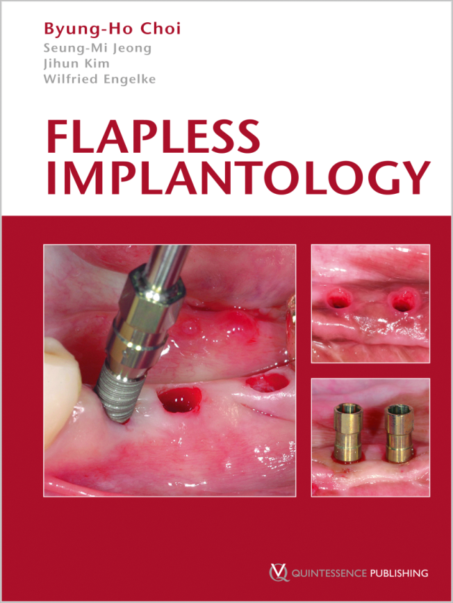
Pages 139-152, Language: GermanChoi, Byung-Ho / Engelke, WilfriedDie Fortschritte bei der Oberflächengestaltung moderner Implantatsysteme haben zu einer deutlich verbesserten Osseointegration geführt. Moderne bildgebende Verfahren ermöglichen, dass die Implantatinsertion heute mit einer wesentlich höheren Präzision geplant werden kann. Deshalb erscheint es bedeutsam, auch die chirurgische Insertionstechnik von Implantaten so weit zu optimieren, dass bei minimiertem chirurgischen Trauma vorhersagbare Ergebnisse für Funktion, Ästhetik und Patientenkomfort erzielt werden können. Bei der Insertion von Implantaten ohne Bildung eines mukoperiostalen Lappens [geschlossene Implantation = flapless implantation (FI)] resultiert eine geringere Narbenbildung der umgebenden Gewebestrukturen im Vergleich zum offenen Vorgehen; es kommt zu einer besseren Vaskularisierung der periimplantären Mukosa mit einer reduzierten Sulkustiefe, und der initiale Knochenabbau fällt geringer aus. Das geschlossene Vorgehen verbessert weiterhin ästhetische Resultate durch den Erhalt der ursprünglichen Form und Dicke der die Implantate umgebenden Weichgewebe. Ein bedeutsamer Nachteil dieser Technik ist der Mangel an Übersicht über die Hartgewebekontur während des Eingriffs. Der vorliegende Artikel beschreibt das operative Vorgehen bei "flapless implantation" und zeigt darüber hinaus Möglichkeiten auf, wie verfahrensbedingte Probleme gelöst werden können. Dies gilt auch für anspruchsvolle klinische Fälle und schwierige Knochensituationen. Die Beobachtung geschlossener Operationsgebiete, wie subgingivaler Räume und interner Knochenstrukturen, mit Hilfe endoskopischer Verfahren kann dazu beitragen, die Therapiequalität der geschlossenen Implantation weiter zu optimieren.
Keywords: Geschlossene Implantation, geschlossene Augmentation, Miniflap, Endoskopie, minimalinvasive Implantation
The International Journal of Oral & Maxillofacial Implants, 6/2005
Pages 891-897, Language: EnglishEngelke, Wilfried G. H. / Capobianco, MercedesPurpose: Sinus floor augmentation has become a routine procedure with predictable results. Flapless implant placement is recommended for a series of indications with sufficient bone volume. Flapless surgery in the atrophic maxilla is presented as a refinement of the endoscopic subantroscopic latero-basal sinus floor augmentation (SALSA) technique.
Materials and Methods: Based on computerized tomography (CT) scans, the site of sinus trephination and implant positions are planned using a commercially available planning program, and surgical templates are fabricated according to the data of the treatment plan. Subantral space is augmented using the SALSA technique without raising a mucoperiosteal flap. Implants are placed transgingivally without raising a mucoperiosteal flap, with endoscopic control of the cover screw at the bone level.
Results: In a case series of 6 patients, 21 implants were placed and augmented simultaneously. The mean augmentation height was 10.7 mm (range, 5.7 to 16.6 mm); the mean residual bone height was 5.1 mm (range, 1.9 to 12.1 mm). Complications such as insufficient primary stability and sinus membrane perforation were treated without changing to an open surgical approach. Discussion and
Conclusion: Flapless sinus augmentation (FSA) can reduce the surgical trauma significantly. The procedure has high acceptance by the patient and less postoperative discomfort. FSA enlarges the spectrum of minimally invasive surgery and may offer better vascularization and less alveolar resorption.
Keywords: endoscopy, minimally invasive surgery, sinus augmentation, 3-dimensional surgical templates
The International Journal of Oral & Maxillofacial Implants, 1/2003
Pages 135-143, Language: EnglishEngelke, Wilfried G. H.Purpose: The aim of the article was to introduce a new subantroscopic laterobasal sinus augmentation (SALSA) technique as a minimally invasive approach to maxillary peri-implant surgery.
Materials and Methods: The SALSA technique consists of the following steps: (1) microsurgical opening of the subantral space (SAS) with detachment of the sinus membrane (SM) under supported videoendoscopy; (2) enlargement of the SAS by laterobasal tunnelling; (3) subantroscopic examination of the SAS with (4) optional reinforcement or repair of the SM; (5) implant site preparation with subantroscopic identification of the cavities; and (6) precise stepwise placement of graft material under endoscopic control.
Results: Since 1996, 118 sinus augmentations have been performed on 83 patients using particulate alloplastic augmentation material (tricalcium phosphate) with various amounts of autogenous bone and blood. Mean augmentation height was 8.6 mm (range, 1 to 15 mm). Twentyeight perforations of sinus mucosa were observed without further complication (1 case of sinusitis was treated and re-augmented endoscopically). Of 211 titanium screw-type implants placed, 11 failures were observed.
Discussion: SALSA is a predictable surgical technique. With this minimally invasive method, adequate bone height can be achieved.
Conclusion: SALSA may offer advantages related to lower morbidity, conservation of bone volume and blood supply, optimized view of the surgical field, and high acceptance by patients.
The International Journal of Oral & Maxillofacial Implants, 5/2002
Pages 703-706, Language: EnglishEngelke, Wilfried G. H.Pathologies of the implant cavity wall currently cannot be diagnosed by direct observation because of the rapid pollution of the optical systems used. A technique to examine prepared implant sites intraoperatively to diagnose possible risk factors for the osseointegration process is presented. Examination of implant cavities is performed with support immersion endoscopy (SIE). Using a specially designed support and irrigation sheath (SIS), a 1.9-mm endoscope can be placed at a certain distance to the underlying bone surface. When immersed in a bleeding implant site, the endoscope window is cleaned by continuous laminar irrigation flow to allow observation of the cavity walls under variable magnification. Cortical and cancellous bone structures can be differentiated in situ and pathologies detected during capillary bleeding. Two case reports citing practical applications are reported. By means of SIE, possible risk factors during and after implant cavity preparation can be detected.


