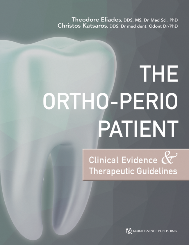
Oral Health and Preventive Dentistry, 1/2022
Open Access Online OnlyOrthodonticsDOI: 10.3290/j.ohpd.b3666501, PubMed ID (PMID): 3650408812. Dec 2022,Pages 517-524, Language: EnglishBernini, Domino A. J. / Eliades, Theodore / Patcas, Raphael / Papageorgiou, Spyridon N. / Koretsi, VasilikiPurpose: To assess mandibular incisor inclination after leveling the curve of Spee (CoS) in patients treated with fixed appliances.
Materials and Methods: This was a retrospective study, which included 80 consecutive patients with a mild CoS treated without extraction but with various biomechanical approaches. The depth of CoS was digitally measured on scanned plaster casts and mandibular incisor inclination was assessed with lateral cephalograms pre- and posttreatment. Patients were treated with 0.018”-slot edgewise fixed appliances and cinched back wires. Data were analyzed using linear regression modeling at 5%.
Results: A total of 80 patients (40% female; mean age 13.8 years) were included with mean ANB = 4.4 ± 1.9°, mean SN/ML = 31.7 ± 4.7°, mean L1/ML = 95.0 ± 7.7°, and a mean depth of CoS = 1.1 ± 0.4 mm. The depth of CoS was leveled by -0.85 ± 0.39 mm to a post-treatment median of 0.18 mm (interquartile range = 0.09 to 0.35 mm). A small mandibular incisor proclination was observed through treatment (2.49 ± 9.1°), but this was not associated with the reduction in the depth of CoS (p > 0.05) and no statistically significant modifying effect from the different treatment mechanics was observed.
Conclusion: Under the limitations of this study, leveling a mild CoS was not associated with mandibular incisor proclination during fixed-appliance treatment.
Keywords: cephalometry, Curve of Spee, fixed appliances, mandibular incisors, proclination
Oral Health and Preventive Dentistry, 1/2022
Open Access Online OnlySystematic ReviewDOI: 10.3290/j.ohpd.b3556039, PubMed ID (PMID): 363463388. Nov 2022,Pages 433-448, Language: EnglishScherer, Isabella / Tzanetakis, Giorgos N. / Eliades, Theodore / Koletsi, DespinaPurpose: To identify and assess any changes in the pulp tissue complex following orthodontic force application.
Materials and Methods: Published and unpublished literature was searched in seven databases until 9 August 2022 for randomised controlled trials (RCTs) and prospective trials (nR-PCT). Representative key words included ‘pulp response’, ‘pulp tissue’, ‘orthodontic force’, and ‘tooth movement’. Study selection, data extraction, risk of bias and certainty of evidence assessment were conducted independently by two reviewers. Random effects meta-analyses with respective confidence intervals (95%CIs) were conducted where applicable.
Results: A total of 363 records were screened, a final number of 24 articles were eligible for qualitative synthesis, while 8 of those contributed to meta-analyses. There was evidence that pulpal blood flow (PBF) decreased after 3 weeks of tooth movement compared to no force application (4 studies, mean difference: -1.68; 95% CI: -3.21, -0.15; p = 0.03). However, this was not the case after 6 months of treatment (p = 0.68). A rise in the activity of aspartate aminotransferase (AST) was detected after 7 days of treatment, but combining 2 studies, this was not statistically significant (p = 0.25). Other outcomes were assessed through single studies. Risk of bias was within the range of ‘some concerns/moderate to high/critical overall’, while certainty of evidence was low to very low according to GRADE.
Conclusions: As a short-term effect, PBF decreased upon initiation of orthodontic force application, while enzymatic and peptide activity within the pulp was transiently affected. Further long-term evidence of improved quality and certainty is needed.
Keywords: orthodontic force, meta-analysis, pulp tissue, systematic review
Oral Health and Preventive Dentistry, 1/2021
Open Access Online OnlySystematic ReviewDOI: 10.3290/j.ohpd.b2403661, PubMed ID (PMID): 348741438. Dec 2021,Pages 659-672, Language: EnglishOikonomou, Elissaios / Foros, Petros / Tagkli, Aikaterini / Rahiotis, Christos / Eliades, Theodore / Koletsi, DespinaPurpose: To identify and assess differences in oral hygiene parameters in patients undergoing orthodontic treatment with clear aligners compared to fixed appliances.
Materials and Methods: Published and unpublished literature was searched in seven databases until May 31st 2021. Representative keywords included ‘orthodontic aligner’, ‘fixed appliance’, ‘oral hygiene’, ‘plaque index’, ‘caries’. Study selection, data extraction, risk of bias and certainty of evidence assessment were undertaken independently by three reviewers. Random effects meta-analyses with respective confidence intervals (95% CI) were conducted, where applicable.
Results: A total of 882 unique records were screened, with a final number of 21 articles being eligible for qualitative synthesis, while 4 of those contributed to meta-analyses. Risk of bias was rated within the range of low to high or serious overall, while certainty of evidence was low to very low according to GRADE. For periodontal parameters, adults undergoing aligner orthodontic treatment presented summary plaque scores 0.58 lower than those treated with fixed appliances, within the first 6 to 12 weeks (4 studies: mean difference: -0.58; 95%CI: -0.82, -0.34; p < 0.001; I2 squared: 71.3%), while no evidence of difference was recorded for inflammation indices. Microbiologic parameters such as presence of S. mutans and lactobacilli were more pronounced in patients with fixed appliances for the first 3 to 6 months (synthesised data from 2 studies).
Conclusions: In the short-term after initiation of orthodontic treatment, patients treated with aligners and no additional attachments/adjuncts presented potentially higher levels of oral health overall. However, the evidence is supported by low to very low certainty.
Keywords: fixed appliances, oral hygiene, orthodontic aligners, systematic review
Oral Health and Preventive Dentistry, 1/2020
Open Access Online OnlySystematic ReviewDOI: 10.3290/j.ohpd.a45404, PubMed ID (PMID): 3321547913. Oct 2020,Pages 873-879, Language: EnglishPapageorgiou, Spyridon N. / Koletsi, Despina / Patcas, Raphael / Will, Leslie A. / Eliades, TheodorePurpose: To assess the knowledge of postgraduate dental students about evidence-based methodology pertaining to the design, conduct, and critical appraisal of clinical trials.
Materials and Methods: Senior postgraduate students were surveyed from the dental schools of three universities in Athens (Greece), Boston (USA), and Zürich (Switzerland). The proportion of students correctly answering each of the 10 questions of the survey, as well as the cumulative scores, were analysed statistically with descriptive statistics and logistic/linear regression analysis at α = 5%.
Results: A total of 96 students with a mean age of 30.0 years attained an overall correct score of 45.6% ± 15.0%, with correct answers to each question ranging from 13.5% to 86.5%. The questions most frequently answered incorrectly pertained to characterising sensitivity/specificity (13.5%), the number needed to treat (14.0%), the credibility of trial synthesis in meta-analysis (23.7%), and publication bias (29.5%). The vast majority of postgraduate students could correctly identify the role of statistical power of a trial (63.8%), random allocation sequence in a randomised trial (76.0%), and blinding in a randomised trial (86.5%). Paediatric dentistry postgraduate students scored better than students from other departments (+15.1%; 95% CI: 3.0% to 27.1%; p = 0.02).
Conclusions: Postgraduate students in orthodontics and other dental specialties possessed moderate knowledge on evidence-based methodology and clinical trials. Efforts should be made to integrate such subjects in university postgraduate curricula, so that future dental specialists can critically appraise such research papers.
Keywords: clinical trial, educational assessment, epidemiologic research design, evidence-based dentistry, questionnaire, survey, systematic review
The Journal of Adhesive Dentistry, 6/2017
DOI: 10.3290/j.jad.a39279, PubMed ID (PMID): 29152622Pages 463-473, Language: EnglishBaumgartner, Stefan / Koletsi, Despina / Verna, Carlalberta / Eliades, TheodorePurpose: To critically appraise the evidence regarding the effect of enamel sandblasting on the bond strength of orthodontic brackets on either the labial or lingual tooth surface.
Materials and Methods: An electronic database search of published and unpublished literature was performed. Search terms included sandblasting, enamel abrasion, tooth surface, bond strength, bond failure, and adhesive remnant; data were extracted in standardized piloted forms. Risk of bias was assessed using the Cochrane risk of bias tool, adapted for in vitro studies where necessary.
Results: Of the 81 articles initially retrieved, 13 were eligible for inclusion in the systematic review. All of the latter were in vitro studies with unclear risk of bias primarily due to unclear reporting of blinding of outcome assessors. Eight studies assessed the combined effect of enamel sandblasting and etching, while only five evaluated the isolated effect of sandblasting on the buccal enamel surface. In view of the apparently heterogeneous study settings, intervention protocols, specimen preparation and storage sequences, only two studies were deemed eligible for quantitative synthesis. Random effects meta-analysis revealed no evidence to support sandblasting prior to etching over etching alone with regard to shear bond strength of orthodontic brackets bonded in vitro to lingual enamel surfaces of extracted premolars (standardized mean difference: 0.36; 95% CI: -0.21, 0.94; p = 0.22).
Conclusions: The findings of the present study cannot support lingual enamel sandblasting prior to etching for augmentation of the bond strength of orthodontic brackets.
Keywords: sandblasting, orthodontic bonding, shear bond strength, lingual orthodontics, brackets


