We use cookies to enable the functions required for this website, such as login or a shopping cart. You can find more information in our privacy policy.
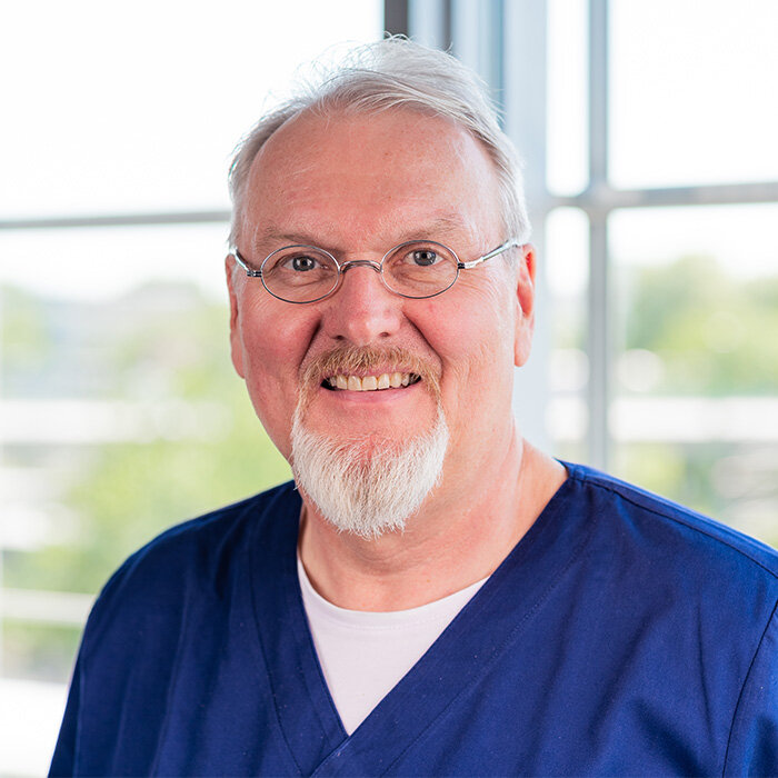
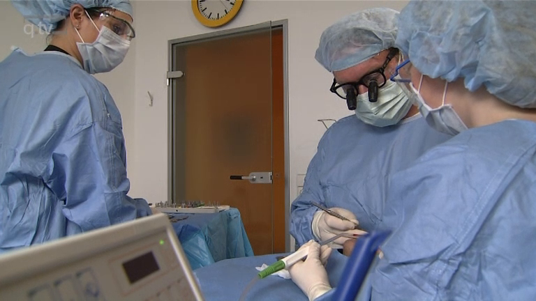
Dr. Detlef Hildebrand is a dentist and dental technician, specializing in oral implantology, navigation, and robotics. After his time at the dental clinics of the universities in Freiburg and Berlin (Charité), he founded the Center of Competence in the heart of Berlin in 1998. Since 2007, he has been leading the dentist lists in FOCUS and STERN and is setting the course for dentistry at the highest level at the dental forum berlin. His specialty is treating anxious patients using the immediate treatment technique. Most treatments are performed under anesthesia with the help of certified anesthetists.
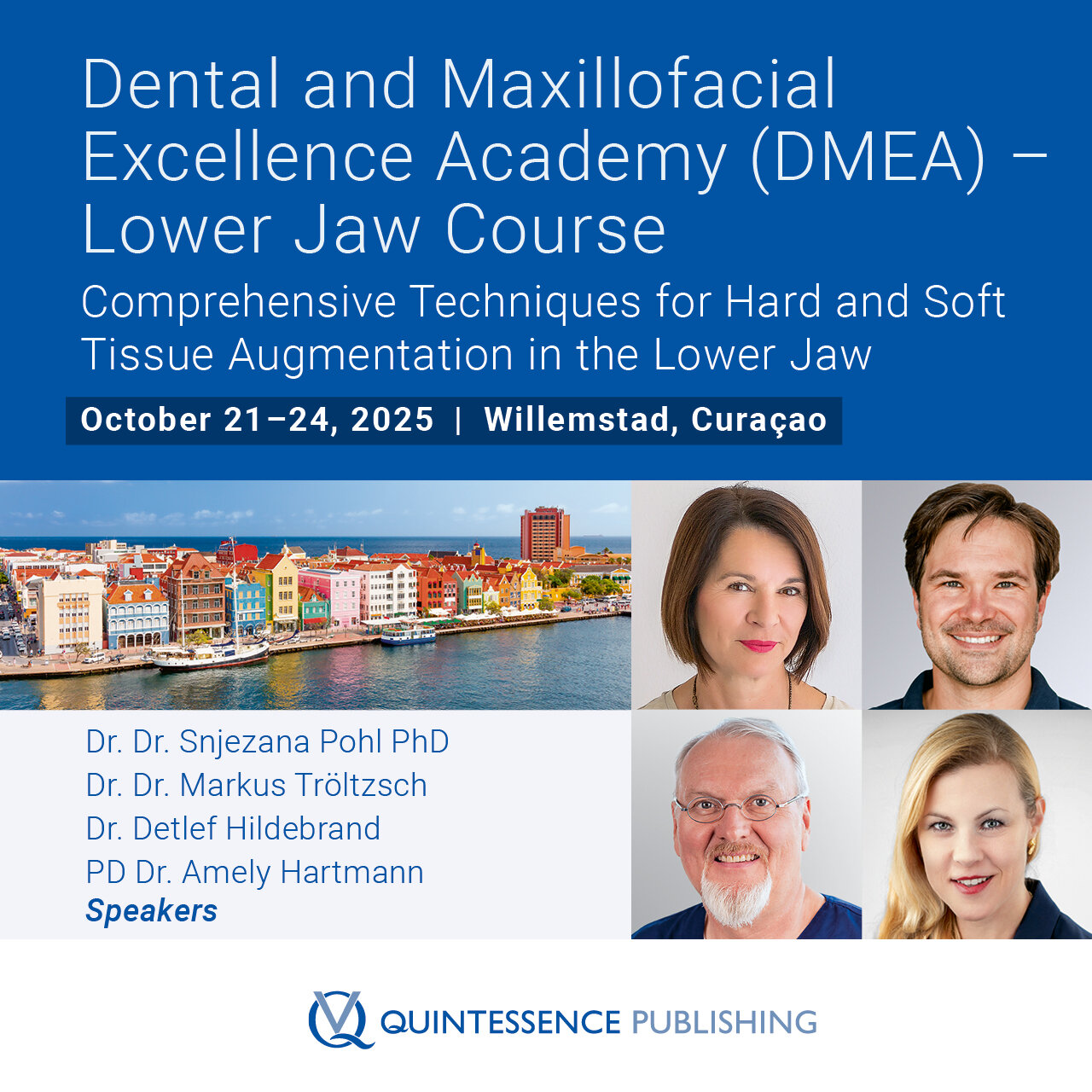
Speakers: Samir Abou-Ayash, Bilal Al-Nawas, Thomas Bernhart, Florian Beuer, Stefan Bienz, Elena Calciolari, Najla Chebib, Andreas Dengel, Vincent Donker, Joke Duyck, Roberto Farina, Gary Finelle, Alberto Fonzar, Tobias Fretwurst, Rudolf Fürhauser, Oscar Gonzalez-Martin, Stefano Gracis, Knut A. Grötz, Christian Hammächer, Lisa J. A. Heitz-Mayfield, Detlef Hildebrand, Norbert Jakse, Jim Janakievski, Tim Joda, Daniel Jönsson, Gregg Kinzer, Vincent G. Kokich, Michael Krimmel, Cecilia Larsson Wexell, Martin Lorenzoni, Georg Mailath-Pokorny, Julia Mailath-Pokorny, Frank Georg Mathers, Gerry McKenna, Henny Meijer, Alberto Monje, Torsten Mundt, Nadja Nänni, David Nisand, Robert Nölken, Nicole Passia, Michael Payer, Christof Pertl, Aušra Ramanauskaitė, Eik Schiegnitz, Martin Schimmel, Ulrike Schulze-Späte, Frank Schwarz, Falk Schwendicke, Robert Stigler, Michael Stimmelmayr, Anette Strunz, Christian Ulm, Stefan Vandeweghe, Kay Vietor, Arjan Vissink, Asaf Wilensky, Stefan Wolfart, Werner Zechner, Anja Zembic, Nicola Zitzmann
European Association for Osseintegration (EAO)
This author's journal articles
International Journal of Computerized Dentistry, 1/2017
The primary objective of implant insertion is optimal prosthetic implant positioning while simultaneously protecting sensitive anatomical structures. In this context, navigated implants show significantly better results than freehand-inserted implants. Computer-assisted navigation, in combination with three-dimensional (3D) imaging by cone beam computed tomography (CBCT), is an ideal way to achieve higher predictability for successful implant therapy. Basically, one can distinguish between static navigation using templates, and direct dynamic navigation using optical transmission systems. Both options demonstrate comparably good results as far as the precision of implant positioning is concerned. Today, the gold standard is digital manufacturing of the template. Direct navigation is the more attractive option, provided acquisition costs can be reduced by simplifying the system. This article presents patient cases that demonstrate different variants of navigated implantology.
Keywords: static navigation, dynamic navigation, digital manufacturing, template, CAD/CAM, reference marker
Purpose: The aim of this study was to evaluate the success rate of 2 different implant systems with sandblasted and acid-etched modified surfaces loaded after reduced healing periods.
Materials and Methods: One-hundred seventeen patients with a mean observation period of 3.75 years (24 to 61 months) were included in this evaluation. Chart reviews of a standardized recall program were evaluated. All 532 placed implants showed an unloaded healing time of 6 weeks in the mandible and 12 weeks in the maxilla. At abutment placement a torque value of 35 Ncm was one of the primary variables, and the success of the implants over time was determined by the criteria of Buser et al. The survival was analyzed using Kaplan-Meier method, and the probability of an event within 1 group independent of time was evaluated using the chi-square test and Fisher exact test.
Results: Of the 532 implants, 235 were placed in female and 297 in male patients; 448 implants were located in the maxilla and 84 in the mandible. Three implants were lost prior to abutment connection in 3 patients. Life table analyses show an overall success rate of 99.4% at 5 years, as no implants were lost after abutment connection. There was no significant association of the implant type (P = .185), gender (P = .99), or jaw (maxilla/mandible; P = .06) and the survival of the implants within this study.
Conclusion: Based on the data found in this investigation, it can be concluded that implants with sandblasted, acid-etched surfaces can be restored after a 6- to 12-week healing period with a high predictability of success.
Keywords: reduced healing period, sand-blasted and acid-etched
In this present study new bone formation of fresh extraction socket afteraugmentation with Bio-Oss Collagen was analyzed after a healing period of 6weeks using histomorphometry Material and methods: Ten patients, referredfor extraction of decayed teeth of all regions, were included in this study.The extraction sockets were instrumented to eliminate all remnants ofperiodontal ligament tissue and showed no defect in extraction site. Eachsocket was grafted with Bio-Oss Collagen without flap management. After a 6weeks healing period, at implant placement, bone biopsy samples wereobtained with a trephine bur and evaluated histomorphometrically, usingMasson's trichrome and Toluidine staining. Quantification of new boneformation and BioOss-remnants was performed using a digital imaging system(AxioVision, Zeiss, Germany). Results and discussion: The values found fornew bone formation ranged from 35% - 59% and 34% for remainingBioOss-particles. This is comparable to the findings of studies with healingperiod of 12 weeks in canines. These results encouraged an early onsetimplantation after healing period of six weeks. The long-term success andsurvival rate of these implants will be the subject of futureinvestigation.
Keywords: extraction socket, augmentation
Purpose: In the present study solid monocortical hipbone onlay grafts of the maxilla were analyzed histologically after a healing period of 3 months. The clinical success of the implants placed in the grafted bone was evaluated.
Materials and Methods: Nineteen patients underwent augmentation with avascular iliac bone. A 2-stage procedure was performed with a 3-month healing period between graft and implant placement. At implant placement bone biopsy samples were taken at the proposed implant sites.
Results: Of the 117 implants placed, 1 was not osseointegrated at the time of abutment connection. No implants were lost after loading during an observation period of up to 3 years. Clinical appearance of the augmented bone after 3 months showed a dense cortical layer with good blood perfusion. Histologic specimens were analyzed quantitatively and showed an average of 43.2% newly formed bone.
Discussion: Histomorphometry showed that the amount of newly formed bone after 3 months was comparable to that found after a healing period of 4.5 months. The clinical success of the implants placed after the shortened healing period was comparable to that found in nonaugmented bone.
Conclusion: This study showed that after avascular iliac bone grafting, 3 months of revascularization was sufficient to ensure the secure placement of dental implants in second-stage surgery for this patient population.
Keywords: dental implants, iliac bone grafting, onlay bone grafting