International Journal of Esthetic Dentistry (DE), 2/2023
Clinical ResearchSeiten: 186-206, Sprache: DeutschMartins da Rosa, José Carlos / Pértile de Oliveira Rosa, Ariádene Cristina
Mit dem Wandel der Erwartungen an zahnärztliche Behandlungen ist die Ästhetik in den vergangenen Jahrzehnten zu einem Schlüsselfaktor bei der Bewertung des Behandlungserfolgs in der Implantattherapie avanciert. Ein stabiles Niveau der periimplantären Mukosa mit natürlicher Farbe und Textur gilt als entscheidend für gute und nachhaltige Behandlungsergebnisse. In diesem Beitrag soll der Zusammenhang zwischen der Implantatposition, dem periimplantären Weichgewebemanagement und der langfristigen Erhaltung der Ergebnisse nach Sofortimplantationen in Extraktionsalveolen aufgezeigt werden. An einer Serie von 12 Fällen mit einer mittleren Nachbeobachtungszeit von 21,91 Monaten wird das Konzept des „magischen Quadrats“ (MQ) vorgestellt. Der Begriff bezeichnet eine gedachte Fläche im Bereich des Implantathalses, die sich im Fall einer idealen Implantatposition ergibt. Diese ideale Position ist durch einen korono-apikalen Abstand von 3 mm zwischen Implantatplattform und Gingivarand, die Erhaltung von 3 mm horizontaler Dicke des vestibulären Knochens (Hartgewebespalt von der vestibulären Implantatoberfläche zur Außenfläche des vestibulären Knochens) sowie eine Weichgewebedicke von ≥ 3 mm auf Höhe des Implantathalses definiert. Die Umsetzung des magischen Quadrats fördert das Weichgewebewachstum und die Bildung eines dicken periimplantären Knochenkamms und stellt damit ein langfristig stabiles Behandlungsergebnis sicher.
International Journal of Esthetic Dentistry (EN), 2/2023
Clinical ResearchPubMed-ID: 37166771Seiten: 180-198, Sprache: EnglischMartins da Rosa, José Carlos / Pértile de Oliveira Rosa, Ariádene Cristina
A case seriesWith changing expectations for dental treatment, esthetics have become an essential factor in defining successful rehabilitation with dental implants. The stability of the gingival contours as well as the color and texture of the surrounding tissue are critical for the long-term maintenance of successful implant treatment outcomes. The aim of the present article is to demonstrate the correlation of the 3D implant position and the adjacent tissue management protocol with the long-term maintenance of immediate implant placement results in postextraction sites. A series of 12 cases with a mean follow-up of 21.91 months is presented to introduce the concept of the ‘magic square’ (MS), denoting the area formed in the cervical implant region when the ideal 3D implant position is achieved. This position is 3-mm coronoapical from the implant platform to the gingival margin, with the maintenance of a 3-mm vestibulopalatine thickness of the buccal bone (ie, hard tissue gap from the buccal implant surface to the outer portion of the buccal bone wall), and a ≥ 3-mm soft tissue gap at the cervical portion of the implant. The achievement of the MS promotes soft tissue growth and the formation of a thicker peri-implant bone ridge, and ensures the stability of treatment outcomes over time.
International Journal of Esthetic Dentistry (EN), 2/2017
PubMed-ID: 28653055Seiten: 258-270, Sprache: EnglischMartins da Rosa, José Carlos / Fadanelli, Marcos Alexandre / Zimmerman, Diego / de Oliveira Rosa, Ariádene Cristina Pértile
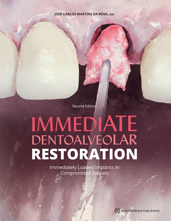
Purpose: This article describes the use of rapid prototyping (RP) for diagnosis, planning, and execution of the reconstruction of hard and soft tissue in socket defects using immediate dentoalveolar restoration (IDR).
Summary: In cases of tissue loss in anterior dental areas, esthetic rehabilitation poses a major challenge with respect to treatment planning with the goal of long-term tissue maintenance. The IDR technique consists of immediate reconstruction in a single procedure of bone and soft tissue around implants placed immediately after extraction, and prosthetic rehabilitation. As this procedure is immediate and flapless, the correct diagnosis of tissue loss and correct graft adaptation are mandatory. RP can increase the precision of the procedure, as demonstrated using a clinical case characterized by total loss of the buccal bone wall and gingival recession. The results were evaluated by clinical assessment, photography, radiography, cone beam computed tomography (CBCT), and prototyping.
Conclusion: The application of RP facilitated the execution of IDR as it enabled more accurate diagnosis of the socket defect and more precise adaptation of the tissue graft. A clinical study should be conducted to evaluate the effects of RP on the clinical results of the IDR technique.