Wir verwenden Cookies ausschließlich zu dem Zweck, technisch notwendige Funktionen wie das Login oder einen Warenkorb zu ermöglichen, oder Ihre Bestätigung zu speichern. Mehr Informationen zur Datenerhebung und -verarbeitung finden Sie in unserer Datenschutzerklärung.
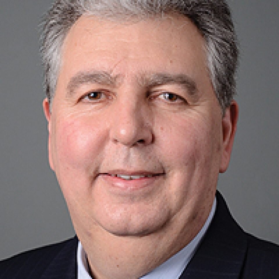
Joseph P. Fiorellini, DMD, DMSc, is a professor in the Department of Periodontics and the director of the Advanced Graduate Program in Periodontics at the University of Pennsylvania School of Dental Medicine, trustee for the American Academy of Periodontology, board director for the Academy of Osseointegration, and a consultant for American Dental Association Commission for Continuing Education Recognition Program. He has published over 100 manuscripts, reviews, and textbook chapters. Dr Fiorellini serves as the associate editor of The International Journal of Oral & Maxillofacial Implants, is a member of several editorial boards, and is the recipient of several awards.
Veranstaltungen
The 14th International Symposium on Periodontics and Restorative Dentistry (ISPRD)
Juni 9, 2022 — Juni 12, 2022Boston Marriott Copley Place, Boston, MA, Vereinigte Staaten von Amerika
Referenten: Tara Aghaloo, Edward P. Allen, Evanthia Anadioti, Wael Att, Vinay Bhide, Markus Blatz, Scotty Bolding, Lorenzo Breschi, Jeff Brucia, Daniel Buser, Luigi Canullo, Daniele Cardaropoli, Stephen J. Chu, Donald Clem, Christian Coachman, Lyndon F. Cooper, Daniel Cullum, Lee Culp, José Carlos Martins da Rosa, Sergio De Paoli, Marco Degidi, Nicholas Dello Russo, Serge Dibart, Joseph P. Fiorellini, Mauro Fradeani, Stuart J. Froum, David Garber, Maria L. Geisinger, William Giannobile, Luca Gobbato, Ueli Grunder, Galip Gürel, Chad Gwaltney, Christoph Hämmerle, Robert A. Horowitz, Marc Hürzeler, David Kim, Gregg Kinzer, Christopher Köttgen, Ina Köttgen, Purnima S. Kumar, Burton Langer, Lydia Legg, Pascal Magne, Kenneth A. Malament, Jay Malmquist, George Mandelaris, Pamela K. McClain, Michael K. McGuire, Mauro Merli, Konrad H. Meyenberg, Craig M. Misch, Julie A. Mitchell, Marc L. Nevins, Myron Nevins, Michael G. Newman, Miguel A. Ortiz, Jacinthe M. Paquette, Stefano Parma-Benfenati, Michael A. Pikos, Giulio Rasperini, Pamela S. Ray, Christopher R. Richardson, Isabella Rocchietta, Marisa Roncati, Marco Ronda, Paul S. Rosen, Maria Emanuel Ryan, Irena Sailer, Maurice Salama, David M. Sarver, Takeshi Sasaki, Todd Scheyer, Massimo Simion, Michael Sonick, Sergio Spinato, Dennis P. Tarnow, Lorenzo Tavelli, Douglas A. Terry, Tiziano Testori, Carlo Tinti, Istvan Urban, Hom-Lay Wang, Robert Winter, Giovanni Zucchelli
Quintessence Publishing Co., Inc. USA
Zeitschriftenbeiträge dieses Autors
The International Journal of Oral & Maxillofacial Implants, Pre-Print
DOI: 10.11607/jomi.11074, PubMed-ID: 39365911Oktober 4, 2024,Seiten: 1-20, Sprache: EnglischFiorellini, Joseph P. / Mojaver, Sean / Kay, George / Chang, Yu Cheng / Brown, Michael / Sarmiento, Hector
The development of a successful bone grafting technology with cohesive and adhesive properties has been an elusive goal for dental and orthopedic researchers. Tetracalcium phosphate combined with phosphoserine (TTCP-PS) is a synthetic, injectable, cohesive, self-setting, mineral-organic wet-field adhesive. The objective of this study was to evaluate four formulations of TTCP-PS in comparison to the conventional grafting materials, Bioglass and deproteinized cancellous bovine bone with a bioresorbable collagen membrane in standardized defects created in the angle of the rat mandible. Microcomputed tomography and histomorphometry were utilized to evaluate bone replacement with each of these materials after in vivo residence of either 4 or 12 weeks. The results of this study demonstrate that specific TTCP-PS formulations can form bone comparable to conventional materials in an osteopromotive mechanism but with the advantage of having cohesive and adhesive properties.
Placement of short implants is a common approach to rehabilitate edentulous areas. The objective of this study was to evaluate the long-term survival of 7.0- and 8.5-mm implants placed in either a delayed or immediate loading protocol. Life table analysis revealed the implants treated with the delayed loading protocol had a 90.9% survival rate and the implants treated with the immediate loading protocol had a survival rate of 92.0%. The results of this 8-year prospective study demonstrate similar survival rates of short, cylindrical threaded implants placed by either a delayed or immediate loading protocol.
The International Journal of Oral & Maxillofacial Implants, 3/2020
DOI: 10.11607/jomi.8114, PubMed-ID: 32406653Seiten: 551-559, Sprache: EnglischFiorellini, Joseph P. / Sourvanos, Dennis / Crohin, Constant C. / Crohin, Michele / Chang, Jane J. / Mattos, Marcelo / Ko, Kang I.
Purpose: Diabetes mellitus has been shown to delay osseointegration of titanium dental implants. This study tested the hypothesis that serum derived from diabetes negatively affects osteoblast adhesion to polystyrene and titanium surfaces, partly through the presence of advanced glycation end products (AGEs).
Materials and Methods: Twenty-four Sprague- Dawley rats were divided into three groups: normoglycemic control, streptozotocin-induced diabetic group, and diabetic group treated with the AGE inhibitor aminoguanidine. Polystyrene or titanium disks were preincubated in serum derived from each group. Human osteoblasts transfected with green fluorescent protein (GFP) were cultured, and the number of adherent osteoblasts was quantified. High-pressure liquid chromatography (HPLC) was used to fractionate eluates, which were further characterized by western blot with AGE antibody and adhesion assays. In parallel, sera derived from healthy patients, patients with controlled diabetes, and patients with uncontrolled diabetes were utilized for osteoblast adhesion assay and western blot.
Results: Diabetic serum significantly reduced the number of adherent osteoblast and osteoblast aggregates on titanium disks, whereas aminoguanidine-treated serum rescued the effect of diabetes on the number of adherent osteoblast aggregates. Fractionated diabetic serum revealed distinct AGE bands at ~100 kDa and 44 kDa, whereas healthy serum did not express any. In human serum samples, both controlled and uncontrolled diabetes led to a significant reduction in the number of adherent osteoblasts on polystyrene and titanium surfaces compared with normoglycemic serum. This correlated with presence of AGEs in western blot in diabetic but not in healthy serum.
Conclusion: Osteoblast adhesion on the titanium surface was greatly reduced by the exposure of serum derived from diabetic rats or humans. Recovery of osteoblast aggregates by aminoguanidine treatment suggests that AGEs played a role in this negative effect. The correlating presence of AGEs from the fractionated sera of diabetic rats or humans and impaired osteoblast adhesion on the titanium surface further supports this role.
Schlagwörter: bone healing, dental implant, diabetes mellitus, hyperglycemia, metabolism, osseointegration, osteoblast, streptozotocin, wound healing
Purpose: The purpose of this review was to explore the available literature and compile studies that discuss the relevance of the biofilm, onset and progression of disease, critical peri-implant pocket depth, frequency of supportive implant therapy, excess cement, and keratinized peri-implant tissues as related to peri-implant disease.
Materials and Methods: PubMed, Cochrane Oral Health Group Specialized Trial Register, and hand searches of related journals were performed in relationship to the focused question. Reports describing techniques, preclinical studies, and case reports were excluded.
Results: Due to the absence of controlled studies, a meta-analysis could not be performed. Summaries of relevant publications were completed for each topic area. Clinical recommendations were developed to provide guidance to the practitioner.
Conclusion: The importance of proper diagnosis, planning, and clinical treatment cannot be overstated. Patient factors including systemic disease, periodontal status, and oral hygiene significantly impact peri-implant health. Clinician factors such as implant position, excess cement, and restorative design can contribute to development of peri-implant disease. Surveillance of implant status is essential and can be assisted by the assessment of risk factors, establishment of a proper recall program, and monitoring changes in bone and peri-implant pocket depths.
Schlagwörter: biofilm, cement, dental implants, inflammation, peri-implant disease, pocket depth, supportive implant therapy
International Journal of Periodontics & Restorative Dentistry, 5/2018
DOI: 10.11607/prd.3639, PubMed-ID: 30113606Seiten: 665-671, Sprache: EnglischSarmiento, Hector L. / Norton, Michael / Korostoff, Jonathan / Ko, Kang I. / Fiorellini, Joseph P.
The objective of this case series was to describe surgical approaches that can be used to efficiently and effectively treat peri-implantitis as measured by positive changes in clinical parameters. A total of 32 patients with 45 implants were treated surgically to eliminate peri-implantitis. Baseline clinical parameters measured prior to surgery were compared to those made 6 months postsurgery to evaluate the efficacy of each procedure. Implants demonstrating signs of peri-implantitis were treated by one of three approaches: (1) regenerative surgery, (2) osseous resective surgery, or (3) apically repositioned flap surgery. In all instances, the exposed implant surfaces were debrided and decontaminated. Relative to baseline values, regenerative surgery yielded statistically significant changes in probing depth (PD) (7.21 ± 0.27 mm to 4.09 ± 0.14 mm) and percentage of sites exhibiting bleeding on probing (BoP) (100.0% ± 0.0% to 10.6% ± 3.3%) as measured at the 6-month recall visit (P ≤ .05). The decrease in probing depth was not dependent on the type of graft material used (P ≤ .05). Resective surgery yielded statistically significant changes in PD (5.86 ± 0.23 mm to 3.63 ± 0.14 mm) and the percentage of sites exhibiting BoP (100.0% ± 0.0% to none) (P ≤ .05). Finally, the implants treated via apically repositioned flap surgery demonstrated statistically significant decreases (P ≤ .05) in both PD (6.79 ± 0.27 mm to 4.32 ± 0.16 mm) and BOP (100.0% ± 0.0% to 14.3% ± 6.7%) (P ≤ .05). Regenerative, resective, and apically positioned flap surgery can be utilized to successfully treat peri-implantitis.
International Journal of Periodontics & Restorative Dentistry, 3/2018
DOI: 10.11607/prd.3397, PubMed-ID: 29444198Seiten: 389-394, Sprache: EnglischFiorellini, Joseph P. / Norton, Michael R. / Luan, Kevin WanXin / Kim, David Minjoon / Wada, Keisuke / Sarmiento, Hector L.
The objective of this study was to evaluate the effectiveness of precise threedimensional hydroxyapatite printed micro- and macrochannel devices for alveolar ridge augmentation in a canine model. All grafts induced minimal inflammatory and fibrotic reactions. Examination of undecalcified sections revealed that both types of grafts demonstrated bone ingrowth. The majority of the bone growth into the block graft was into the channels, though a portion grew directly into the construct in the form of small bony spicules. In conclusion, bone ingrowth was readily demonstrated in the middle of the implanted printed devices.
Treatments for enhancing surface energy were studied in an effort to create a more favorable environment for cell adhesion. Cold argon plasma (CAP) is able to improve titanium-cell contact, producing hydrophilic surfaces with higher wettability. The aim of this in vitro study was to estimate the early cell morphology after CAP treatment of different commercially available titanium surfaces. Surface wettability was significantly augmented in all the treated samples. The authors investigated how CAP affected the behavior of osteoblasts by evaluating the cell morphology outcome. Cell surface areas differed in a statistically significant way when plasma-treated samples were compared to the untreated ones. The positive effect of CAP was shown on smooth, moderately rough, and rough implant surfaces.
The maxillary sinus augmentation procedure has become a predictable treatment to regenerate bone for implant placement. The purpose of this study was to evaluate the effect of recombinant human platelet-derived growth factor BB (rhPDGF-BB) combined with a deproteinized cancellous bovine bone graft for sinus augmentation. The lateral window approach was used for maxillary sinuses with minimal residual bone. After a healing period of 4 months, dental implants were placed and then restored following a 2-month osseointegration period. The result demonstrated increased bone height and ISQ values and a 100% survival rate. This study indicates that the addition of rhPDGF-BB to deproteinized cancellous bovine bone accelerated the healing period in maxillary sinuses with minimal native bone.
Osteopontin (OPN) and an OPN-derived synthetic peptide, OC-1016, have demonstrated their potential to enhance osseointegration in vitro. The purpose of this study was to evaluate bone-to-implant contact (BIC) and surrounding bone density (BD) of implants coated with either recombinant human OPN (rhOPN) or OC-1016 as compared with noncoated titanium plasma sprayed (TPS) surface in a canine model. Histomorphometric analysis revealed that at 4 weeks, %BIC and %BD of coated implants were significantly higher than those of noncoated TPS implants. At 12 weeks, %BIC of coated implants was also significantly higher than that of noncoated implants; however, there was no statistically significant difference in %BD. The rhOPN and OC-1016 were concluded to be capable of significantly accelerating the early stage of osseointegration and bone healing around implants.
Peri-implant bone levels are influenced by pathologic and nonpathologic conditions. The understanding of peri-implant disease has evolved over the past several decades, and the classification of peri-implantitis has been limited to descriptions of disease progression or those involving soft and/or hard tissues (peri-implant mucositis or peri-implantitis). However, no classification system has been established based on etiology. The objective of this study was to identify various etiologies for peri-implantitis and to establish a classification system based on the pathogenesis. The results indicate that the majority of bone loss was related to biofilm, followed by iatrogenic factors, exogenous irritants, absence of keratinized tissue, and extrinsic pathology. The proposed classification system will allow the clinician to properly diagnose peri-implant diseases in relation to etiology. These conditions may respond differently to applied therapies.