Wir verwenden Cookies ausschließlich zu dem Zweck, technisch notwendige Funktionen wie das Login oder einen Warenkorb zu ermöglichen, oder Ihre Bestätigung zu speichern. Mehr Informationen zur Datenerhebung und -verarbeitung finden Sie in unserer Datenschutzerklärung.
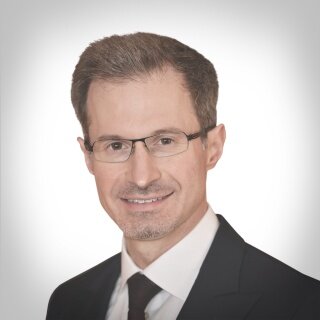
Jim Janakievski erhielt seinen D.D.S. von der University of Toronto und absolvierte anschließend eine allgemeine Praxisresidenz. Nach mehreren Jahren in der Allgemeinpraxis absolvierte er seine postgraduale Ausbildung an der University of Washington, wo er ein Zertifikat in Parodontologie mit einem M.S.D.-Abschluss und ein Fellowship in Prothetik erwarb. Er begann seine akademische Laufbahn an der University of Washington als stellvertretender Direktor für postgraduale Parodontologie und ist derzeit außerordentlicher assoziierter Professor. Dr. Janakievski ist Diplomat des American Board of Periodontology und Fellow der American Academy of Esthetic Dentistry. Er ist Gutachter für mehrere zahnmedizinische Fachzeitschriften und hat auf dem Gebiet der interdisziplinären Zahnmedizin, zahnärztlichen Implantaten und autotransplantierten Zähnen veröffentlicht. Dr. Janakievski unterrichtet an der Spear Education in Scottsdale, Arizona. Darüber hinaus beteiligt er sich an praxisbasierten klinischen Forschungen durch das McGuire Institute. Er führt eine Privatpraxis in Tacoma, Washington.
30th EAO Annual Scientific Meeting / 37th DGI Annual Congress
Berlin reloadedSeptember 28, 2023 — September 30, 2023CityCube Berlin, Berlin, Deutschland
Referenten: Samir Abou-Ayash, Bilal Al-Nawas, Thomas Bernhart, Florian Beuer, Stefan Bienz, Elena Calciolari, Najla Chebib, Andreas Dengel, Vincent Donker, Joke Duyck, Roberto Farina, Gary Finelle, Alberto Fonzar, Tobias Fretwurst, Rudolf Fürhauser, Oscar Gonzalez-Martin, Stefano Gracis, Knut A. Grötz, Christian Hammächer, Lisa J. A. Heitz-Mayfield, Detlef Hildebrand, Norbert Jakse, Jim Janakievski, Tim Joda, Daniel Jönsson, Gregg Kinzer, Vincent G. Kokich, Michael Krimmel, Cecilia Larsson Wexell, Martin Lorenzoni, Georg Mailath-Pokorny, Julia Mailath-Pokorny, Frank Georg Mathers, Gerry McKenna, Henny Meijer, Alberto Monje, Torsten Mundt, Nadja Nänni, David Nisand, Robert Nölken, Nicole Passia, Michael Payer, Christof Pertl, Aušra Ramanauskaitė, Eik Schiegnitz, Martin Schimmel, Ulrike Schulze-Späte, Frank Schwarz, Falk Schwendicke, Robert Stigler, Michael Stimmelmayr, Anette Strunz, Christian Ulm, Stefan Vandeweghe, Kay Vietor, Arjan Vissink, Asaf Wilensky, Stefan Wolfart, Werner Zechner, Anja Zembic, Nicola Zitzmann
European Association for Osseintegration (EAO)
Zeitschriftenbeiträge dieses Autors
International Journal of Periodontics & Restorative Dentistry, 6/2022
Online OnlyDOI: 10.11607/prd.6124Seiten: e161-e174, Sprache: EnglischVelasquez, Diego / Araújo, Mauricio G / Clem, Donald S / Gunsolley, John C / Heard, Rick H / Janakievski, Jim / McClain, Pamela K / McGuire, Michael K / Misch, Craig M / Nevins, Marc / Pickering, Steve / Pope, Bryan / Richardson, Chris / Santarelli, Greg / Scheyer, E Todd / Schallhorn, Rachel / Toback, Gregory
Postextraction bone grafting and implant placement help preserve alveolar bone volume. Collagen wound dressings and soft tissue graft substitutes may help protect extraction socket bone grafts and provide better gingival contours. This randomized, controlled, multicenter, and double-blinded study was conducted to compare a control (wound dressing) and a test (soft tissue graft) substitute in nearly intact extraction sockets. Both test and control sockets were grafted with a xenogeneic bone graft. Graft containment, extraction socket soft tissue gap closure, gingival contour, and gingival thickness were examined over 16 weeks, at which time implants were placed. Healing was uneventful for both groups, and there was no significant difference (P < .05) between the times required to close the extraction socket soft tissue gap (~80% of sites closed by 8 weeks). Bone grafts were covered and contained longer in the test group (~4 weeks vs ~2 weeks), with less contour disruption out to 4 weeks; however, at implant placement, soft tissue contours in both groups were comparable, and soft tissue thicknesses were not significantly different.
Purpose: To study bone healing at implant sites in simulated extraction sockets with 1-mm marginal defects and compare healing around a turned surface (T) to that around a porous oxide surface prepared by anodic oxidation (AO) with or without the use of an autogenous bone graft.
Materials and Methods: All mandibular premolars and first molars were extracted from 10 mongrel dogs. After 9 weeks, four sites were prepared on both sides of all mandibles. Each osteotomy was widened in the coronal 5 mm to create a marginal defect of 1 mm around the implants. Autogenous bone was collected during the drilling procedure. The sites were randomized to receive implants with a T or an AO surface, with or without bone grafting. The animals were sacrificed 4 months after implant placement for histologic analysis.
Results: Clinically, all sites healed with complete bone fill. The combination of an AO implant and a bone graft resulted in a significantly greater percentage of bone-to-implant contact (BIC) (P .05) versus all other groups. The highest point of BIC was achieved with the AO group, which was significantly greater than the lowest group (T). No significant differences between groups were found when the apical 4 mm (nongap areas) were compared (P = .65).
Conclusions: Studies have demonstrated that bone can fill in a marginal defect around a titanium implant with varied histologic BIC, depending on implant surface type and defect dimensions. Based upon this animal study using 10 mongrel dogs, marginal circumferential defects of 1 mm showed significantly higher BIC values for implants that were prepared by AO compared to implants with a turned surface. The addition of autogenous bone grafts further enhanced the degree of BIC.
Schlagwörter: anodic oxidation, autogenous, bone graft, histomorphometry, osseointegration