Wir verwenden Cookies ausschließlich zu dem Zweck, technisch notwendige Funktionen wie das Login oder einen Warenkorb zu ermöglichen, oder Ihre Bestätigung zu speichern. Mehr Informationen zur Datenerhebung und -verarbeitung finden Sie in unserer Datenschutzerklärung.
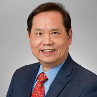
Hom-Lay Wang, DDS., MSD., Ph D, Professor and Director of Graduate Periodontics at the University of Michigan.He has co-editor two textbooks, published more than 50 book chapters/invited reviews and more than 750 peer-reviewed scientific articles. Dr. Wang serves as a Co-Editor-in-Chief for Clinical Implant Dentistry and Related Research, an Associate Editor for The IJOMI and IJOI.Dr. Wang is the recipient of many awards/honors including AAP Outstanding Educator Award (2017), AAP Distinguished Scientist Award (2017),AAP Master Clinician Award (2019), AAP Distinguished Service Award (2021), and AAP Clinical Research Award (2021).
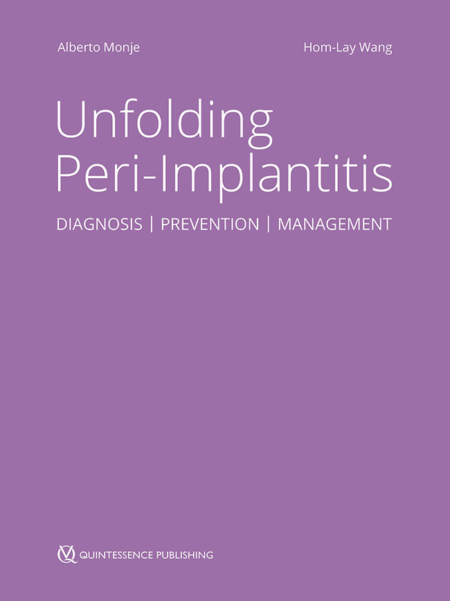
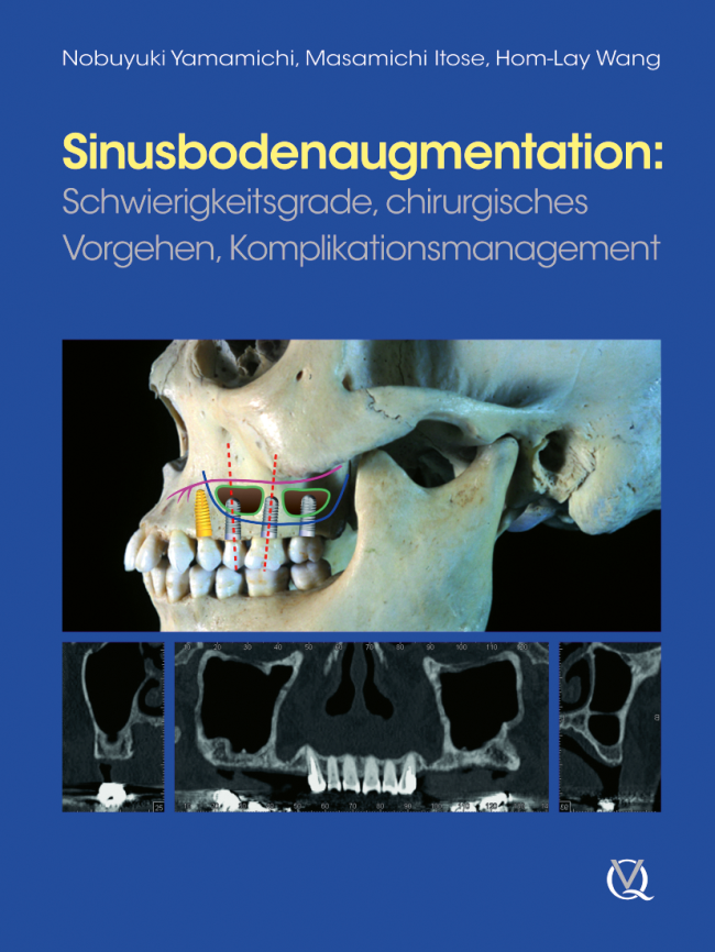
2. Auflage 2024 Buch Hardcover; 21 x 28 cm, 836 Seiten, 1800 Abbildungen Sprache: Englisch Kategorien: Implantologie, Parodontologie ISBN 978-1-78698-147-9 QP Deutschland
Details make perfectionOktober 24, 2024 — Oktober 26, 2024MiCo - Milano Convention Centre, Milano, Italien
Referenten: Bilal Al-Nawas, Gil Alcoforado, Federico Hernández Alfaro, Sofia Aroca, Wael Att, Gustavo Avila-Ortiz, Kathrin Becker, Anne Benhamou, Juan Blanco Carrión, Dieter Bosshardt, Daniel Buser, Francesco Cairo, Paolo Casentini, Raffaele Cavalcanti, Tali Chackartchi, Renato Cocconi, Luca Cordaro, Luca De Stavola, Nuno Sousa Dias, Egon Euwe, Vincent Fehmer, Alberto Fonzar, Helena Francisco, Lukas Fürhauser, German O. Gallucci, Oscar Gonzalez-Martin, Dominik Groß, Robert Haas, Alexis Ioannidis, Simon Storgård Jensen, Ronald Jung, France Lambert, Luca Landi, Georg Mailath-Pokorny jun., Silvia Masiero, Iva Milinkovic, Carlo Monaco, Jose Nart, José M. Navarro, Katja Nelson, Manuel Nienkemper, David Nisand, Michael Payer, Sergio Piano, Bjarni E. Pjetursson, Sven Reich, Isabella Rocchietta, Giuseppe Romeo, Irena Sailer, Mariano Sanz, Ignacio Sanz Martín, Frank Schwarz, Shakeel Shahdad, Massimo Simion, Ralf Smeets, Benedikt Spies, Bogna Stawarczyk, Martina Stefanini, Hendrik Terheyden, Tiziano Testori, Daniel Thoma, Ana Torres Moneu, Piero Venezia, Lukas Waltenberger, Hom-Lay Wang, Stefan Wolfart, Giovanni Zucchelli, Otto Zuhr
European Association for Osseintegration (EAO)
4th Urban Regeneration Symposium
Timelessness in RegenerationOktober 18, 2024 — Oktober 19, 2024Vigadó Concert Hall (Pesti Vigadó), Budapest, Ungarn
Referenten: Edward P. Allen, Matteo Chiapasco, Lisa J. A. Heitz-Mayfield, Giulio Rasperini, Massimo Simion, Istvan Urban, Hom-Lay Wang, Giovanni Zucchelli
Urban Regeneration Institute
The 3 Masters: Esthetic Masters
Februar 22, 2024 — Februar 26, 2024Kuala Lumpur Convention Centre, Kuala Lumpur, Federal Territory of Kuala Lumpur, Malaysia
Referenten: Nuno Sousa Dias, Gustavo Giordani, Nazariy Mykhaylyuk, Hom-Lay Wang, Giovanni Zucchelli, Otto Zuhr
The 14th International Symposium on Periodontics and Restorative Dentistry (ISPRD)
Juni 9, 2022 — Juni 12, 2022Boston Marriott Copley Place, Boston, MA, Vereinigte Staaten von Amerika
Referenten: Tara Aghaloo, Edward P. Allen, Evanthia Anadioti, Wael Att, Vinay Bhide, Markus B. Blatz, Scotty Bolding, Lorenzo Breschi, Jeff Brucia, Daniel Buser, Luigi Canullo, Daniele Cardaropoli, Stephen J. Chu, Donald Clem, Christian Coachman, Lyndon F. Cooper, Daniel Cullum, Lee Culp, José Carlos Martins da Rosa, Sergio De Paoli, Marco Degidi, Nicholas Dello Russo, Serge Dibart, Joseph P. Fiorellini, Mauro Fradeani, Stuart J. Froum, David Garber, Maria L. Geisinger, William Giannobile, Luca Gobbato, Ueli Grunder, Galip Gürel, Chad Gwaltney, Christoph Hämmerle, Robert A. Horowitz, Marc Hürzeler, David Kim, Gregg Kinzer, Christopher Köttgen, Ina Köttgen, Purnima S. Kumar, Burton Langer, Lydia Legg, Pascal Magne, Kenneth A. Malament, Jay Malmquist, George Mandelaris, Pamela K. McClain, Michael K. McGuire, Mauro Merli, Konrad H. Meyenberg, Craig M. Misch, Julie A. Mitchell, Marc L. Nevins, Myron Nevins, Michael G. Newman, Miguel A. Ortiz, Jacinthe M. Paquette, Stefano Parma-Benfenati, Michael A. Pikos, Giulio Rasperini, Pamela S. Ray, Christopher R. Richardson, Isabella Rocchietta, Marisa Roncati, Marco Ronda, Paul S. Rosen, Maria Emanuel Ryan, Irena Sailer, Maurice Salama, David M. Sarver, Takeshi Sasaki, Todd Scheyer, Massimo Simion, Michael Sonick, Sergio Spinato, Dennis P. Tarnow, Lorenzo Tavelli, Douglas A. Terry, Tiziano Testori, Carlo Tinti, Istvan Urban, Hom-Lay Wang, Robert Winter, Giovanni Zucchelli
Quintessence Publishing Co., Inc. USA
Zeitschriftenbeiträge dieses Autors
International Journal of Periodontics & Restorative Dentistry, Pre-Print
DOI: 10.11607/prd.7332, PubMed-ID: 39241216September 6, 2024,Seiten: 1-15, Sprache: EnglischAlrmali, Abdusalam E. / Misch, Jonathan / Melker, Daniel / Al yosuf, Hasan / Chen, Zhaozhao / Wang, Hom-Lay
This paper introduces the Continuous Crossing Mattress Periosteal Suture Technique (CMPST), designed to enhance flap adaptation, and maintain the established apical displacement of the flap during various periodontal resective surgeries, particularly restorative-driven (or functional) crown lengthening in multiple teeth at anterior or posteriors areas. This suture technique securely positions the flap apically, just coronal to the bone, anchored at the periosteum, combining the advantages of both continuous and cross-compression sutures without compromising the esthetic results. By eliminating flap mobility, it ensures stable and intimate contact with underlying tissues. Further controlled studies are needed to assess the clinical applicability of this technique compared to other suture techniques.
Schlagwörter: Suture techniques, crown lengthening, esthetics, prosthodontics
Despite the various barrier membranes proposed, one of the main challenges for guided bone regeneration (GBR) is space maintenance for large defects as well as ensure adequate blood supply. The presented feasibility case series aims to introduce an original titanium frame (TF) design, customized for each defect, as a modification of well-known principles and materials for GBR, for an enhanced and more predictable horizontal and vertical bone augmentation. Three patients with significant horizontal defects were treated with pre-trimmed TFs to create needed space, a 50%-50% mixture of autograft and bovine xenograft was placed, and then covered with collagen membrane. After 8 months of healing, the sites were reopened, the titanium screws were removed with the frame. An average of 8.0 ± 1.0mm horizontal and 3.0 ± 0.0mm vertical bone gain was achieved at the time of re-entry and implant placement surgery. Bone core biopsy was obtained during the implant placement. Histomorphometric analysis revealed that 42.8% of the sample was new vital bone, 18.8% was residual bone graft particles, and 38.4% was bone marrow like structures. After 3-4 months from implant placement, the implants were restored with provisional crowns and then finalized with zirconia screw-retained crowns. This case series suggests that GBR utilizing TFs with or without collagen membranes can be considered a suitable approach for horizontal and vertical bone augmentation. However, based on only three reported cases, the result should be carefully interpreted.
Peri-implantitis, a common complication among patients receiving implant-supported restorative
therapy, often requires surgical intervention for effective treatment. Understanding the specific
configuration of peri-implant bony defect and adjacent bone peaks is crucial for tailoring treatment
strategies and improving outcomes. A decision tree for reconstructive peri-implantitis therapy has been
developed based on the new classification of defect configurations (Class I to V), guiding clinicians in
selecting treatment options, including biomaterials, techniques, and healing approaches. Furthermore,
clinicians are encouraged to consider various factors such as local predisposing factors (such as soft
tissue characteristics, prosthetic design, and implant position in three-dimensional perspective), clinical
factors (surgeon skill and experience), and patient-related factors (such as local and systemic health,
preferences, and cost) when evaluating reconstructive therapy options.
Schlagwörter: bone regeneration, dental implants, peri-implant defect, peri-implantitis, reconstructive therapy
Successful bone augmentation relies on primary wound closure. Labial frenum is a soft tissue that connects the lip to alveolar mucosa or gingiva. However, frenum may exert biomechanical forces to the wound edge, causing wound instability. The aim of this study is to (1) review the frenum composition and classifications; (2) understand the significance of frenum in wound stability upon bone regeneration. An electronic search was conducted through the three online databases together with manual search on studies published until September 2022. A total of 300 articles were identified and 11 studies were included in this review. Two of the included six studies discovered that 35-37.5% of the labial frenum had muscle fibers. Other studies showed that labial frenum was mainly composed of connective tissue with elastic fibers. There are two widely used classifications for frenum based on morphology and position of attachment. No studies specifically evaluated the impact of frenum on bone regeneration. Frenum location intercorrelated with the amount of keratinized tissue, which could influence wound stability. A modified frenum classification for the edentulous ridge and a decision diagram to manage the frenum is proposed for research and evidenced practice.
Purpose: This systematic review and meta-analysis aimed to investigate the clinical outcomes of xenogeneic bone blocks (XBB) used for lateral ridge augmentation, specifically focusing on bone gain, graft survival, and implant survival. Materials and Methods: Data search was conducted in Pubmed, Embase, and ClinicalTrial.gov for randomized controlled trials (RCTs), and prospective cohort studies up to March 1, 2024. Horizontal bone gain (HBG), horizontal bone resorption (HBR), graft survival rate, and implant survival rate were analyzed. Cochrane Risk of Bias Tool 2 and Newcastle-Ottawa Scale were applied to assess the quality and risks of the included studies. Results: Four RCTs and five prospective cohortstudies, comprised 120 graft sites and 141 implants in total were included for the meta- analysis. Non-comparative analysis resulted in a weighted mean horizontal bone gain (HBG) of 4.38 mm and horizontal bone resorption (HBR) of 0.85 mm. Comparative analysis with data from 4 RCTs that paired xenogeneic bone block (XBB) with autogenous bone block (ABB) exhibited a statistically significant greater HBG in XBB, with a mean difference of 0.72mm (95% CI=0.067 to 1.382, p=0.031, I2=28.2%). The weighted graft survival rate for XBB was 91.3% (95% CI = 76.6% to 97.1%, I2 = 58.0 %), and the weighted implant survival rate was 84.3% (95% CI = 72.6% to 91.6%, I2 = 31.6 %). Histologically, mean percentage of mineralized vital bone in XBB ranged from 11.6% to 29.8%, and the resorption rate ranged from 7.3% to 21%. Conclusion: The utilization of xenogeneic bone block for lateral ridge augmentation demonstrates an acceptable survival rate and yields an adequate volume of bone for subsequent implant therapy. Nonetheless, the survival rate of implants placed in ridges augmented with xenogeneic blocks is less favorable when compared to those augmented with autogenous block grafts.
Implantoplasty involves mechanically modifying the exposed implant surface by removing macro- and micro-structures and is typically considered a resective approach. The potential for bone regeneration with implantoplasty alone has not yet been studied. This research aimed to evaluate long-term changes in peri-implant bone levels without using regenerative materials. 56 patients (70 implants) were included in the study and were followed for up to 10 years, with a mean observation time of 59 months (~5 years). The implants were evaluated retrospectively through a radiographic, as well as clinical (bleeding on probing and suppuration) analysis. Out of the 70 implants, 11 (15.7%) were lost due to disease recurrence and additional bone loss. One implant (1.4%) experienced up to 1.0 mm of bone loss, 15 implants (21.4%) showed no change in marginal bone levels (MBL), and 43 implants (61.5%) gained bone up to 4.8 mm, all without the use of any graft material or membrane. The overall mean MBL change for the remaining 59 implants at the end of the follow-up period (84.3%) was a gain of 1.27 mm. Implantoplasty alone without osseous surgery, flap repositioning, or using graft material or membrane; may be a viable option for managing peri-implantitis defects. However, future randomized clinical trials with larger sample sizes are necessary to validate current findings.
Schlagwörter: Dental implant, peri-implantitis, implantoplasty, peri-implant marginal bone level, bone regeneration
Background: Tooth autotransplantation is a reliable procedure with high long-term success and survival rates, showing many advantages over dental implants, particularly for young patients in craniofacial development. Nonetheless, insufficient bone availability at the recipient site is a key limitation. This study aims to establish the feasibility of tooth autotransplantation combined with staged or simultaneous bone regeneration to address complex cases. Material & Methods: Two young patients with missing teeth due to hypodontia or trauma, deemed unsuitable for dental implants, were treated. Both complex cases were addressed through autologous tooth autotransplantation, either in conjunction with simultaneous guided bone regeneration or into a previously regenerated crest. Results: Five-year follow-up for the autotransplanted cuspid and more than 20-year follow-up for the premolar revealed satisfactory results for both autotransplanted teeth. These teeth played pivotal roles in restoring both function and aesthetics in young patients. Additionally, tooth vitality was preserved throughout the follow-up period, with no complications noted. Conclusion: Tooth autotransplantation in combination with previous or simultaneous guided bone regeneration proved to be effective in restoring function and aesthetics in complex situations in young patients.
Schlagwörter: guided bone regeneration, impacted tooth, tooth autotransplantation
Robotic systems have revolutionized various industries, and dentistry is no exception. Recently, due to the robust advancements in artificial intelligence and technology, there has been a significant evolution of dental robotic systems, ranging from surgeon-controlled and robot-assisted operations to more autonomous processes. The present clinical case report describes a 1-year follow-up of the successful use of an autonomous dental implant robot system with an osseodensification protocol for implant osteotomy preparation, maxillary sinus elevation, and simultaneous implant placement at the maxillary second premolar site. A prefabricated provisional prosthesis was delivered immediately after implant placement, with final prosthesis delivery at 3 months. The findings from this report demonstrate the integration and clinical augmentation of more autonomous protocols in the field of implant dentistry using dental robots.
Schlagwörter: case report, dental implant, maxillary sinus, osseodensification, robotics surgery
Purpose: To conduct a systematic review and meta-analysis to examine the impact of titanium (Ti) base–supported single-implant restorations on peri-implant conditions. Materials and Methods: Six randomized controlled trials (RCTs) comprising 274 implants met the inclusion criteria and were chosen for data analysis. A random-effects model was employed for the meta-analysis. Results: Data from this study revealed that the Ti-basegroup exhibited a small but statistically significant increase in peri-implant marginal bone loss (MBL; mean difference = 0.088 mm; 95% CI = 0.003 to 0.17; P = .041) compared to the one-piece abutment group. These effects were consistent in the subgroup analysis of regularly threaded implants compared to the microthreaded subgroup. No significant differences were observed between the Ti-base group and the abutment group concerning probing depth (PD), bleeding on probing (BoP), and the risk of prosthetic-related complications. Conclusions: The use of a Ti-base in a single implant-supported restorationis associated with a slight increase in peri-implant MBL, while other peri-implant health parameters show no significant correlation. Therefore, the evidence of the impact that Ti-bases have on the peri-implant conditions of single implant-supported restorations is insufficient based on the findings of the present meta-analysis.
Schlagwörter: clinical trials, meta-analysis, peri-implant conditions systematic review, Ti-base, titanium base, single-implant restorations
Implants with deficient papillae and black triangles are common findings. The treatment of these esthetic complications is considered to be challenging with limited predictability. Therefore, the present report aims to describe a novel technique for papilla augmentation: the “iceberg” connective tissue graft (iCTG) after extraction and interproximal bone reconstruction in the anterior region. A 35-year-old patient presented with a hopeless tooth with interproximal clinical attachment loss extending to the apical third of the adjacent tooth. Interproximal bone reconstruction was performed through alveolar ridge preservation by directly applying recombinant human platelet-derived growth factor-BB (rhPDGF-BB) to the exposed root surface of the adjacent tooth. A mixture of autogenous bone chips (obtained from the ramus) and bovine bone xenograft particles (previously mixed with the growth factor) was also used. The patient was able to return for implant therapy only 2 years later, at which time an incomplete regeneration of the interproximal bone was observed. Therefore, to compensate the interproximal deficiency, the iCTG approach was utilized, involving a double layer of CTG with different origins. Two small grafts from the tuberosity were sutured to the mesial and distal ends of a wider CTG harvested from the palate, aiming to gain additional volume at the interproximal sites. The composite graft was then sutured on top of the implant platform, and the flap was then released and closed by primary intention. After conditioning the peri-implant tissues, the case was finalized with a satisfactory outcome. The described iCTG could be an effective approach for reconstructing peri-implant papillae following interproximal bone reconstruction.