Wir verwenden Cookies ausschließlich zu dem Zweck, technisch notwendige Funktionen wie das Login oder einen Warenkorb zu ermöglichen, oder Ihre Bestätigung zu speichern. Mehr Informationen zur Datenerhebung und -verarbeitung finden Sie in unserer Datenschutzerklärung.
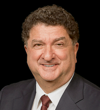
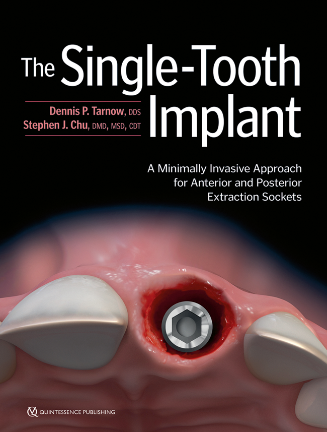
Dennis P. Tarnow, DDS, ist Klinikprofessor für Parodontologie und Leiter der Fachausbildung für Implantologie an der Columbia School of Dental Medicine. Er wurde mit dem Master Clinician Award der American Academy of Periodontology, dem Teacher of the Year Award der New York University und dem Distinguished Lecturer Award des American College of Prosthodontists ausgezeichnet. Er hat über 200 Artikel zur Perio-Prothetik und Implantologie veröffentlicht und ist Koautor von vier Lehrbüchern, darunter The Single-Tooth Implant (Quintessence Publishing, 2020) mit Dr. Stephen J. Chu. Dr. Tarnow betreibt eine Privatpraxis in New York City und hielt bereits Vorträge in über 45 Ländern.
A Minimally Invasive Approach for Anterior and Posterior Extraction Sockets
1st Edition 2019 Buch Hardcover, 21,6 x 27,9 cm, 240 Seiten, 1117 Abbildungen Sprache: Englisch Kategorien: Implantologie, Oralchirurgie, Prothetik Artikelnr.: B7710 ISBN 978-0-86715-771-0 QP USA
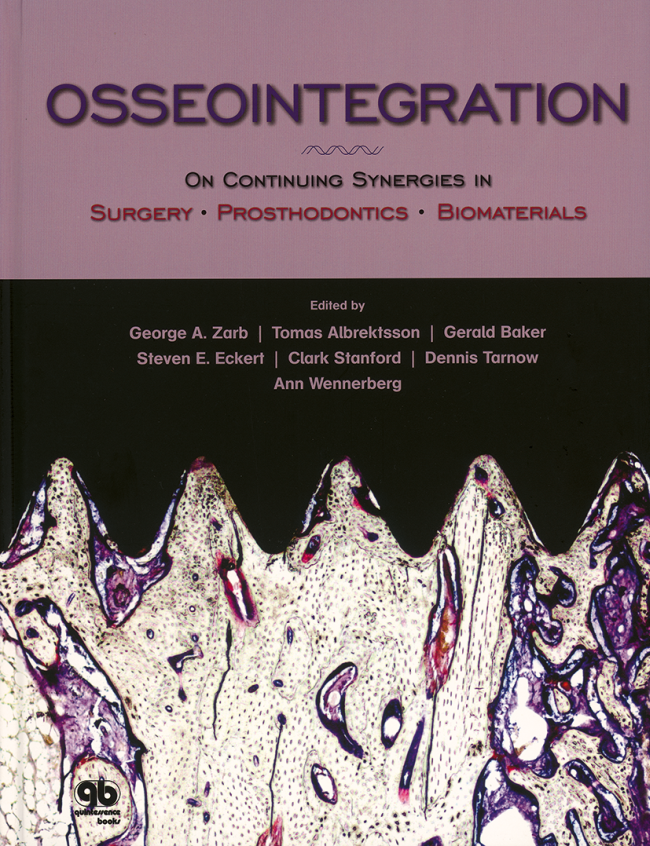
On Continuing Synergies in Surgery, Prosthodontics, and Biomaterials
1st Edition 2008 Buch Hardcover, 200 Seiten, 491 Abbildungen Sprache: Englisch Kategorie: Implantologie ISBN 978-0-86715-479-5 QP USA
Produkt vergriffen.
vergriffen
Veranstaltungen
SEPES – 50+ 1 Annual Congress
Oktober 13, 2022 — Oktober 15, 2022ExpoMeloneras, Maspalomas, Las Palmas, Spanien
Referenten: Nitzan Bichacho, Markus B. Blatz, Christian Coachman, Luca De Stavola, Mirela Feraru, Gustavo Giordani, Galip Gürel, Ronald Jung, Nazariy Mykhaylyuk, Nelson R. F. A. Silva, Dennis P. Tarnow
SEPES - Sociedad Española de Prótesis Estomatologías y Estética
The 14th International Symposium on Periodontics and Restorative Dentistry (ISPRD)
Juni 9, 2022 — Juni 12, 2022Boston Marriott Copley Place, Boston, MA, Vereinigte Staaten von Amerika
Referenten: Tara Aghaloo, Edward P. Allen, Evanthia Anadioti, Wael Att, Vinay Bhide, Markus B. Blatz, Scotty Bolding, Lorenzo Breschi, Jeff Brucia, Daniel Buser, Luigi Canullo, Daniele Cardaropoli, Stephen J. Chu, Donald Clem, Christian Coachman, Lyndon F. Cooper, Daniel Cullum, Lee Culp, José Carlos Martins da Rosa, Sergio De Paoli, Marco Degidi, Nicholas Dello Russo, Serge Dibart, Joseph P. Fiorellini, Mauro Fradeani, Stuart J. Froum, David Garber, Maria L. Geisinger, William Giannobile, Luca Gobbato, Ueli Grunder, Galip Gürel, Chad Gwaltney, Christoph Hämmerle, Robert A. Horowitz, Marc Hürzeler, David Kim, Gregg Kinzer, Christopher Köttgen, Ina Köttgen, Purnima S. Kumar, Burton Langer, Lydia Legg, Pascal Magne, Kenneth A. Malament, Jay Malmquist, George Mandelaris, Pamela K. McClain, Michael K. McGuire, Mauro Merli, Konrad H. Meyenberg, Craig M. Misch, Julie A. Mitchell, Marc L. Nevins, Myron Nevins, Michael G. Newman, Miguel A. Ortiz, Jacinthe M. Paquette, Stefano Parma-Benfenati, Michael A. Pikos, Giulio Rasperini, Pamela S. Ray, Christopher R. Richardson, Isabella Rocchietta, Marisa Roncati, Marco Ronda, Paul S. Rosen, Maria Emanuel Ryan, Irena Sailer, Maurice Salama, David M. Sarver, Takeshi Sasaki, Todd Scheyer, Massimo Simion, Michael Sonick, Sergio Spinato, Dennis P. Tarnow, Lorenzo Tavelli, Douglas A. Terry, Tiziano Testori, Carlo Tinti, Istvan Urban, Hom-Lay Wang, Robert Winter, Giovanni Zucchelli
Quintessence Publishing Co., Inc. USA
Zeitschriftenbeiträge dieses Autors
International Journal of Periodontics & Restorative Dentistry, 1/2024
DOI: 10.11607/prd.6733, PubMed-ID: 37552170Seiten: 115-126, Sprache: EnglischGiglio, Graziano D. / Giglio, Ana Becil / Tarnow, Dennis P.
The use of conventional scan bodies (SBs) with an intraoral scanner (IOS) to capture the position of a complete arch of dental implants has proven to be challenging. The literature is unclear about the accuracy of intraoral scanning techniques using SBs that are connected vertically to multiunit abutments (MUAs) for numerous adjacent implants in the same arch. Recently, there has been a paradigm shift from vertical SBs to horizontal SBs, which are positioned perpendicular to the long axis of the MUAs. Most IOSs available today can capture these horizontal SBs, called scan gauges (SGs), with better accuracy and consequently acquire the position of multiple adjacent implants using an effective scan path, thus reducing stitching and the number of images. The key to implementing this novel technology is to strategically arrange the SGs to optimize horizontal overlap of multiple adjacent SGs without touching each other. By superimposing two high-resolution intraoral scans of the SGs, an artificial intelligence (AI) algorithm is employed to produce a calibrated digital best-fit model on which a passive complete-arch prosthesis can be designed and fabricated. The advantages and disadvantages of SBs and SGs are discussed, and a case report using a digital workflow is presented.
International Journal of Periodontics & Restorative Dentistry, 4/2020
DOI: 10.11607/prd.4440, PubMed-ID: 32559033Seiten: 509-517, Sprache: EnglischChu, Stephen J. / Saito, Hanae / Östman, Pär-Olov / Levin, Barry P. / Reynolds, Mark A. / Tarnow, Dennis P.
Immediate tooth replacement therapy (ITRT), ie, immediate implant placement and provisional restoration in postextraction sockets, has been shown to achieve favorable outcomes in reference to soft tissue stability and esthetics. However, avoiding socket perforation with uniaxial implants in the anterior maxilla can be challenging due to the inherent anatomy. Dual or co-axis subcrestal angle correction (SAC) implants have been developed to change the restorative angle of the clinical crown restoration subcrestally at the implant-abutment interface to enhance the incidence of screw-retained definitive restorations. An additional benefit of this macrodesign implant feature is variable platform switching (VPS) that increases soft tissue gap distance above the implant platform. The purpose of this prospective study on ITRT in maxillary anterior postextraction sockets was to investigate the effect of SAC with VPS (SAC/VPS) compared to conventional platform-switch–design implants (PS) relative to ridge dimension stability and peri-implant soft tissue thickness. A total of 29 patients had undergone ITRT and received either a PS or SAC/VPS implant; previously described measurements were made compared to the contralateral natural tooth sites. When the comparison of buccal soft tissue thickness was made, SAC/VPS showed a greater increase compared to PS (3.12 mm vs 2.39 mm, respectively) with statistical significance (P = .05). The increase was independent from periodontal phenotype. Therefore, SAC/VPS may increase peri-implant soft tissue thickness and help minimize recession following ITRT.
International Journal of Periodontics & Restorative Dentistry, 7/2018
SupplementSeiten: 9-15, Sprache: EnglischSaito, Hanae / Chu, Stephen J. / Zamzok, Jonathan / Brown, Marion / Smith, Richard / Sarnachiaro, Guido / Hochman, Mark / Fletcher, Paul / Reynolds, Mark A. / Tarnow, Dennis P.
This paper presents the results of a prospective study on the clinical effect of grafting the buccal gap with platform switching, following flapless tooth extraction and immediate implant placement and provisional restoration, on (1) alterations in buccolingual ridge dimension and (2) midfacial peri-implant soft tissue thickness. Fifty-six patients were placed in one of four treatment groups: (1) a non-platform-switching implant (Non-PS, n = 14); (2) a non-platform-switching implant and bone graft (BG/Non-PS, n = 14); (3) a platform-switching implant (PS, n = 15); and (4) a platform-switching implant and bone graft (BG/PS, n = 13). Buccolingual ridge dimensional change and peri-implant soft tissue thickness at 2 mm below the free gingival margin were measured. Both PS and BG/PS groups showed an increase in buccolingual dimension compared to the contralateral natural tooth site as well as peri-implant soft tissue thickness of > 2.5 mm, clinically. Platform switching was associated with significantly greater midfacial soft tissue thickness than bone grafting (mean ± standard deviation: 2.17 ± 0.04 for Non-PS and 3.55 ± 0.14 for PS). Using platform-switched implants, in conjunction with immediate implant placement and immediate provisional restoration, was associated with a significantly greater stability of ridge dimension and peri-implant soft tissue thickness than using non-platform-switched implants when measured 12 months after placement of the final restoration. Platform switching may help achieve preservation of ridge dimension and enhance the stability of peri-implant soft tissue following immediate implant placement and provisional restoration without bone grafting. Further research is required to assess the long-term outcome.
International Journal of Periodontics & Restorative Dentistry, 7/2018
SupplementSeiten: 17-27, Sprache: EnglischChu, Stephen J. / Östman, Pär-Olov / Nicolopoulos, Costa / Yuvanoglu, Petros / Chow, James / Nevins, Myron / Tarnow, Dennis P.
A prospective cohort clinical study was performed to evaluate the concept and design of a novel macro hybrid implant placed into maxillary anterior postextraction sockets. Thirty-three patients with an equal number of hybrid implants were used to replace nonrestorable single anterior teeth with immediate tooth replacement therapy (immediate implant placement and immediate provisional restoration). The macro features of this hybrid implant are unique in geometry, as it combines two different shapes-a cylindrical coronal and tapered apical portion-into a singular body design, each comprising roughly half of the implant length. The hybrid design of this platform-switched implant also has a subcrestal angle correction, or Co-Axis feature, that facilitates screw-retained restorations. Mean implant survival at 1 year relative to primary stability, labial bone plate thickness with socket grafting at two reference points (L1 and L2), tooth-to-implant interproximal bone crest thickness, and pink esthetic score (PES) were evaluated. A mean insertion torque value of 65 Ncm (range 45 to 100 Ncm) was reached with the use of the tapered apical half of the implant body. No implants failed during an average healing period of 1 year. A labial plate dimension between 1.8 and 2.1 mm was attained immediately posttreatment and remained stable over time. A toothto- implant interdental bone crest distance and dimension of 2.3 to 2.6 mm was reached; it was also sustained at the 1-year follow-up. The average PES was 12.5 (range 9.0 to 14.0), with nearly 90% of treated sites with an "almost perfect" score. This macro hybrid implant in concept and design may be useful in immediate tooth replacement therapy of maxillary anterior postextraction sockets to achieve successful implant survival and esthetic outcomes, specifically labial plate and papilla preservation without midfacial or interdental tissue loss and discoloration.
Porous tantalum trabecular-structured metal (PTTM) has been applied to titanium orthopedic and dental implants. This study evaluated the healing pattern of bone growth into experimental PTTM cylinders (N = 24; 3.0 × 5.0 mm) implanted in the partially edentulous jaws of 23 healthy volunteers divided into four groups. Six PTTM cylinders per group were explanted, prepared, and analyzed histologically/metrically after 2, 3, 6, and 12 weeks of submerged healing. PTTM implant osseoincorporation resulted from the formation of an osteogenic tissue network that over the course of 12 weeks resulted in vascular bone volume levels in PTTM that are comparable to clinically observed mean trabecular volumes in edentulous posterior jaws.
Purpose: Maxillary sinus augmentation has been a predictable procedure. However, in-depth analysis of tissue healing after sinus grafting with simultaneous implant placement is limited. This study aimed to compare histologic outcomes after sinus grafting with a synthetic bone graft compared with a xenograft.
Materials and Methods: A randomized controlled split-mouth study was conducted to compare bone formation around microimplants (2.00 mm, Dentium) placed at the time of maxillary sinus augmentation with a synthetic material (Osteon, Dentium) (OST) and deproteinized bovine bone (Bio-Oss) (BIO) as the control group. Four microimplants per subject (n = 13) were placed bilaterally for intrasubject comparison (two implants per side/patient). Bone cores with osseointegrated microimplants were harvested for histomorphometric analysis 6 to 8 months after sinus augmentation surgery.
Results: Histologic analysis revealed newly formed bone deposited on the microimplant surface and bridging to bone graft material in both groups. Further, there was no histologic evidence of signs of inflammation in all specimens. In general, bone-to-implant contact was comparable and ranged from 6.1% to 67.0% with a mean of 38.4% ± 11.61% in OST and from 10.5% to 57.0% with a mean of 34.58% ± 12.55% in BIO. However, a significantly higher percentage of bone-to-implant contact in the first four threads of the grafted area was noted in OST compared with BIO (P = .016).
Conclusion: The synthetic OST was found to be equivalent to BIO in new bone formation and clinical success after sinus augmentation in conjunction with microimplant placement. Although there are some statistically significant differences in the histologic outcomes, the clinical relevance of these needs to be further evaluated. Nevertheless, the findings of this study indicate that this synthetic alloplast would be a viable alternative to an allograft material.
Schlagwörter: allografts, dental implants, sinus floor augmentation, treatment outcome, xenografts
International Journal of Periodontics & Restorative Dentistry, 4/2018
DOI: 10.11607/prd.3571, PubMed-ID: 29889914Seiten: 509-516, Sprache: EnglischChu, Stephen J. / Saito, Hanae / Salama, Maurice A. / Garber, David A. / Salama, Henry / Sarnachiaro, Guido O. / Reynolds, Mark A. / Tarnow, Dennis P.
This article presents the results of a soft tissue color study on flapless immediate implant therapy from a sample of 23 patients who received either a provisional restoration alone or with bone grafting. The gingival color in clinical photographs was measured for the implant and for the contralateral tooth site at 2.0 and 5.0 mm below the free gingival margin using Photoshop software (Lightroom CC, Adobe). The average color difference (ΔE) values for the two groups were 2.6 and 2.4 at 2.0 mm and 1.9 and 2.5 at 5.0 mm from the free gingival margin, respectively. Approximately 80% of the sites were below the visibly perceptible threshold (ΔE = 3.1 ± 1.5) and not detectable by the human eye. The use of provisional restorations has shown positive outcomes on the stability of peri-implant soft tissue thickness and lower ΔE values. Further research is required to assess esthetic outcomes inclusive of color change relative to the clinical treatment rendered.
International Journal of Periodontics & Restorative Dentistry, 4/2017
DOI: 10.11607/prd.3037, PubMed-ID: 28196152Seiten: 498-508, Sprache: EnglischFletcher, Paul / Deluiz, Daniel / Tinoco, Eduardo M. B. / Ricci, John L. / Tarnow, Dennis P. / Tinoco, Justine Monnerat
The treatment of peri-implant disease is one of the most controversial topics in implant dentistry. The multifactorial etiology and the myriad proposed techniques for managing the problem make successful decontamination of an implant surface affected by peri-implantitis one of the more unpredictable challenges dental practitioners have to face. This article presents the first known published case report demonstrating human histologic evidence of reosseointegration using a plastic curette for mechanical debridement and dilute sodium hypochlorite, hydrogen peroxide, and sterile saline for chemical detoxification. Guided bone regeneration in the infrabony component of the peri-implantitis lesion was accomplished using calcium sulfate and bovine bone as grafting materials and a porcine collagen barrier for connective tissue and epithelial exclusion.