Implantologie, 2/2009
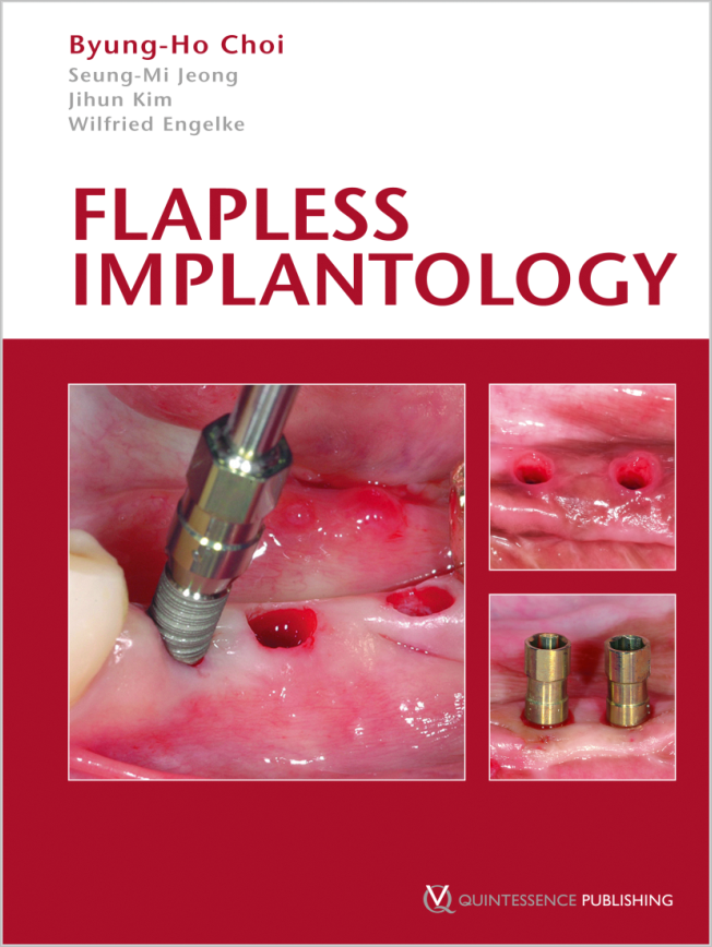
Seiten: 139-152, Sprache: DeutschChoi, Byung-Ho / Engelke, WilfriedDie Fortschritte bei der Oberflächengestaltung moderner Implantatsysteme haben zu einer deutlich verbesserten Osseointegration geführt. Moderne bildgebende Verfahren ermöglichen, dass die Implantatinsertion heute mit einer wesentlich höheren Präzision geplant werden kann. Deshalb erscheint es bedeutsam, auch die chirurgische Insertionstechnik von Implantaten so weit zu optimieren, dass bei minimiertem chirurgischen Trauma vorhersagbare Ergebnisse für Funktion, Ästhetik und Patientenkomfort erzielt werden können. Bei der Insertion von Implantaten ohne Bildung eines mukoperiostalen Lappens [geschlossene Implantation = flapless implantation (FI)] resultiert eine geringere Narbenbildung der umgebenden Gewebestrukturen im Vergleich zum offenen Vorgehen; es kommt zu einer besseren Vaskularisierung der periimplantären Mukosa mit einer reduzierten Sulkustiefe, und der initiale Knochenabbau fällt geringer aus. Das geschlossene Vorgehen verbessert weiterhin ästhetische Resultate durch den Erhalt der ursprünglichen Form und Dicke der die Implantate umgebenden Weichgewebe. Ein bedeutsamer Nachteil dieser Technik ist der Mangel an Übersicht über die Hartgewebekontur während des Eingriffs. Der vorliegende Artikel beschreibt das operative Vorgehen bei "flapless implantation" und zeigt darüber hinaus Möglichkeiten auf, wie verfahrensbedingte Probleme gelöst werden können. Dies gilt auch für anspruchsvolle klinische Fälle und schwierige Knochensituationen. Die Beobachtung geschlossener Operationsgebiete, wie subgingivaler Räume und interner Knochenstrukturen, mit Hilfe endoskopischer Verfahren kann dazu beitragen, die Therapiequalität der geschlossenen Implantation weiter zu optimieren.
Schlagwörter: Geschlossene Implantation, geschlossene Augmentation, Miniflap, Endoskopie, minimalinvasive Implantation
The International Journal of Oral & Maxillofacial Implants, 3/2007
PubMed-ID: 17622008Seiten: 417-422, Sprache: EnglischYou, Tae-Min / Choi, Byung-Ho / Zhu, Shi-Jiang / Jung, Jae-Hyung / Lee, Seoung-Ho / Huh, Jin-Young / Lee, Hyun-Jung / Li, JingxuPurpose: The aim of this study was to compare the effects of platelet-enriched fibrin glue and platelet-rich plasma (PRP) on the repair of bone defects adjacent to titanium dental implants.
Materials and Methods: In 6 mongrel dogs, 3 screw-shaped titanium dental implants per dog were placed into the osteotomy sites in the tibia. Before implantation, a standardized gap (2.0 mm) was created between the implant surface and the surrounding bone walls. Six gaps were left empty (control group), 6 gaps were filled with autogenous particulate bone mixed with PRP (PRP group), and 6 gaps were filled with autogenous particulate bone mixed with platelet-enriched fibrin glue (fibrin glue group).
Results: After 6 weeks, the bone-implant contact was 59.7% in the fibrin glue group, 29.2% in the PRP group, and 10.2% in the control defects; this difference was statistically significant (P .05).
Discussion and Conclusion: Greater bone-implant contact was achieved with platelet-enriched fibrin glue than with PRP. The results indicate that platelet-enriched fibrin glue can induce a stronger peri-implant bone reaction than PRP in the treatment of bone defects adjacent to titanium dental implants.
Schlagwörter: bone grafting, bone regeneration, dental implants, fibrin glue, platelet-rich plasma
The International Journal of Oral & Maxillofacial Implants, 2/2000
Seiten: 193-196, Sprache: EnglischChoi, Byung-HoThe aim of this study was to investigate whether a new periodontal ligament attachment will form on titanium implants when they are implanted with cultured periodontal ligament cells. Periodontal ligament cells obtained from the teeth of 3 dogs were cultured and attached to the surface of titanium implants. The implants with the cultured autologous periodontal ligament cells were placed in the mandibles of the dogs. After 3 months of healing, histologic examination revealed that, on some implant surfaces, a layer of cementum-like tissue with inserting collagen fibers had been achieved. These results demonstrated that cultured periodontal ligament cells can form tissue resembling a true periodontal ligament around implants.
Schlagwörter: cell culture, implants, periodontal ligament, titanium implants


