ENDO, 4/2017
Seiten: 243-248, Sprache: EnglischWarnsinck, Jan / Koutris, Michail / Shemesh, Hagay / Lobbezoo, FrankPain in the dentoalveolar region is a common symptom. This usually involves acute pain, meaning the correct treatment can be rapidly applied. However, for certain types of persistent pain in this region, the aetiology is difficult to determine. Previously, there has been confusion regarding the diagnosis and classification of this type of persistent pain, which was often designated under different terms, such as atypical odontalgia, phantom pain or deafferentation pain. Recently, a classification system was developed for these types of conditions and the following term was agreed: persistent dentoalveolar pain. This new term was the first step in arriving at an improvement in the taxonomy: three criteria were established to lead to advancements in the fields of clinical research and treatment.
Schlagwörter: neuropathic pain, non-odontogenic pain
ENDO, 3/2017
Seiten: 167-171, Sprache: EnglischWarnsinck, Jan / Shemesh, HagayPeriapical lesions regularly occur in untreated teeth, as well as in teeth that have already been subjected to endodontic treatment. There are various known factors that lead to effective endodontic treatment where the clinical symptoms disappear and the periapical lesion disappears or decreases in size. The risk of painful inflammation alongside a persistent periapical lesion is small, even if the periapical lesion has increased in size. The survival of a tooth with a periapical lesion that has undergone root canal treatment is 87% after 10 years. In cases where an endodontically treated tooth has to be extracted, a restorative problem is often the reason for extraction, while a persistent periapical lesion has a limited effect on tooth loss. There are insufficient data available on the effect of a periapical lesion on general health.
Schlagwörter: effectiveness, general health, pain, periapical lesion, survival
Quintessence International, 8/2015
DOI: 10.3290/j.qi.a34396, PubMed-ID: 26185797Seiten: 657-668, Sprache: EnglischCohenca, Nestor / Shemesh, HagayPart 2: Applications associated with advanced endodontic problems and complicationsThe use of cone beam computed tomography (CBCT) in endodontics has been extensively reported in the literature. Compared with the traditional spiral computed tomography, limited field of view (FOV) CBCT results in a fraction of the effective absorbed dose of radiation. The purpose of this manuscript is to review the application and advantages associated with advanced endodontic problems and complications, while reducing radiation exposure during complex endodontic procedures. The benefits of the added diagnostic information provided by intraoperative CBCT images in select cases justify the risk associated with the limited level of radiation exposure.
Schlagwörter: cone beam computed tomography, dental trauma, intraoperative, outcome, root resorption
Quintessence International, 6/2015
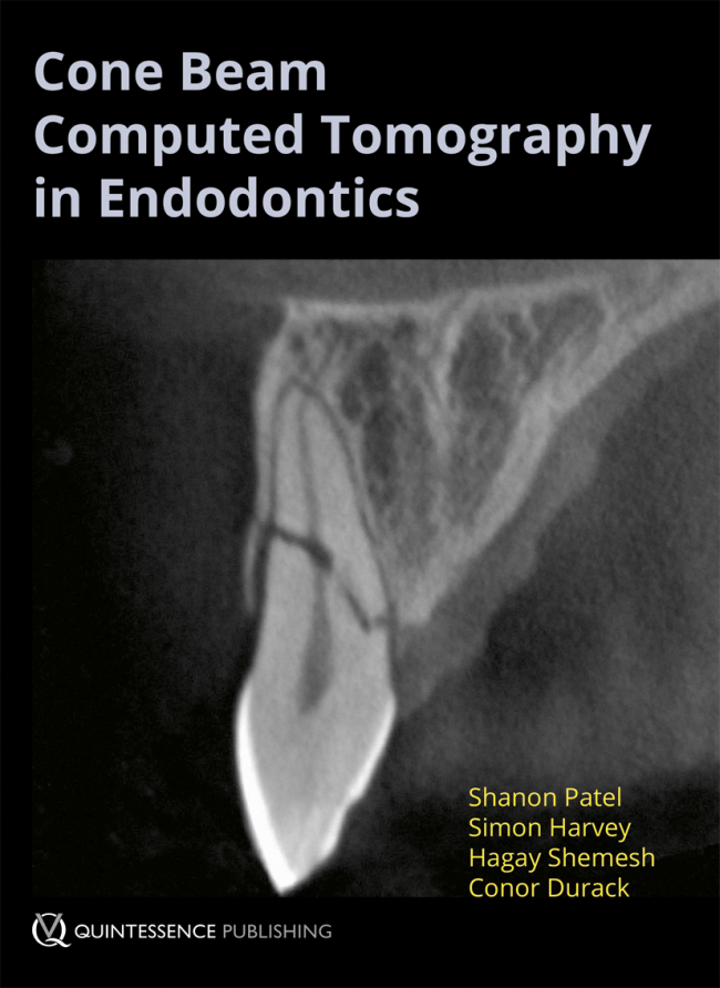
DOI: 10.3290/j.qi.a33990, PubMed-ID: 25941678Seiten: 465-480, Sprache: EnglischCohenca, Nestor / Shemesh, HagayPart 1: Applications associated with endodontic treatment and diagnosisCone beam computed tomography (CBCT) is a new technology that produces three-dimensional (3D) digital imaging at reduced cost and less radiation for the patient than traditional CT scans. It also delivers faster and easier image acquisition. By providing a 3D representation of the maxillofacial tissues in a cost- and dose-efficient manner, a better preoperative assessment can be obtained for diagnosis and treatment. This comprehensive review presents current applications of CBCT in endodontics. Specific case examples illustrate the difference in treatment planning with traditional periapical radiography versus CBCT technology.
Schlagwörter: 3D, cone beam computed tomography, endodontics, root canal therapy
The Journal of Adhesive Dentistry, 6/2014
DOI: 10.3290/j.jad.a33200, PubMed-ID: 25516883Seiten: 567-574, Sprache: EnglischMoinzadeh, Amir T. / Mirmohammadi, Hesam / Veenema, Tjibbe / Kleverlaan, Cornelis J. / Wesselink, Paul R. / Wu, Min-Kai / Shemesh, HagayPurpose: To investigate whether the placement of a methacrylate root canal sealer or a conventional epoxy root canal sealer in two steps increases their dislocation resistance when compared to a one-step placement procedure.
Materials and Methods: Eighty single-rooted teeth were randomly allocated to 4 groups (n = 20). All canals were instrumented to size 40, 0.06 taper and irrigated according to a standardized protocol. Root canal filling was conducted as follows: group 1: methacrylate sealer placed in two steps; group 2: methacrylate sealer placed in one step; group 3: epoxy sealer placed in two steps; group 4: epoxy sealer placed in one step. After setting, thin slices at different root levels were obtained and submitted to push-out testing. Results were analyzed with non-parametric tests to compare the two-step procedures to their one-step counterparts. Failure modes were determined by stereomicroscopy. Random untested methacrylate sealer specimens were also examined with scanning electron microscopy.
Results: At each root level, dislocation resistance was significantly higher for the two-step procedure than for the one-step procedure using the methacrylate sealer (p = 0.003, p = 0.005, p 0.001) but not the epoxy sealer (p = 0.83, p = 0.1, p = 0.06). Among root levels, there were no significant differences in dislocation resistance in the methacrylate sealer two-step group, while all other groups showed differences.
Conclusion: A two-step placement procedure resulted in significantly higher dislocation resistance for the methacrylate sealer but not for the epoxy sealer.
Schlagwörter: adhesion, configuration factor, dislocation resistance, methacrylate resin, polymerization shrinkage, root canal sealer
ENDO, 4/2013
Seiten: 275-280, Sprache: EnglischMarques, Miguel / Shemesh, Hagay / van der Sluis, LucAfter pulp exposure, direct pulp capping can help preserve pulp vitality. As reported in the related literature on direct pulp capping recently, good results have been obtained when MTA was used as capping material. Before the placement of the MTA, all infected dentine was removed. In this article, three case reports are presented describing this treatment procedure, combined with a follow-up of the treatment.
Schlagwörter: deep caries lesion, direct pulp capping, MTA, pulp vitality preservation
Endodontie, 4/2011
Seiten: 399-401, Sprache: DeutschWu, Min-Kai / Shemesh, Hagay / Wesselink, Paul R.Epidemiologische Untersuchungen versetzen uns in die Lage, potenzielle Verbindungen zwischen periapikalen Entzündungen endodontischen Ursprungs und systemischen Erkrankungen zu entdecken. Diese Verbindungen belegen die Notwendigkeit einer effektiven endodontischen Behandlung für die Allgemeingesundheit und das Wohlbefinden der Patienten. Gut geplante und durchgeführte prospektive klinische Studien sind notwendig, um das Ergebnis endodontischer Behandlung zu ermitteln. Die Resultate klinischer Studien zur Erfolgsquote erlauben es uns, die Prognose unterschiedlicher Therapieverfahren abzuschätzen und den Patienten in seiner Therapieentscheidung so zu unterstützen, dass er oder sie auf der Grundlage valider Informationen die beste Therapie für sein spezifisches Problem auswählen kann (Informed Consent).


