Wir verwenden Cookies ausschließlich zu dem Zweck, technisch notwendige Funktionen wie das Login oder einen Warenkorb zu ermöglichen, oder Ihre Bestätigung zu speichern. Mehr Informationen zur Datenerhebung und -verarbeitung finden Sie in unserer Datenschutzerklärung.
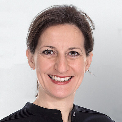
Prof. Dr. Irena Sailer promovierte an der Universität Tübingen in Deutschland (1997/1998). Sie erhielt eine Assistenzprofessur an der Klinik für festsitzende und herausnehmbare Prothetik und zahnärztliche Materialkunde in Zürich, Schweiz (2003), wo sie ab 2010 als außerordentliche Professorin tätig war. Im Jahr 2007 war Prof. Dr. Sailer Visiting Scholar am Department of Biomaterials and Biomimetics am Dental College der New York University, USA. Seit 2009 ist sie Gastprofessorin am Department of Preventive and Restorative Sciences, Robert Schattner Center, University of Pennsylvania in Philadelphia, USA (Leitung: Prof. Dr. M. B. Blatz). Prof. Dr. Sailer ist Direktorin der Abteilung für festsitzende Prothetik und Biomaterialien an der Universität Genf, Schweiz. Im Jahr 2019 erhielt sie eine Honorarprofessur an der Universität Aarhus, Dänemark. Sie ist Spezialistin für Prothetik (Schweizerische Gesellschaft für Rekonstruktive Zahnmedizin) und besitzt ein Zertifikat für besondere Aktivitäten in der dentalen Implantologie (WBA) der Schweizerischen Gesellschaft für Zahnmedizin. Sie ist Mitglied des Vorstands der European Association of Osseointegration (EAO), Vizepräsidentin der European Academy of Esthetic Dentistry (EAED), Mitglied der Schweizerischen Gesellschaft für Rekonstruktive Zahnmedizin, des Education Committee des International Team for Implantology (ITI) und der Greater New York Academy of Prosthodontics (GNYAP) sowie Chefredakteurin des International Journal of Prosthodontics und hat zahlreiche Publikationen veröffentlicht.
Bridging the Gap Between Science and Clinical Practice
Erscheinungsweise: zweimonatlich Sprache: Englisch Impact Faktor: 2.1 (2023) Kategorie: Prothetik Schriftleitung / Chefredaktion: Prof. Dr. Irena Sailer QP USA
Details make perfectionOktober 24, 2024 — Oktober 26, 2024MiCo - Milano Convention Centre, Milano, Italien
Referenten: Bilal Al-Nawas, Gil Alcoforado, Federico Hernández Alfaro, Sofia Aroca, Wael Att, Gustavo Avila-Ortiz, Kathrin Becker, Anne Benhamou, Juan Blanco Carrión, Dieter Bosshardt, Daniel Buser, Francesco Cairo, Paolo Casentini, Raffaele Cavalcanti, Tali Chackartchi, Renato Cocconi, Luca Cordaro, Luca De Stavola, Nuno Sousa Dias, Egon Euwe, Vincent Fehmer, Alberto Fonzar, Helena Francisco, Lukas Fürhauser, German O. Gallucci, Oscar Gonzalez-Martin, Dominik Groß, Robert Haas, Alexis Ioannidis, Simon Storgård Jensen, Ronald Jung, France Lambert, Luca Landi, Georg Mailath-Pokorny jun., Silvia Masiero, Iva Milinkovic, Carlo Monaco, Jose Nart, José M. Navarro, Katja Nelson, Manuel Nienkemper, David Nisand, Michael Payer, Sergio Piano, Bjarni E. Pjetursson, Sven Reich, Isabella Rocchietta, Giuseppe Romeo, Irena Sailer, Mariano Sanz, Ignacio Sanz Martín, Frank Schwarz, Shakeel Shahdad, Massimo Simion, Ralf Smeets, Benedikt Spies, Bogna Stawarczyk, Martina Stefanini, Hendrik Terheyden, Tiziano Testori, Daniel Thoma, Ana Torres Moneu, Piero Venezia, Lukas Waltenberger, Hom-Lay Wang, Stefan Wolfart, Giovanni Zucchelli, Otto Zuhr
European Association for Osseintegration (EAO)
35th EAED Spring Open Meeting
35 years of Esthetic Dentistry - The masters, the fundamentals, today's trendsMai 23, 2024 — Mai 25, 2024The Baron's Hall & Gallery, Vila Nova Gaia, Portugal
Referenten: Luís Azevedo, Urs C. Belser, Nitzan Bichacho, Markus B. Blatz, Jorge Cardoso, Nuno Sousa Dias, Petra Gierthmühlen, Aiste Gintaute, Oscar Gonzalez-Martin, Stefano Gracis, Ueli Grunder, Arndt Happe, Marc Hürzeler, France Lambert, Amélie Mainjot, Alexandros Manolakis, Konrad H. Meyenberg, Nazariy Mykhaylyuk, José M. Navarro, Gaetano Paolone, Stavros Pelekanos, Roberto Perasso, João Pitta, Pablo Ramírez, Andrea Ricci, Giano Ricci, Giuseppe Romeo, Irena Sailer, David Winkler, Yu Zhang, Giovanni Zucchelli
73rd Annual Scientific Session of the American Academy of Fixed Prosthodontics
Navigating Expectations in Fixed ProsthodonticsFebruar 23, 2024 — Februar 24, 2024Chicago Marriott Downtown Magnificent Mile, Chicago, Vereinigte Staaten von Amerika
Referenten: Karen Baker, Lino Calvani, Stephen J. Chu, Arian Deutsch, Terry E. Donovan, Nicholas L. Egbert, German O. Gallucci, Sarit Kaplan, Sonia Leziy, Mariam Malament, Radi Masri, Bradley A. Purcell, Irena Sailer
American Academy of Fixed Prosthodontics
5th ITI Congress Middle East
Innovations and trends in the digital eraOktober 18, 2023 — Oktober 20, 2023Hilton Dead Sea Resort & Spa, Sweimeh , Jordanien
Referenten: Motasum Abu-Awwad, Esem Alem, Rola Alhabashneh, Arwa Alsayed, Kai-Hendrik Bormann, Hossam Elsabagh, Vincent Fehmer, German O. Gallucci, Ahmad Hamdan, Najla Kasabreh, Stephanie Khalaf, Lina Khasawneh, Carla Maria Kheirallah, Stephanie Mrad, Dalia Nourah, Mario Roccuzzo, Moustapha Saad, Irena Sailer, Anton Sculean, Mahmoud Shalash, Hani Tohme, Fahad Umer, Ronald Younes
ITI International Team for Implantology
The Buser & Belser Master Course on Esthetic Implant Dentistry
Referenten: Samir Abou-Ayash, Urs C. Belser, Dieter Bosshardt, Daniel Buser, Jordi Caballé-Serrano, Stephen Chen, Vincent Fehmer, Manrique Fonseca, Ronald Jung, Irena Sailer, Anton Sculean
Buser & Sculean Academy
The 9th World Dental Meeting in Japan 2023
No Dentistry, No Wellness!September 29, 2023 — Oktober 1, 2023Pacifico Yokohama Conference Center, Yokohama, Japan
Referenten: Alessandro Agnini, Andrea Mastrorosa Agnini, Wael Att, Gustavo Avila-Ortiz, Markus B. Blatz, Victor Clavijo, Karim Dada, Glécio Vaz de Campos, Vincent Fehmer, Naoki Hayashi, Mario Imburgia, Guillaume Jouanny, Sérgio Kahn, Bertrand Khayat, Sandra Khong Tai, Christopher Köttgen, Stefen Koubi, Tomas Linkevičius, Nazariy Mykhaylyuk, Ravindra Nanda, Andreas Nolte, Léon Parienté, Jose Manuel Reuss , Domenico Ricucci, Isabella Rocchietta, Irena Sailer, Todd R. Schoenbaum, Werner Schupp, Istvan Urban, Eric Van Dooren, Débora R. Vilaboa, Otto Zuhr
Quintessence Publishing Co. Ltd. Japan
Zeitschriftenbeiträge dieses Autors
The International Journal of Prosthodontics, 7/2024
Open Access Supplement Online OnlyDOI: 10.11607/ijp.8538, PubMed-ID: 38489221Seiten: s55-s62, Sprache: EnglischWan, Qiucen / Lee, Jae-Hyun / Daher, René / Karasan, Duygu / Myagmar, Gerelmaa / Sailer, Irena
Purpose: To evaluate the wear resistance of a printed interim resin manufactured with different printing and postpolymerization parameters. Materials and Methods: Overall, 130 rectangular resin specimens (15 × 10 × 10 mm) were 3D-printed. Among the specimens, 60 were printed with different printing orientations (0, 45, and 90 degrees) and layer thicknesses (50 and 100 μm) to create six groups to investigate the effects of the printing parameters (n = 10 per group). The remaining 70 specimens were used to evaluate the effects of postpolymerization; for this, seven groups were created as follows (n = 10 per group): nonpostpolymerized; postpolymerized for 5, 15, and 30 minutes with an ultraviolet light–emitting diode (LED) device; and postpolymerized for 5, 15, and 30 minutes with an ultraviolet light bulb device. After masticatory simulation, the wear volume loss was calculated with 3D metrology software. One-way and two-way ANOVA were used for intergroup comparisons (α = .05). Results: The group printed with a build angle of 45 degrees showed lower wear volume loss than the 0- and 90-degree groups (P < .01). The wear volume loss in the ultraviolet LED group was significantly greater than that in the ultraviolet light bulb group (P = .04). No significant difference was observed in the wear volume loss of the printed resin with respect to the layer thickness and polymerization time (P > .05). However, the non-postpolymerized group showed significantly greater wear volume loss than the other groups (P < .001). Conclusions: The printed resin showed greater wear resistance when it was printed at a build angle of 45 degrees and postpolymerized with an ultraviolet light bulb device.
Purpose: To evaluate the accuracy of complete-arch digital implant impressions using different intraoral scan body (ISB) materials and intraoral scanners (IOSs). Materials and Methods: The mandibular dental cast of an edentulous patient with six tissue-level dental implants was used as the master cast. Two types of ISBs—polyether ether ketone (PEEK) and plasma-coated medical titanium—were used with five IOSs: TRIOS 4 (T4), Virtuo Vivo (VV), Medit i700 (Mi700), iTero 5D (i5D), and Primescan (PS). To assess accuracy, digital impressions (n = 10) with each IOS and ISB were compared to two reference models obtained by digitizing the master cast with each ISB type using a desktop scanner (IScan4D LS3i) and importing the scan data into metrology software (Geomagic Control X). Root mean square (RMS) error was employed to evaluate overall deviation values (trueness), while precision was determined using the SD of RMS values. Statistical significance was set at P < .05. Kruskal-Wallis test was used, followed by the pairwise comparison method with Bonferroni correction (α = .05). Results: An interaction between ISB material and IOS was found (P = .001). Plasma-coated medical titanium ISBs demonstrated significantly higher trueness and precision compared to PEEK ISBs with T4 (P = .001), Mi700 (P = .001; P = .004), and i5D (P = .001). Conversely, VV exhibited higher trueness and precision values with PEEK ISBs (P = .005; P = .003). PS provided the highest trueness and precision regardless of the ISB material (P = .912). T4 showed the lowest accuracy for PEEK ISBs, and VV showed the lowest accuracy for plasma-coated medical titanium ISBs. Conclusions: Except for PS, all IOSs showed significant differences between ISB materials. PS demonstrated the highest accuracy with both ISB materials, whereas T4 had the lowest accuracy for PEEK ISBs, and VV showed the lowest accuracy for plasma-coated medical titanium ISBs.
Purpose: To measure the surface temperature distribution after using a CO2 laser to heat titanium dental implants via different power settings, application intervals, and irradiation times. Materials and Methods: A total of 10 tissue-level titanium implants (Screw-Line Promote Plus, Camlog; 4.3 × 11 mm) were placed (EpoFix, Struers) and irradiated with a CO2 laser (Denta 2, Lutronic) with a wavelength of 10.6 μm at power levels of 4 watts (Group 1), 6 watts (Group 2), 8 watts (Group 3), and 10 watts (Group 4). A continuous beam mode (setting I) and noncontinuous beam modes with 5-second (setting II) and 10-second (setting III) pause intervals were used. For each setting, a total irradiation time of 50 seconds was used and repeated 10 times. The temperature was measured using external thermocouple (Testo) in contact with the implant surface at the implant shoulder, middle, and apex. A linear regression model was used to analyze the data (P = .05). Results: Setting I demonstrated the most rapid increase in implant surface temperature in all three test sites as well as the greatest total temperature at 50 seconds of irradiation time. The greater the pause interval (settings II and III) during the 50 seconds of irradiation, the lower the rate of temperature increase as well as the total temperature in all three test sites and with all power levels. The average temperature difference between the apex and shoulder site was significant for setting III for all groups, but not for any groups in settings I and II. Conclusions: Heating the internal aspect of an implant with a CO2 laser produces different temperature distribution profiles depending on the laser power level and the application interval. Laser-beam irradiation leads to a temperature gradient, which is greatest at the implant apex and smallest at the implant shoulder.
The International Journal of Prosthodontics, 2/2024
DOI: 10.11607/ijp.8852, PubMed-ID: 38270461Seiten: 221-224, Sprache: EnglischRevilla-León, Marta / Barmak, Basir A. / Sailer, Irena / Kois, John C. / Att, Wael
Purpose: To compare the performance of licensed dentists and two software versions (3.5 legacy and 4.0) of an artificial intelligence (AI)-based chatbot (ChatGPT) answering the exam for the 2022 Certification in Implant Dentistry of the European Association for Osseointegration (EAO). Materials and Methods: The 50-question, multiple-choice exam of the EAO for the 2022 Certification in Implant Dentistry was obtained. Three groups were created based on the individual or program answering the exam: licensed dentists (D group) and two software versions of an artificial intelligence (AI)-based chatbot (ChatGPT)—3.5 legacy (ChatGPT-3.5 group) and the 4.0 version (ChatGPT-4.0 group). The EAO provided the results of the 2022 examinees (D group). For the ChatGPT groups, the 50 multiple-choice questions were introduced into both ChatGBT versions, and the answers were recorded. Pearson correlation matrix was used to analyze the linear relationship among the subgroups. The inter- and intraoperator reliability was calculated using Cronbach’s alpha coefficient. One-way ANOVA and Tukey post-hoc tests were used to examine the data (α = .05). Results: ChatGPT was able to pass the exam for the 2022 Certification in Implant Dentistry of the EAO. Additionally, the software version of ChatGPT impacted the score obtained. The 4.0 version not only pass the exam but also obtained a significantly higher score than the 3.5 version and licensed dentists completing the same exam. Conclusions: The AIbased chatbot tested not only passed the exam but performed better than licensed dentists.
Purpose: To validate an innovative 3D volumetric method of evaluating tissue changes proposed by Lee et al in 2020 by comparing the results of this method—in which the scanned peri-implant surfaces were transformed, visualized, and analyzed as 3D objects—to the results reported by an existing method based on calculation of the mean distance between measured surfaces. The null hypothesis was that there was no statistically significant difference between the two methods. Additionally, the present study evaluated peri-implant tissue changes 5 years after single implant placement in the esthetic zone. Materials and Methods: Both methods were applied to 11 oral implant site casts (6 maxillary central incisor sites, 5 maxillary lateral incisor sites) taken from 11 patients at crown placement and at follow-up examinations 5 years later. The methods are based on digital workflows in which the reference and 5-year casts are scanned and the resulting STL files are superimposed and analyzed for three regions of interest (mesial papilla, central area, and distal papilla). The volumetric changes reported by the Lee et al method and the mean distance method were calculated and compared using the Spearman rank correlation coefficient (P < .01) and the Wilcoxon signed-rank test (P < .05). Results: The correlation between the two sets of measurements was very high (Spearman rank correlation coefficient = 0.885). The new volumetric method indicated a mean volume loss of 2.82 mm3 (SD: 5.06), while the method based on the measurement of mean distance showed a mean volume loss of 2.92 mm3 (SD: 4.43; Wilcoxon signed-rank test result: P = .77). No statistically significant difference was found. The two methods gave equivalent results, and the null hypothesis was accepted. Conclusions: The new volumetric method was validated and can be considered a trustworthy tool.
Schlagwörter: volumetric analysis, digital workflow, peri-implant tissue, esthetic zone
Eine retrospektive Studie mit bis zu 27 Jahren Nachbeobachtung
Ziel: In dieser retrospektiven Studie sollte die (technische und biologische) Langzeitbewährung ausgedehnter keramischer Oberkieferfrontzahnveneers mit einer inzisalen Dicke von mehr als 2 mm untersucht werden.
Material und Methoden: Patienten, die zwischen 1990 und 2003 an der Universitätszahnklinik Genf von demselben Zahnarzt mit ausgedehnten Keramikveneers versorgt worden waren, wurden zu einer Nachuntersuchung eingeladen. Von den 37 identifizierten Patienten erklärten sich zehn Patienten mit 50 Veneers zu der Nachuntersuchung bereit und wurden inkludiert. Die Überlebensraten sowie die technischen und biologischen Ergebnisse wurden durch eine klinische Untersuchung bestimmt (anhand der modifizierten United States Public Health Services-Kriterien). Zusätzlich wurden die Patientenakten ausgewertet, um die Patientendaten und alle Komplikationsereignisse zu erfassen. Schließlich wurden Patient-R eported Outcomes (PRO) zu den Aspekten Ästhetik, Funktion und Phonetik, Kaufunktion, Zahnempfindlichkeit und Bereitschaft zu einer Ersatzversorgung im Fall eines Restaurationsversagens mithilfe visueller Analogskalen erhoben. Die Daten wurden deskriptiv ausgewertet. Die Analyse des Restaurationsüberlebens und der Komplikationsereignisse erfolgte mittels Kaplan-Meier-Schätzer.
Ergebnisse: Die Überlebensrate der Veneers betrug 96 % nach im Mittel 20,7 ± 3,7 Jahren in Funktion. Die Rate technischer Komplikationen lag bei 30 %. Diese Komplikationen bestanden in zwei Veneerverlusten, neun reparablen Frakturen, drei Rissbildungen und einer Veneerverlagerung durch Zahnunfall. Es fanden sich weder über Kreideflecken hinausgehende Kariesläsionen noch endodontische Komplikationen dokumentiert. Die PRO bezüglich Zufriedenheit mit der Ästhetik und der Phonetik waren sehr gut.
Schlussfolgerungen: Im Rahmen dieser retrospektiven Studie erwiesen sich ausgedehnte Keramikveneers, gemessen an den klinischen Ergebnissen und der Patientenzufriedenheit, als langfristig erfolgreiche Versorgungsmöglichkeit.
Schlagwörter: adhäsive Zahnmedizin, Ästhetik, Keramik, Prothetik, restaurative Zahnmedizin, Veneers