Wir verwenden Cookies ausschließlich zu dem Zweck, technisch notwendige Funktionen wie das Login oder einen Warenkorb zu ermöglichen, oder Ihre Bestätigung zu speichern. Mehr Informationen zur Datenerhebung und -verarbeitung finden Sie in unserer Datenschutzerklärung.
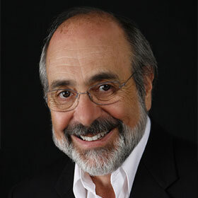
David A. Garber, DMD, is a past president of the American Academy of Esthetic Dentistry (AAED) and has served on the boards of the AAED and the American Academy of Fixed Prosthodontics. He is dual trained clinician and professor in the Department of Periodontics as well as in the Department of Oral Rehabilitation at the Medical College of Georgia. He is a clinical professor in the Department of Prosthodontics at Louisiana State University and in the Department of Restorative Dentistry at the University of Texas in San Antonio.
Veranstaltungen
12th World Congress of Esthetic Dentistry – IFED 2022
Oktober 27, 2022 — Oktober 29, 2022Conrad Abu Dhabi Etihad Towers, Abu Dhabi, Vereinigte Arabische Emirate
Referenten: Gil Alcoforado, Andreas Bindl, Renato Cocconi, Luca Cordaro, David De Franco, Didier Dietschi, Vincent Fehmer, Stefano Gracis, Dan Grauer, Sonia Leziy, Feng Liu, Baldwin Marchack, Francesco Mintrone, Robert Relle, Giovanni Zucchelli
International Federation of Esthetic Dentistry
The 14th International Symposium on Periodontics and Restorative Dentistry (ISPRD)
Juni 9, 2022 — Juni 12, 2022Boston Marriott Copley Place, Boston, MA, Vereinigte Staaten von Amerika
Referenten: Tara Aghaloo, Edward P. Allen, Evanthia Anadioti, Wael Att, Vinay Bhide, Markus B. Blatz, Scotty Bolding, Lorenzo Breschi, Jeff Brucia, Daniel Buser, Luigi Canullo, Daniele Cardaropoli, Stephen J. Chu, Donald Clem, Christian Coachman, Lyndon F. Cooper, Daniel Cullum, Lee Culp, José Carlos Martins da Rosa, Sergio De Paoli, Marco Degidi, Nicholas Dello Russo, Serge Dibart, Joseph P. Fiorellini, Mauro Fradeani, Stuart J. Froum, David Garber, Maria L. Geisinger, William Giannobile, Luca Gobbato, Ueli Grunder, Galip Gürel, Chad Gwaltney, Christoph Hämmerle, Robert A. Horowitz, Marc Hürzeler, David Kim, Gregg Kinzer, Christopher Köttgen, Ina Köttgen, Purnima S. Kumar, Burton Langer, Lydia Legg, Pascal Magne, Kenneth A. Malament, Jay Malmquist, George Mandelaris, Pamela K. McClain, Michael K. McGuire, Mauro Merli, Konrad H. Meyenberg, Craig M. Misch, Julie A. Mitchell, Marc L. Nevins, Myron Nevins, Michael G. Newman, Miguel A. Ortiz, Jacinthe M. Paquette, Stefano Parma-Benfenati, Michael A. Pikos, Giulio Rasperini, Pamela S. Ray, Christopher R. Richardson, Isabella Rocchietta, Marisa Roncati, Marco Ronda, Paul S. Rosen, Maria Emanuel Ryan, Irena Sailer, Maurice Salama, David M. Sarver, Takeshi Sasaki, Todd Scheyer, Massimo Simion, Michael Sonick, Sergio Spinato, Dennis P. Tarnow, Lorenzo Tavelli, Douglas A. Terry, Tiziano Testori, Carlo Tinti, Istvan Urban, Hom-Lay Wang, Robert Winter, Giovanni Zucchelli
Quintessence Publishing Co., Inc. USA
Zeitschriftenbeiträge dieses Autors
International Journal of Periodontics & Restorative Dentistry, 4/2018
DOI: 10.11607/prd.3571, PubMed-ID: 29889914Seiten: 509-516, Sprache: EnglischChu, Stephen J. / Saito, Hanae / Salama, Maurice A. / Garber, David A. / Salama, Henry / Sarnachiaro, Guido O. / Reynolds, Mark A. / Tarnow, Dennis P.
This article presents the results of a soft tissue color study on flapless immediate implant therapy from a sample of 23 patients who received either a provisional restoration alone or with bone grafting. The gingival color in clinical photographs was measured for the implant and for the contralateral tooth site at 2.0 and 5.0 mm below the free gingival margin using Photoshop software (Lightroom CC, Adobe). The average color difference (ΔE) values for the two groups were 2.6 and 2.4 at 2.0 mm and 1.9 and 2.5 at 5.0 mm from the free gingival margin, respectively. Approximately 80% of the sites were below the visibly perceptible threshold (ΔE = 3.1 ± 1.5) and not detectable by the human eye. The use of provisional restorations has shown positive outcomes on the stability of peri-implant soft tissue thickness and lower ΔE values. Further research is required to assess esthetic outcomes inclusive of color change relative to the clinical treatment rendered.
International Journal of Periodontics & Restorative Dentistry, 6/2015
DOI: 10.11607/prd.2178, PubMed-ID: 26509983Seiten: 802-809, Sprache: EnglischChu, Stephen J. / Salama, Maurice A. / Garber, David A. / Salama, Henry / Sarnachiaro, Guido O. / Sarnachiaro, Evangelina / Gotta, Sergio Luis / Reynolds, Mark A. / Saito, Hanae / Tarnow, Dennis P.
This article presents the results of evaluating the changes in peri-implant soft tissue dimensions associated with immediate implant placement into anterior postextraction sockets for four treatment groups: no BGPR (no bone graft, no provisional restoration), PR (no bone graft, provisional restoration), BG (bone graft, no provisional restoration), and BGPR (bone graft, provisional restoration). The vertical distance of the peri-implant soft tissue was greater for grafted sites than for nongrafted ones (2.72 mm vs 2.29 mm, P .06). The facial soft tissue thickness at the gingival third also was greater for grafted than for nongrafted sites (2.90 mm vs 2.28 mm, P .008) and for sites with provisional restorations compared to sites without them (2.81 mm vs 2.37 mm, P .06), respectively. The net gain in soft tissue height and thickness was about 1 mm. The increases in vertical and horizontal dimensions for grafted sites were between 0.5 and 1.0 mm, as compared to sites with no bone graft and no provisional restoration.
The aim of this study was to present in detail the clinical steps of the rootmembrane technique. This technique combines the benefits of conventional root submergence via intentional maintenance of a root fragment for ridge preservation with those of immediate implant placement for functional rehabilitation of the treated site. A case study of a tooth diagnosed with a horizontal root fracture is used to illustrate this technique step by step. The clinical application of the root-membrane technique not only allowed for immediate placement in a site with compromised buccal plate but also facilitated excellent clinical stability of soft tissue contours during the 3 years of follow-up.
Purpose: To clinically evaluate immediate implant placement with simultaneous intentional retention of the buccal aspect of the root and to report longitudinal data on survival of implants placed with the use of this novel technique.
Materials and Methods: A retrospective case series of implants placed with the root-membrane technique in the maxillary anterior region of adult patients was conducted. Clinical and radiographic analysis was performed to assess implant success and to evaluate the survival of the retained root fragment based on predetermined criteria. A Kaplan-Meier method analysis was used to estimate the 5-year success rate of implants placed with this technique.
Results: Data from 46 patients (median follow-up time, 40 months; range, 24 to 60 months) were evaluated. Each patient contributed one implant site in this study. All implants successfully maintained osseointegration at the end of the follow-up period for a 100% cumulative survival rate, based on clinical and radiographic criteria. Radiographic examination revealed good crestal bone stability with mean crestal bone loss on the mesial and distal aspects of the implants estimated to be 0.18 ± 0.09 mm and 0.21 ± 0.09 mm, respectively. The only complication noted in this patient cohort was apical root resorption of a single retained root fragment that did not interfere with the osseointegration of the implant.
Conclusion: The intentional retention of the buccal aspect of the root with its periodontal apparatus during immediate implant placement can lead to predictable and sustainable osseointegration of implants placed in the maxillary anterior region of healthy adults.
Schlagwörter: alveolar bone preservation, dental implant, dentin fragment, esthetics, immediate implant placement, root retention
International Journal of Periodontics & Restorative Dentistry, 3/2014
DOI: 10.11607/prd.1821, PubMed-ID: 24804283Seiten: 322-331, Sprache: EnglischTarnow, Dennis P. / Chu, Stephen J. / Salama, Maurice A. / Stappert, Christian F. J. / Salama, Henry / Garber, David A. / Sarnachiaro, Guido O. / Sarnachiaro, Evangelina / Gotta, Sergio Luis / Saito, Hanae
The dental literature has reported vertical soft tissue changes that can occur with immediate implant placement, bone grafting, and provisional restoration ranging from a gain or loss of 1.0 mm. However, little is known of the effects of facial-palatal collapse of the ridge due to these clinical procedures. Based upon treatment modalities rendered, an ensuing contour change can occur with significant negative esthetic consequences. The results of a retrospective clinical cohort study evaluating the change in horizontal ridge dimension associated with implant placement in anterior postextraction sockets are presented for four treatment groups: (1) group no BGPR = no bone graft and no provisional restoration; (2) group PR = no bone graft, provisional restoration; (3) group BG = bone graft, no provisional restoration; and (4) group BGPR = bone graft, provisional restoration. Bone grafting at the time of implant placement into the gap in combination with a contoured healing abutment or a provisional restoration resulted in the smallest amount of ridge contour change. Therefore, it is recommended to place a bone graft and contoured healing abutment or provisional restoration at the time of flapless postextraction socket implant placement.
The International Journal of Oral & Maxillofacial Implants, 2/2011
Online OnlyPubMed-ID: 21483873Seiten: 439, Sprache: EnglischEvian, Cyril I. / Waasdorp, Jonathan A. / Mandracchia, Martine / Garber, David / Rosenberg, Edwin S.
The facial soft tissue form of dental implants is influenced by a variety of biologic, surgical, and restorative factors. Implants and/or restorative components that are positioned facially present a challenge to created ideal facial soft tissue contours. The following case reports display how modification of the restorative components, namely, abutment and crown, will influence the soft tissue height and contour when minor tissue discrepancies exist due to nonideal implant placement.
Schlagwörter: abutment, dental implant, malposed, recession, soft tissue form
Achieving an esthetic outcome in tooth replacement and implant treatment requires a proper tooth shape and stable surrounding soft tissue profiles. Bone augmentation is considered vital to support the esthetic soft tissue profile around definitive restorations. To prevent recession of the peri-implant soft tissue in cases with multiple implants, buccal bone augmentation of more than 2 mm from the implant platform is necessary to overcome the normal pattern of bone remodeling. Drawing an imaginary horizontal line spanning the space between the remaining healthy interproximal bone peaks is the most reliable vertical augmentation target to create esthetic papillae around an implant prosthesis. Provided that the adjacent bone peaks are at an ideal height and the bone is augmented vertically up to this line, the accepted general guideline of 2 to 3 mm of interproximal vertical bone augmentation from ideally placed implant platforms will invariably also be achieved. In addition, placing pontics in strategic positions to avoid consecutively placed implants has been suggested to facilitate vertical bone height preservation after bone augmentation. Even with esthetically successful results, there have been very few long-term studies on compromised cases with multiple implants. This will become more and more critical over time and must be remedied.
Part 1 of the present series presented a rationale for including prosthetic gingiva in the planning of a fixed restoration to ensure an esthetic result for patients with severe horizontal and vertical ridge deficiencies. The second part focused on the diagnostic and treatment planning aspects of the use of artificial gingiva. This third and final installment in the series focuses on the laboratory and clinical procedures involved in fabricating a prosthesis with artificial gingiva and provides information on proper maintenance of these restorations.
This second article in a three-part series on the use of artificial gingiva in fixed partial restorations focuses on diagnosis and treatment planning. Often, traditional treatment of complicated situations can result in a definitive restoration that is less than esthetic. With appropriate planning that incorporates thorough clinical examinations, three-dimensional imaging, diagnostic wax-ups, and consideration of patient expectations, a modified approach to ridge augmentation, implant placement, and prosthetic reconstruction can lead to an ideal restoration that incorporates artificial gingiva.
International Journal of Periodontics & Restorative Dentistry, 6/2009
PubMed-ID: 20072735Seiten: 583-591, Sprache: EnglischNevins, Myron / Garber, David / Hanratty, James J. / McAllister, Bradley S. / Nevins, Marc L. / Salama, Maurice / Schupbach, Peter / Wallace, Steven / Bernstein, Simon M. / Kim, David M.
The objective of this proof-of-principle study was to examine the potential for improved bone regenerative outcomes in maxillary sinus augmentation procedures when recombinant human platelet-derived growth factor BB (0.3 mg/mL) is combined with particulate anorganic bovine bone mineral. The surgical outcomes in all treated sites were uneventful at 6 to 8 months, with sufficient regenerated bone present to allow successful placement of maxillary posterior implants. Large areas of dense, well-formed lamellar bone were seen throughout the intact core specimens in more than half of the grafted sites. Abundant numbers of osteoblasts were noted in concert with significant osteoid in all sites, indicating ongoing osteogenesis. A number of cores demonstrated efficient replacement of the normally slowly resorbing anorganic bovine bone mineral matrix particles with newly formed bone when the matrix was saturated with recombinant human platelet-derived growth factor BB.