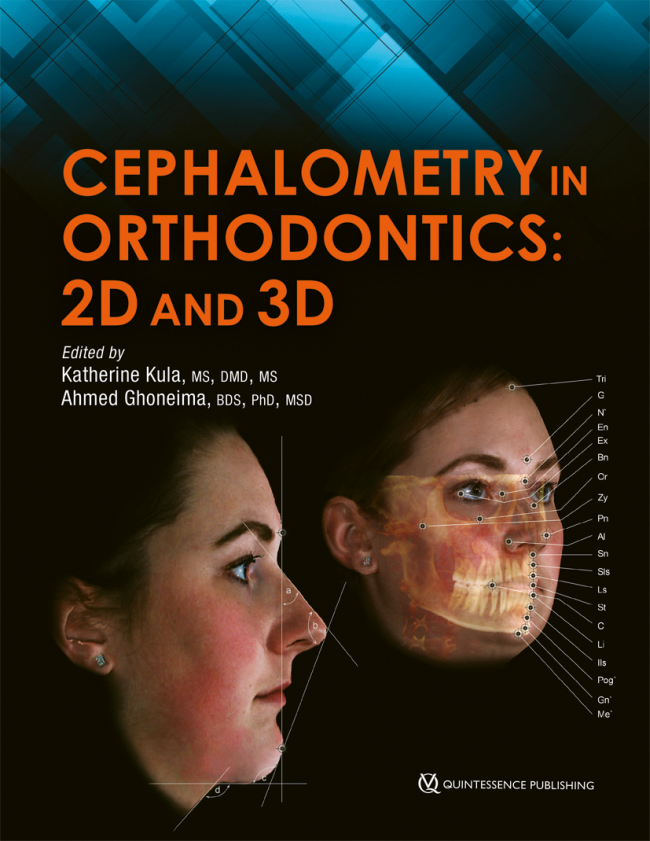
Cephalometrics has been used for decades to diagnose orthodontic problems and evaluate treatment. However, the shift from 2D to 3D radiography has left some orthodontists unsure about how to use this method effectively. This book defines and depicts all cephalometric landmarks on a skull or spine in both 2D and 3D and then identifies them on radiographs. Each major cephalometric analysis is described in detail, and the linear or angular measures are shown pictorially for better understanding. Because many orthodontists pick specific measures from various cephalometric analyses to formulate their own analysis, these measures are organized relative to the skeletal or dental structure and then compared or contrasted relative to diagnosis, growth, and treatment. Cephalometric norms (eg, age, sex, ethnicity) are also discussed relative to treatment and esthetics. The final chapter shows the application of these measures to clinical cases to teach clinicians and students how to use them effectively. As radiology transitions from 2D to 3D, it is important to evaluate the efficacy and cost-effectiveness of each in diagnosis and treatment, and this book outlines all of the relevant concerns for daily practice.
Contents
Chapter 01. Introduction to the Use of Cephalometrics
Chapter 02. 2D and 3D Radiography
Chapter 03. Skeletal Landmarks and Measures
Chapter 04. Frontal Cephalometric Analysis
Chapter 05. Soft Tissue Analysis
Chapter 06. A Perspective on Norms and Standards
Chapter 07. The Transition from 2D to 3D Cephalometrics: Understanding the Problems of Landmarks and Measures
Chapter 08. Cephalometric Airway Analysis
Chapter 09. Radiographic Superimposition: From 2D to 3D
Chapter 10. Growth and Treatment Predictions: Accuracy and Reliability
Chapter 11. Measuring Bone with CBCT
Chapter 12. Common Pathologic Findings in Cephalometric Radiology
Chapter 13. The Cost of 2D Versus 3D Radiology
Chapter 14. Clinical Cases


