0,00 €
Zum Warenkorb
  • Quintessence Publishing USA
Filter
Ralf J. Radlanski / Karl H. Wesker

The Face

Pictorial Atlas of Clinical Anatomy

2nd revised Edition 2015
Book
Hardcover, 24 x 30 cm, 368 pages, 380 illus
Language: English
Categories: Anatomy, Dermatology, Oral/Maxillofacial Surgery

Stock No.: BG116
ISBN 978-1-85097-289-1
QP Deutschland

$178.00
1

The indicated prices are net prices and do not yet include sales tax. This will be added subsequently depending on the country of delivery. Prices for products in this shop do not include postage and packaging - a shipping fee will be added. Please note if a shipment if being sent outside of the US there may be a customs charge for duties and taxes due at the time of delivery. If you have any questions please contact us at service@quintbook.com. For student-priced subscriptions, we require a letter on letterhead from the school's dean or admissions office indicating the student's current enrollment status. Product specifications may change without prior notice. Please read our General Terms and Conditions and take notice of our Privacy Policy.

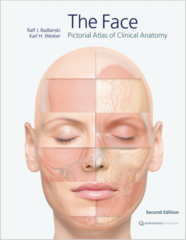
For the first time, the highly complex topographic-anatomical relationships of facial anatomy are depicted layer by layer using extremely detailed anatomical illustrations with a three-dimensional aspect. Important landmarks, anatomical details, and clinically relevant constellations of hard and soft tissues, as well as of nerves and blood vessels, have been detailed. Another important feature is that the point of view is maintained throughout while moving through the different layers of preparation. While the accompanying text and figure captions highlight specific issues, the images remain in the foreground. The elaborate illustrations are based mainly on live anatomy and corresponding images obtained from magnetic resonance imaging, with some support from anatomical preparations.

Contents
Chapter 1. The Face
• Introduction
• The face in anterior view
• The face in lateral view
• The head in vertical view
• The head in dorsal view
• The neck
• Facial expression
• The facial skeleton
• Sectional anatomy
• Schematic representations of pathways in the face

Chapter 2. The Orbital Region
• Surface topography of the orbital region
• Preseptal muscles and fat layers
• The orbital septum and the eyeball
• Vascular and nerve supply in the orbital region
• Vascular and nerve supply in the orbital region in relation to the muscles
• Sectional anatomy of the orbital region

Chapter 3. The Nasal and Midfacial Region
• Surface topography of the nasal region
• The nose in anterior view
• The nose in lateral view
• The nose in caudal view
• The nasal cavity
• The sinuses

Chapter 4. The Mouth
• Extraoral topography of the oral region
• Topographical anatomy of the oral region
• Vascular and nerve supply of the oral region
• The oral cavity
• Anatomy of the lips, teeth, periodontium and alveolar bone in sections
• The anterior oral vestibule
• Anatomy in the area around the mandibular ramus
• The temporomandibular joint
• Anatomy of the oral region in sections
• Pathways of odontogenic spread of infections

Chapter 5. The Ear
Chapter 6. The Skin and Aging of the Face

alt
Author

Prof. Dr. Dr. Ralf J. Radlanski

Germany, Berlin

Ralf J. Radlanski is Director of the Department of Craniofacial Developmental Biology at Charité, the Center for Dental and Craniofacial Sciences. As an anatomist, an internationally esteemed scientist, and as a practicing orthodontist, he works at the interface between biology-anatomy on the one hand and esthetic clinical requirements on the other, a crucial area of interaction in many clinical disciplines.


alt
Author

Karl H. Wesker

Germany

Karl H. Wesker is an artist and an illustrator. He has worked for many years on the visualization and didactic presentation of complex structures. He has developed new methods that produce highly detailed and yet esthetically fascinating images of human anatomy. These methods form the foundation of the three-volume atlas of anatomy Prometheus, which is being published by Thieme and for which he has created most of the illustrations.

PreviewDescriptionFile typeDownload
Cover, Radlanski/Wesker: The Face

Our recommendations

  
Jörg Neugebauer / Joachim E. Zöller

Cone Beam Volumetric Imaging in Dental, Oral and Maxillofacial Medicine

Fundamentals, Diagnostics and Treatment Planning

2nd completely revised and expanded Edition 2014
Book
Hardcover incl. DVD-ROM, 288 pages, 523 illus colored
Language: English
Categories: Interdisciplinary, Radiology and Photography, General Dentistry

Stock No.: BG110
ISBN 978-1-85097-269-3
QP Deutschland

Product out of print.