Utilizamos cookies para habilitar las funciones necesarias para este sitio web, como el inicio de sesión o la cesta de la compra. Puede encontrar más información en nuestra política de privacidad.
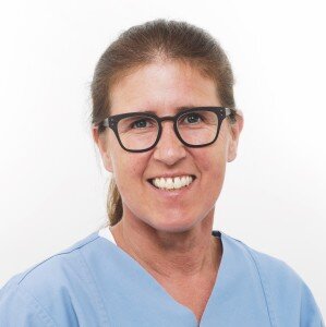
Specialist training in Oral Surgery at the Clinic for Oral and Maxillofacial Surgery, Virchow Clinic, Berlin. Studies and doctorate, Free University of Berlin, Habilitation Humboldt University of Berlin. Research focus: Bone physiology, implant hardware, digital implantology. Clinical focus: Aesthetic and complex implant rehabilitations, augmentations. Since 2003, head of the Department of Implantology and Special Prosthodontics, Clinic for Oral and Maxillofacial Surgery, Humboldt University. 2010 - 2017 head of the Implantology Section, Clinic for Oral and Maxillofacial Surgery, University Hospital Freiburg. In 2017, appointed to the W3 professorship in Translational Implantology, University Hospital Freiburg.
1st Edition 2025 Libro Softcover, 16,8 x 24 cm, 120 páginas, 147 ilustración Idioma: Alemán Categoría: Implantología ISBN 978-3-86867-586-3 QP Deutschland
Eventos
31st EAO Annual Scientific Meeting
Details make perfectionoctubre 24, 2024 — octubre 26, 2024MiCo - Milano Convention Centre, Milano, Italia
Ponentes: Bilal Al-Nawas, Gil Alcoforado, Federico Hernández Alfaro, Sofia Aroca, Wael Att, Gustavo Avila-Ortiz, Kathrin Becker, Anne Benhamou, Juan Blanco Carrión, Dieter Bosshardt, Daniel Buser, Francesco Cairo, Paolo Casentini, Raffaele Cavalcanti, Tali Chackartchi, Renato Cocconi, Luca Cordaro, Luca De Stavola, Nuno Sousa Dias, Egon Euwe, Vincent Fehmer, Alberto Fonzar, Helena Francisco, Lukas Fürhauser, German O. Gallucci, Oscar Gonzalez-Martin, Dominik Groß, Robert Haas, Alexis Ioannidis, Simon Storgård Jensen, Ronald Jung, France Lambert, Luca Landi, Georg Mailath-Pokorny jun., Silvia Masiero, Iva Milinkovic, Carlo Monaco, José Nart, José M. Navarro, Katja Nelson, Manuel Nienkemper, David Nisand, Michael Payer, Sergio Piano, Bjarni E. Pjetursson, Sven Reich, Isabella Rocchietta, Giuseppe Romeo, Irena Sailer, Mariano Sanz, Ignacio Sanz Martín, Frank Schwarz, Shakeel Shahdad, Massimo Simion, Ralf Smeets, Benedikt Spies, Bogna Stawarczyk, Martina Stefanini, Hendrik Terheyden, Tiziano Testori, Daniel Thoma, Ana Torres Moneu, Piero Venezia, Lukas Waltenberger, Hom-Lay Wang, Stefan Wolfart, Giovanni Zucchelli, Otto Zuhr
European Association for Osseintegration (EAO)
Artículos de este autor en revistas
The International Journal of Prosthodontics, 7/2021
SuplementoPáginas s8-s20, Idioma: InglésSchwarz, Frank / Schär, Alex / Nelson, Katja / Fretwurst, Tobias / Flügge, Tabea / Ramanauskaite, Ausra / Trimpou, Georgina / Sailer, Irena / Karasan, Duygu / Fehmer, Vincent / Guerra, Fernando / Messias, Ana / Nicolau, Pedro / Chochlidakis, Konstantinos / Tsigarida, Alexandra / Kernen, Florian / Taylor, Thomas / Vazouras, Konstantinos / Herklotz, Insa / Sader, Robert
The tasks of Working Groups 1 to 6 at the 4th Consensus Meeting of the Oral Reconstruction Foundation were to elucidate clinical recommendations for implant-supported full-arch rehabilitations in edentulous patients. Six systematic/ narrative reviews were prepared to address the following subtopics: (1) the influence of medical and geriatric factors on implant survival; (2) the prevalence of peri-implant diseases; (3) the influence of material selection, attachment type, interarch space, and opposing dentition; (4) different interventions for rehabilitation of the edentulous maxilla; (5) different interventions for rehabilitation of the edentulous mandible; and (6) treatment choice and decision-making in elderly patients. Consensus statements, clinical recommendations, and implications for future research were determined based on structured group discussions and plenary session approval.
Purpose: To provide an overview of the influence of medical and geriatric factors on implant survival in order to form clinical recommendations for the practitioner.
Materials and Methods: This narrative literature review was performed to address the following questions: (1) Is age (> 75 years) a risk factor for implant survival?; (2) Is diabetes mellitus a risk factor for implant survival?; and (3) Is antiresorptive therapy a risk factor for implant survival? The PubMed, Web of Knowledge (Thomson Reuters), and Google Scholar databases were searched for systematic reviews and research papers of evidence level II and above that were published up to February 2019 for each topic.
Results: (1) Age > 75 years does not affect implant survival according to short-term follow up (1 to 5 years). However, polypharmacy should be considered in this patient group. (2) Diabetes mellitus is not a risk factor for implant survival in the short term, but there is no information on appropriate perioperative treatment and wound closure. There is little evidence in the literature on the success of bone grafting and progressive loading protocols in diabetic patients. (3) Implant therapy cannot be recommended in patients under high-dose bisphosphonate and antibody therapy. Bone grafting should be avoided under antiresorptive therapy. There are no treatment regimens available for patients with periimplantitis receiving antiresorptive medication.
Conclusion: This review suggests that the risk assessment for an implant patient should not be based on age, but rather on the patient’s specific risk factors, such as former and current diseases and medication.
Der Beitrag stellt zunächst die Kasuistik eines frühen Implantatverlustes bei einer Patientin mit Vitamin-D-Insuffizienz vor und gibt danach einen Einblick in den aktuellen Stand der einschlägigen Forschung. Welche Grenzwerte sind derzeit für Vitamin D vorgegeben? Wann liegt eine Vitamin-D-Insuffizienz, wann ein Vitamin-D-Mangel vor? Abschließend wird die Frage geklärt, ob vor einer Implantation der Vitamin-D-Spiegel überprüft werden sollte und ob es bei einem niedrigen Wert ratsam ist, eine standardmäßige Substitution durchzuführen.
Palabras clave: Vitamin-D-Mangel, Vitamin-D-Insuffizienz, Implantation, früher Implantatverlust, Periimplantitis
DOI: 10.3290/j.qi.a43949, ID de PubMed (PMID): 32020132Páginas 220-228, Idioma: InglésSpanou, Alexandra / Nelson, Katja / Ermer, Michael Andreas / Steybe, David / Poxleitner, Philipp / Voss, Pit Jacob
Objectives: Tooth extractions are suspected to be a major trigger for the development of bisphosphonate-related osteonecrosis of the jaw (BRONJ). Preventive measures like perioperative antibiotic therapy and primary wound closure have been found to be effective in preventing the development of BRONJ following tooth extraction. The aim of this study was to present long-term results of a treatment protocol for patients under bisphosphonate therapy requiring tooth extraction.
Method and materials: Between January 2008 and December 2012, 232 tooth extractions were performed in 84 patients under current or previous bisphosphonate treatment. Extractions were performed applying an atraumatic technique under prolonged intravenous antibiotic therapy. The bony edges were rounded off meticulously and the extraction sockets were covered with a mucoperiosteal flap.
Results: Out of 84 patients, two developed osteonecrosis of the jaw (ONJ), resulting in an incidence-rate of 2.4%. Both ONJ patients had received radiation therapy to the head and neck region following tooth extraction. The remaining 82 patients had uneventful healing and did not present any signs or symptoms of BRONJ during the follow-up period of 41.5 months.
Conclusion: Tooth extractions, if performed under certain circumstances, would not seem to constitute a major risk factor for the development of BRONJ. The treatment protocol presented in this article seems to be highly effective in preventing development of ONJ after tooth extraction in patients under current or previous bisphosphonate therapy.
Palabras clave: bisphosphonate, bisphosphonate-related osteonecrosis of the jaw (BRONJ), tooth extraction
Purpose: The purpose of this study was to visualize the mode and impact of force transmission in narrowdiameter implants with different implant-abutment designs and material properties and to quantify the displacement of the abutment.
Materials and Methods: Narrow-diameter implants from two manufacturers were examined: Astra 3.0-mm-diameter implants (Astra OsseoSpeed TX; n = 2) and Straumann Bone Level implants with a 3.3-mm diameter made of commercially pure titanium (cpTi) Gr. 4 (n = 2) and 3.3-mm TiZr-alloy (n = 2; Bone Level, Straumann) under incremental force application using synchrotron radiography (absorption and inline x-ray phase-contrast) and tomography.
Results: During loading (250 N), Astra 3.0 and Bone Level 3.3- mm implants showed a deformation of the outer implant shoulder of 61.75 to 95 μm independent of the implant body material; the inner implant diameter showed a deformation of 71.25 to 109.25 μm. A deformation of the implant shoulder persisted after the removal of the load (range, 42.75 to 104.5 μm). An angulated intrusion of the abutment (maximum, 140 μm) into the implant body during load application was demonstrated; this spatial displacement persisted after removal of the load.
Conclusion: This study demonstrated a deformation of the implant shoulder and displacement of the abutment during load application in narrow-diameter implants.
Palabras clave: abutment intrusion, implant diameter, implant shoulder deformation, narrow diameter, overload, synchrotron radiography and tomography
Die vorliegende Kasuistik einer 64-jährigen Patientin illustriert die potenziellen Gefahren und Folgen im Rahmen von implantologischen Eingriffen, welche von Anamnesenlücken bei Patienten mit antiresorptiver Therapie ausgehen können. Zusammenfassend zeigte sich, dass der Patientin unter, vermutlich nicht bekannter, i. v. Bisphosphonatgabe sowohl ein Implantat alio loco inseriert, als auch dieses wieder entfernt wurde. Ferner, dass bei der initialen Drainage und dem Wunddebridement in domo die Bisphosphonateinnahme trotz gezielter Anamneseerhebung unbekannt geblieben ist. Die Wichtigkeit des interdisziplinären Austauschs in der zahnärztlichen Chirurgie und Implantologie mit (allgemein-)medizinischen Fachdisziplinen soll mit diesem Fallbericht betont werden. Bei frühem Implantatverlust, massiver Progredienz einer Periimplantitis oder Wundheilungsstörungen nach Explantation - trotz gründlichem Debridement der Explantationswunde - sollte an eine (bisher nicht bekannte) Antiresorptivatherapie in der Anamnese des Patienten gedacht werden.
Palabras clave: Wundheilungsstörung, Implantation, Explantation, paramandibulärer Abszess, Bisphosphonate, Zolendronsäure, monoklonale Antikörper, Denosumab, Antiresorptiva, antiresorptivaassoziierte Kiefernekrosen
Purpose: The digitization of scanbodies on dental implants is required to use computer-aided design/computer-assisted manufacture processes for implant prosthetics. Little is known about the accuracy of scanbody digitization with intraoral scanners and dental lab scanners. This study aimed to examine the precision of different intraoral digital impression systems as well as a dental lab scanner using commercially available implant scanbodies.
Materials and Methods: Two study models with a different number and distribution of dental implant scanbodies were produced from conventional implant impressions. The study models were scanned using three different intraoral scanners (iTero, Cadent; Trios, 3Shape; and True Definition, 3M ESPE) and a dental lab scanner (D250, 3Shape). For each study model, 10 scans were performed per scanner to produce repeated measurements for the calculation of precision. The distance and angulation between the respective scanbodies were measured. The results of each scanning system were compared using analysis of variance, and post hoc Tukey test was conducted for a pairwise comparison of scanning devices.
Results: The precision values of the scanbodies varied according to the distance between the scanbodies and the scanning device. A distance of a single tooth space and a jaw-traversing distance between scanbodies produced significantly different results for distance and angle measurements between the scanning systems (P .05).
Conclusion: The precision of intraoral scanners and the dental lab scanner was significantly different. The precision of intraoral scanners decreased with an increasing distance between the scanbodies, whereas the precision of the dental lab scanner was independent of the distance between the scanbodies.
The influence of repeated system-specific torque tightening on the position stability of the abutment after de- and reassembly of the implant components was evaluated in six dental implant systems with a conical implant-abutment connection. An established experimental setup was used in this study. Rotation, vertical displacement, and canting moments of the abutment were observed; they depended on the implant system (P = .001, P .001, P = .006, respectively). Repeated torque tightening of the abutment screw does not eliminate changes in position of the abutment.
Meta-Analyse und Systematik Review etablierter Behandlungsmodalitäten
Einleitung: Der Sinuslift ist eine der meist genutzten Techniken um im atrophierten Oberkiefer ein suffizientes knöchernes Implantatlager zu schaffen. Zahlreiche chirurgische Methoden wurden hierzu in der Literatur beschrieben und verglichen. Dennoch fand der Einfluss von Studiendesign-Inkonsistenz sowie der reziproke Einfluss einzelner Faktoren, wie chirurgische Methode, Augmentationsmaterial oder Implantattyp, bisher keine Berücksichtigung bei der Beurteilung des Implantatüberlebens.
Ziel der vorliegenden Studie war es die unterschiedlichen Co-Faktoren von Sinuslift und Implantation und deren reziproker Einfluss aufeinander sowie insbesondere auf das Implantatüberleben zu evaluieren.
Material und Methoden: Diese PRISMA konforme Meta-Analyse berücksichtigt alle Publikationen klinischer Studien (01/1980-01/2013) mit mindestens 10 Patienten, Sinuslift von intern oder extern, Augmentation und Implantation sowie nachuntersuchter Implantat-Belastungszeit von mindestens 6 Monaten. Eingeschlossen wurden 122 Publikationen mit insgesamt 16268 osseointegrierten Implantaten im augmentierten Sinus. Die statistische Auswertung erfolgte mittels einer non-parametrischen univariaten Kaplan-Meier Analyse sowie eines speziell adaptierten Bayes'schen multivariaten, intervall-zensierten Cox'schen Regressionsmodells.
Ergebnisse: Die Therapieparameter Restknochenhöhe, chirurgische Methode, Augmentationsmaterial und Implantat Typ zeigten keine selektive Präferenz. Im Gegensatz hierzu zeigte die Anwendung von Membranen im Sinuslift eine signifikante Reduktion des relativen Risikos unabhängig von allen anderen Co-Faktoren.
Konklusion: Es wird nicht ausgeschlossen das Faktoren, wie beispielsweise die Erfahrung des Operateurs, einen positiven Effekt auf das Langzeitüberleben von Implantaten im augmentierten Sinus haben können. Jedoch zeigte ausschließlich die Anwendung von Membranen einen in jedem Fall signifikanten wirkungsvollen Faktor.
Purpose: To report a rare and dramatic complication following immediate dental implant placement in a heavy smoker, who had a delayed diagnosis of uncontrolled diabetes.
Materials and methods: In this case report we present the dramatic course of a 64-year old female patient treated with five immediate post-extractive dental implants in the mandible, who developed osteomyelitis, which manifested initially as local peri-implant inflammation and progressed into a spontaneous jaw fracture, despite repeated surgical interventions and antibiotic courses over a 3-year period, until diabetes was diagnosed.
Results: A symptom-free status could be achieved only after partial mandibulectomy, treatment of diabetes and reconstruction with a microvascular fibula free flap.
Conclusion: In the presence of mandibular osteomyelitis refractory to therapy, yet undiagnosed underlying pathologies, such as diabetes, should be investigated and treated urgently.
Palabras clave: fibula free flap, immediate dental implant placement, osteomyelitis, uncontrolled diabetes