Implantologie, 4/2024
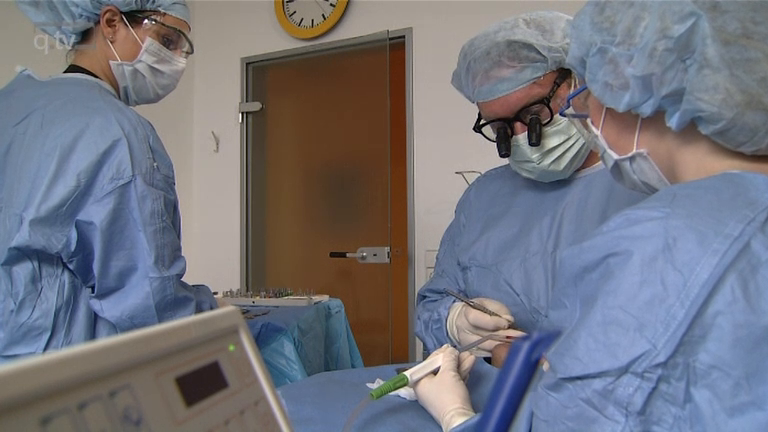
Seiten: 415-432, Sprache: DeutschHildebrand, Detlef / Gärtner, Florian / Rosinski, Andrea / Jäckel, Timo / Siebert, Heiko / Kunz, Andreas / Beuer, FlorianEin Überblick aus der PraxisDie Behandlung prospektiv zahnloser Patienten mit dentalen Implantaten stellt die Zahnärzte auch heute vor große Herausforderungen vor, während und nach der chirurgischen Phase. Die Behandlungsplanung und deren Umsetzung sind von essenzieller Bedeutung, um den klinischen Behandlungserfolg zu garantieren. Anhand einer fundierten Analyse der Ausgangssituation werden im vorliegenden Beitrag die notwendigen Behandlungsschritte vollumfänglich skizziert, in den klinischen Ablauf integriert und umgesetzt. Dabei spielen für die Behandlung des zahnlosen Kiefers grundsätzliche Entscheidungen eine große Rolle: (1) „In welchem Umfang müssen Extraktionen erfolgen?“, (2) „Kann eine sofortige Implantation (Sofortimplantation) erfolgen?“ und (3) „Kann eine zeitgleiche prothetische Versorgung (Sofortversorgung) umgesetzt werden?“. Das Behandlungsteam, bestehend aus Implantologe/Chirurg, Prothetiker und Zahntechniker, funktioniert umso besser und effektiver, je besser die Behandlungsabläufe analog und digital miteinander abgestimmt sind und diese im Sinne eines zielgerichteten Workflows funktionieren. In diesem Artikel werden digitale Hilfsmittel im Bereich der Planung, Platzierung und Weiterentwicklungen moderner Implantatsysteme beleuchtet, die das implantologische Team unterstützen können. Neue Implantate mit speziellen Schraubendesigns helfen nicht nur, die Primärstabilität nach Einbringung in den Knochen vor der initialen Sofortbelastung zu verbessern, sondern ermöglichen mit kurzen bzw. extrakurzen Implantatlängen (8 mm) auch im atrophierten Seitenzahnbereich zahnloser Ober- und Unterkiefer, die klassischen „All-on-X“-Konzepte („All-on-4“) zu erweitern bzw. zu vereinfachen („Fix-on-X“). Anhand von beispielhaften Patientenfällen werden diese aktuellen Konzepte veranschaulicht und anhand der klinischen Daten diskutiert.
Schlagwörter: All-on-4, Fix-on-X, Sofortimplantation, Sofortbelastung, Sofortversorgung, Implantate mit aggressivem Gewinde, Primärstabilität, kurze Implantate, beschleunigte Regeneration
International Journal of Computerized Dentistry, 1/2017
PubMed-ID: 28294202Seiten: 9-19, Sprache: Englisch, DeutschHerklotz, Insa / Beuer, Florian / Kunz, Andreas / Hildebrand, Detlef / Happe, ArndtDas zentrale Ziel der Implantatinsertion ist die optimale prothetische Position des Implantates bei gleichzeitigem Schutz sensibler anatomischer Strukturen. Diesbezüglich zeigen navigiert gesetzte Implantate signifikant bessere Werte im Vergleich zu freihand gesetzten Implantaten. Die computergestützte Navigation in Kombination mit der dreidimensionalen Bildgebung mittels digitaler Volumentomografie ist ein ideales Mittel, um die Vorhersagbarkeit einer erfolgreichen Implantattherapie zu erhöhen. Grundsätzlich kann die statische Navigation mittels Führungsschablonen von der dynamischen Navigation unter Verwendung von optischen Übertragungssystemen unterschieden werden. Beide zeigen bezüglich der Präzision der Implantatposition ähnlich gute Ergebnisse. Aufgrund der verkürzten Arbeitsschritte ist heutzutage die digitale Schablonenherstellung im Gegensatz zur analogen Herstellung Standard. Werden die Investitionskosten der navigierten direkten Technik durch Vereinfachung der Systeme verringert, ist sie auf längere Sicht die attraktivere Wahl in der navigierten Implantologie. Innerhalb dieses Beitrags werden anhand von Kasuistiken die unterschiedlichen Varianten der navigierten Implantologie aufgezeigt.
Schlagwörter: statische Navigation, dynamische Navigation, digitale Schablonenherstellung, CAD/CAM, Referenzmarker
The International Journal of Oral & Maxillofacial Implants, 4/2008
PubMed-ID: 18807571Seiten: 726-732, Sprache: EnglischNelson, Katja / Semper, Wiebke / Hildebrand, Detlef / Özyuvaci, HakanPurpose: The aim of this study was to evaluate the success rate of 2 different implant systems with sandblasted and acid-etched modified surfaces loaded after reduced healing periods.
Materials and Methods: One-hundred seventeen patients with a mean observation period of 3.75 years (24 to 61 months) were included in this evaluation. Chart reviews of a standardized recall program were evaluated. All 532 placed implants showed an unloaded healing time of 6 weeks in the mandible and 12 weeks in the maxilla. At abutment placement a torque value of 35 Ncm was one of the primary variables, and the success of the implants over time was determined by the criteria of Buser et al. The survival was analyzed using Kaplan-Meier method, and the probability of an event within 1 group independent of time was evaluated using the chi-square test and Fisher exact test.
Results: Of the 532 implants, 235 were placed in female and 297 in male patients; 448 implants were located in the maxilla and 84 in the mandible. Three implants were lost prior to abutment connection in 3 patients. Life table analyses show an overall success rate of 99.4% at 5 years, as no implants were lost after abutment connection. There was no significant association of the implant type (P = .185), gender (P = .99), or jaw (maxilla/mandible; P = .06) and the survival of the implants within this study.
Conclusion: Based on the data found in this investigation, it can be concluded that implants with sandblasted, acid-etched surfaces can be restored after a 6- to 12-week healing period with a high predictability of success.
Schlagwörter: reduced healing period, sand-blasted and acid-etched
International Poster Journal of Dentistry and Oral Medicine, 4/2007
Poster 379, Sprache: EnglischHildebrand, Detlef/Bassem, Al-Chawaf/Nelson, KatjaIn this present study new bone formation of fresh extraction socket afteraugmentation with Bio-Oss Collagen was analyzed after a healing period of 6weeks using histomorphometry Material and methods: Ten patients, referredfor extraction of decayed teeth of all regions, were included in this study.The extraction sockets were instrumented to eliminate all remnants ofperiodontal ligament tissue and showed no defect in extraction site. Eachsocket was grafted with Bio-Oss Collagen without flap management. After a 6weeks healing period, at implant placement, bone biopsy samples wereobtained with a trephine bur and evaluated histomorphometrically, usingMasson's trichrome and Toluidine staining. Quantification of new boneformation and BioOss-remnants was performed using a digital imaging system(AxioVision, Zeiss, Germany). Results and discussion: The values found fornew bone formation ranged from 35% - 59% and 34% for remainingBioOss-particles. This is comparable to the findings of studies with healingperiod of 12 weeks in canines. These results encouraged an early onsetimplantation after healing period of six weeks. The long-term success andsurvival rate of these implants will be the subject of futureinvestigation.
Schlagwörter: extraction socket, augmentation
The International Journal of Oral & Maxillofacial Implants, 3/2006
PubMed-ID: 16796281Seiten: 392-398, Sprache: EnglischNelson, Katja / Özyuvaci, Hakan / Bilgic, Bilge / Klein, Martin / Hildebrand, DetlefPurpose: In the present study solid monocortical hipbone onlay grafts of the maxilla were analyzed histologically after a healing period of 3 months. The clinical success of the implants placed in the grafted bone was evaluated.
Materials and Methods: Nineteen patients underwent augmentation with avascular iliac bone. A 2-stage procedure was performed with a 3-month healing period between graft and implant placement. At implant placement bone biopsy samples were taken at the proposed implant sites.
Results: Of the 117 implants placed, 1 was not osseointegrated at the time of abutment connection. No implants were lost after loading during an observation period of up to 3 years. Clinical appearance of the augmented bone after 3 months showed a dense cortical layer with good blood perfusion. Histologic specimens were analyzed quantitatively and showed an average of 43.2% newly formed bone.
Discussion: Histomorphometry showed that the amount of newly formed bone after 3 months was comparable to that found after a healing period of 4.5 months. The clinical success of the implants placed after the shortened healing period was comparable to that found in nonaugmented bone.
Conclusion: This study showed that after avascular iliac bone grafting, 3 months of revascularization was sufficient to ensure the secure placement of dental implants in second-stage surgery for this patient population.
Schlagwörter: dental implants, iliac bone grafting, onlay bone grafting