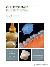
DOI: 10.3290/j.qi.a44705, PubMed ID (PMID): 32500860Pages 525-526, Language: EnglishMupparapu, MelDOI: 10.3290/j.qi.a44496, PubMed ID (PMID): 32368766Pages 528-536, Language: EnglishMeller, Christian / Walker, UweThe rehabilitation of patients with wear on multiple teeth, in which a recovery of the occlusal/incisal vertical dimension is required to acquire satisfactory levels of functionality and esthetics, is often a difficult procedure even for experienced dental practitioners. Based on wax-ups simulating an ideal structural/functional reconstruction of the teeth, the use of shaping indices is a reliable way to plan predictable patient-oriented results when using resin-based composites. The proposal to combine shaping indices with PTFE-based stamp techniques also helps clinicians to easily achieve satisfactory modeling results giving them the unique opportunity to eliminate the presence of composite over-contours before polymerization. By using PTFE-based stamps the requirement to use conventional, strictly transparent indices becomes superfluous. In a clinical case with multiple caries lesions and tooth wear, the advantage of several simple operative measures including PTFE-based stamp procedures are presented as a feasibility example of this noninvasive or minimally invasive restorative approach performed by an undergraduate dental student.
Keywords: composite restoration, direct/indirect restoration, no-prep restoration, occlusal/incisal vertical dimension increase, shaping index, Teflon stamp technique
DOI: 10.3290/j.qi.a44635, PubMed ID (PMID): 32500861Pages 538-544, Language: EnglishLegaz, Juan / Karasan, Duygu / Fehmer, Vincent / Sailer, IrenaThe prototyping protocol to evaluate and make the potential adjustments prior to finalization of the monolithic restorations was described by two clinical situations. In the first case report, following the digital impressions using an intraoral scanner (3Shape Trios, 3Shape) for an implant-supported four-unit fixed dental prosthesis, a digital design (3Shape Dental System, 3Shape) was performed and a prototype using subtractive CAM (milling) (PMMA, Telio CAD, Ivoclar Vivadent) was fabricated. The second case highlights the 3D-printed prototyping (additive CAM) (Sheraprint Model Plus UV, Shera) following digital impressions using an intraoral scanner and digital design in a patient requiring two opposing open-end three-unit fixed dental prostheses. By means of prototyping, the esthetic, fitting, and functional properties could be tested and the adjustments were completed on the prototypes. It is suggested that prototyping is an efficient tool that minimizes the clinical adjustment need for the final restoration while improving the communication between the dental practitioner and the technician.
Keywords: CAD/CAM, ceramics, diagnostic procedure, digital workflow, prosthodontics
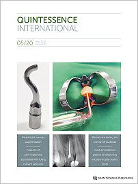
DOI: 10.3290/j.qi.a44629, PubMed ID (PMID): 32500862Pages 546-553, Language: EnglishEmanuel, Noam / Machtei, Eli E. / Reichart, Malka / Shapira, LiorObjectives: In the present pilot, multicenter, randomized, single-blinded, controlled study, surgical treatment with or without the administration of D-PLEX500 (a biodegradable prolonged release local doxycycline formulated with β-tricalcium phosphate bone graft) was accessed for the treatment of peri-implantitis.
Method and materials: Subjects undergoing surgical treatment for intrabony peri-implantitis defects after flap elevation were randomly assigned, to adjunct D-PLEX500 placement group or to control group. Clinical and radiographic parameters were measured at 6 and 12 months.
Results: Twenty-seven subjects (average age: 64.81 ± 7.61 years) were enrolled; 14 patients (18 implants) were randomized to the test group and 13 (14 implants) to the control group. There was no difference in plaque scores between the groups. There was no difference in the changes of mean periodontal probing depth between the test and control groups between baseline and the 6-month follow-up, whereas statistically significant difference was observed after 12 months' follow-up when analyzed for all sites averaged. There was a statistically significant difference in the changes of clinical attachment levels and radiographic bone levels between the groups between baseline and 12 months. These improvements were demonstrated when analyzed at both implant and subject levels. Only D-PLEX500 treatment led to improved bone levels at both time points. The improvement in bone levels was significant in the D-PLEX500 treatment group already after 6 months, and further improved over the 12-month follow-up. Implants were lost only in the control group (14%).
Conclusions: D-PLEX500 sustained release local antibiotic formulated with bone filler showed promising results in enabling healing of peri-implantitis lesions. The antibacterial component of the bone graft material might create favorable conditions that enable implant surface decontamination and soft and hard tissue healing over a prolonged period.
Keywords: bacteria, bone augmentation, dental implants, periodontitis, plaque
DOI: 10.3290/j.qi.a44630, PubMed ID (PMID): 32500863Pages 554-565, Language: EnglishMousa, Mohammed A. / Lynch, Edward / Kielbassa, Andrej M.Objectives: To assess the relationship between the development of denture-related stomatitis (DRS) and the identification of commonly isolated yeast species, and to evaluate various predisposing factors in Saudi participants wearing new removable dental prostheses.
Method and materials: A total of 75 edentulous male participants were recruited, and 64 patients finished the present case-series. All participants received new conventional complete dentures. Colonization of Candida species was assessed, and species were identified by means of the VITEK 2 (bioMérieux) laboratory components.
Results: The most prevalent type of Candida at baseline was C albicans, followed by non-C albicans species (C glabrata). Counts of Candida species significantly increased from the day of insertion to the first month (P . 05), but there were no significant changes between the first and second month (P > . 05). On the day of insertion, C tropicalis, C dubliniensis, and C krusei were extracted from few subjects only, with no significant changes over the first and second month (P > .05). Patients revealing habits of sleeping with their dentures were found to frequently suffer from DRS; development of the latter was rapid, and mixed Candida biofilms (with high CFU/mL counts), along with inadequate oral and denture hygiene, turned out to be contributing factors (P .05).
Conclusion: DRS can develop faster than previously reported, even with new dentures; continued denture wearing and poor cleaning of dentures revealed a considerable impact on DRS onset. In the present cohort, C albicans was the most identified kind of yeast, and was followed by C glabrata infection in cases with DRS.
Keywords: Candida species, complete denture, denture hygiene, denture stomatitis, denture-related stomatitis, sleeping with dentures, VITEK 2 Compact system, yeast species
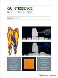
DOI: 10.3290/j.qi.a44631, PubMed ID (PMID): 32500864Pages 566-576, Language: EnglishStrasding, Malin / Sebestyén-Hüvös, Eszter / Studer, Stephan / Lehner, Christian / Jung, Ronald E. / Sailer, IrenaObjectives: Long-term retrospective evaluation of the survival rate and the technical and biologic outcomes of all-ceramic inlays and onlays in premolars and molars.
Method and materials: Fifty-four patients treated as part of a prospective clinical trial and having received 157 inlays and 27 onlays made out of a leucite-reinforced glass-ceramic (IPS Empress) in premolars and molars, were invited to the present follow-up examination. The survival of the restorations was evaluated. The biologic outcomes were assessed by measuring the pocket probing depth (PPD), the Plaque Index (PI), and the Sulcus Bleeding Index (SBI). The technical behavior was evaluated using modified US Public Health Service criteria (modUSPHS). Finally, patient satisfaction was recorded with a questionnaire. Data of patients and restored teeth were analyzed descriptively, and continuous variables were given in mean values and standard deviations. For the analysis of the restoration survival over time, the Kaplan-Meier survival estimate was calculated. The level of statistical significance was set at P .05.
Results: Thirty-six patients (20 women, 16 men; mean age 50.9 years) with 132 restorations, 107 inlays and 25 onlays, were examined after a mean observation time of 11.2 ± 4.3 years. The overall 11-year survival rate of the 132 restorations was 80.3%. Inlays exhibited an 11-year survival rate of 80.4% and onlays of 80.0%. Twenty-two technical complications occurred. Ceramic fractures (10.6%) and chipping (2.3%) were the most frequent complications. Six biologic complications occurred (4.5%).
Conclusion: Glass-ceramic inlays and onlays presented favorable long-term clinical survival and success rates. Technical complications were predominant, and biologic problems remained rare. More clinical long-term data are needed.
Keywords: adhesive cementation, all-ceramic restoration, glass-ceramic, long-term, minimally invasive
DOI: 10.3290/j.qi.a44632, PubMed ID (PMID): 32500865Pages 578-584, Language: EnglishKhehra, Anahat / Levin, LiranAn edentulous posterior maxilla can present a challenge for placement of dental implants due to the proximity of the maxillary sinus. Sinus augmentation is a surgical bone grafting procedure aimed to increase the bone height for implant support. A number of sinus augmentation techniques have been presented and the outcomes show good implant success rates. In order to achieve the desirable outcomes, it is important to gain knowledge of the maxillary sinus anatomy and complete a thorough preoperative evaluation. Being aware of the location of vasculature, nerves, and the presence of septa will help reduce the risk of intraoperative and postoperative complications. This review provides a narrative clinical overview related to the anatomy, preoperative evaluation, contraindications, techniques, postoperative care, outcome measures, and complications of sinus augmentation procedures.
Keywords: bone graft, dental implants, maxilla, sinus elevation, success, survival
DOI: 10.3290/j.qi.a44633, PubMed ID (PMID): 32500866Pages 586-597, Language: EnglishBhattarai, Bishwa Prakash / Bijukchhe, Sanjeev Man / Reduwan, Nor HidayahObjective: This study was conducted to compare the anesthetic and analgesic efficacy of bupivacaine with other local anesthetic agents routinely used for mandibular third molar surgery.
Method and materials: Four electronic databases (PubMed, Scopus, Cochrane, and Web of Science) were explored to isolate randomized controlled trials up to 10 February 2019. The anesthetic and analgesic efficacies were assessed using six evaluation outcomes: onset of anesthesia, success of anesthesia, duration of anesthesia, duration of analgesia, pain score on the fourth postoperative hour, and number of analgesics consumed. Stata software (version 13, StataCorp) was used to analyze the data.
Results: Fourteen studies met the specified criteria. The sample consisted of 1,078 mandibular third molar surgeries performed in 858 patients. Bupivacaine, lidocaine/lignocaine, articaine, etidocaine, levobupivacaine, and carbonated bupivacaine were the local anesthetics used. Compared with other anesthetic agents, bupivacaine showed longer duration of anesthesia (weighted mean difference [WMD] 123.431 minutes; 95% confidence interval [CI] 34.01 to 212.851; P = .007), lower pain score at the fourth and eighth postoperative hours (4 hr-WMD 2.757; 95% CI 0.893 to 4.62; P = .004; 8 hr-WMD 1.697; 95% CI 1.178 to 2.216; P .001), and lower number of analgesics requirement (WMD 0.663; 95% CI 0.258 to 1.067; P = .001). The onset of anesthesia was slower for bupivacaine (WMD 0.865 minutes; 95% CI 0.799 to 0.931; P .001). However, for success of anesthesia (risk ratio 1.003; 95% CI 0.972 to 1.035; P = .831) and duration of analgesia (WMD 45.285 minutes; 95% CI −48.021 to 138.537; P = .342), the local anesthetic agents showed no significant differences.
Conclusions: Except for the onset of anesthesia, bupivacaine showed better anesthetic and analgesic properties than other local anesthetic agents for mandibular third molar surgery.
Keywords: bupivacaine, local anesthesia, mandibular third molar surgery, systematic review
DOI: 10.3290/j.qi.a44634, PubMed ID (PMID): 32500867Pages 598-602, Language: EnglishMatsuda, Shinpei / Yoshida, Hisato / Ohta, Keiichi / Ryoke, Takashi / Yoshimura, HitoshiIntraoral hemorrhage is an undesirable and emergency condition, and it can also occur spontaneously. Clinicians sometimes face difficulty in identifying the hemorrhage points and the causes of hemorrhage, as well as difficulty in the hemostatic procedures. Here, the authors present two rare cases of spontaneous intraoral hemorrhage related to dental calculus. The hemorrhage points and causes of hemorrhage were determined after removing the removable intraoral components, including the dental calculus.
Keywords: dental calculus, dentures, emergency treatment, intraoral hemorrhage, periodontology, prosthodontics


