We use cookies to enable the functions required for this website, such as login or a shopping cart. You can find more information in our privacy policy.
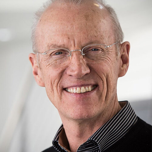
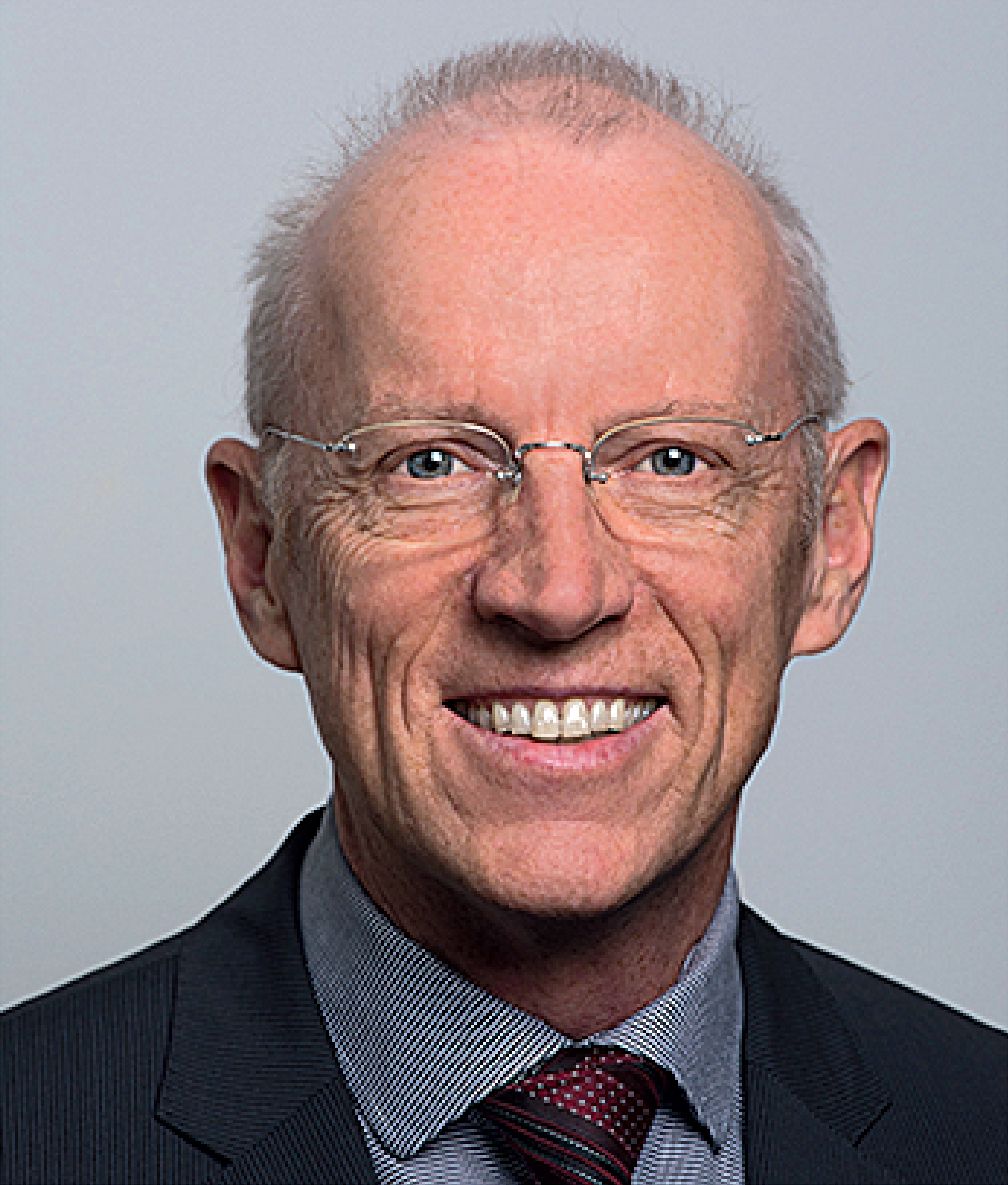
Christoph H. F. Hämmerle, Prof Dr med dent, is the department chairman of the Clinic of Fixed and Removable Prosthodontics in Dental Materials Science and the Division of Implant Dentistry at the University of Zurich in Switzerland. He currently serves or has served as a board member of several scientific organizations, notably the European Association for Osseointegration, Osteology Foundation, Swiss Society for Reconstructive Dentistry, Continental European Division of the International Association for Dental Research, International Team for Implantology Switzerland, and Dental Campus Association. Dr Hämmerle has published more than 300 scientific articles and has lectured internationally.
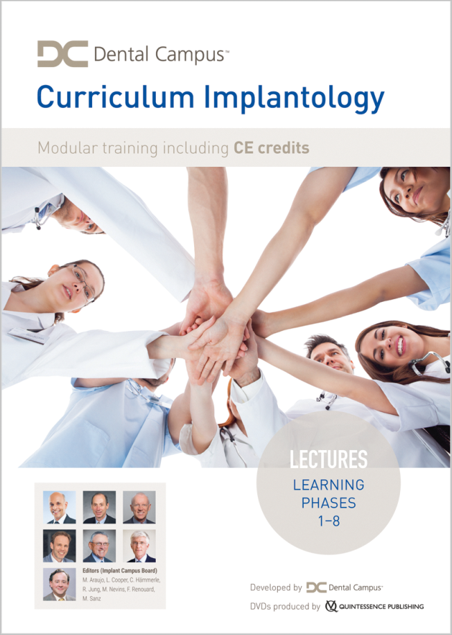
1st Edition 2020 DVD 17-Volume Set; NTSC/PAL; Runtime: 44 h 14 min Language: English Category: Implantology Stock No.: 7669 ISBN 978-1-78698-102-8 QP Deutschland
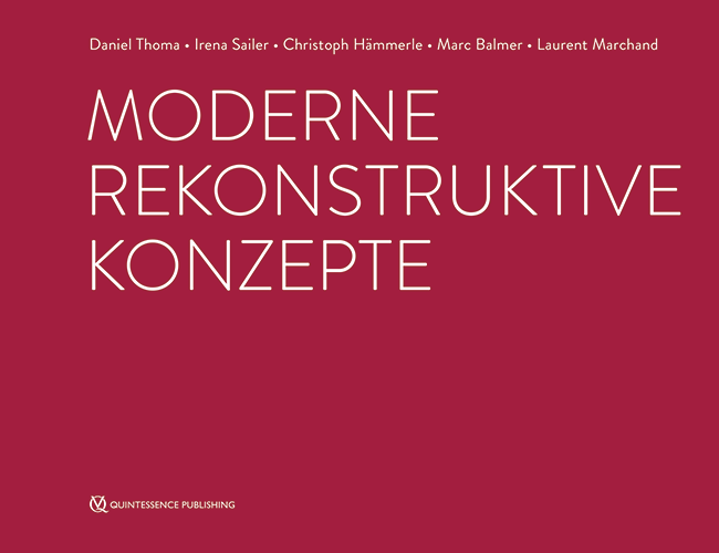
1. Auflage 2008 Book Hardcover, 136 pages, 218 illus Language: German Categories: Prosthodontics, Dental Technology ISBN 978-3-938947-69-2 QP Deutschland
Events
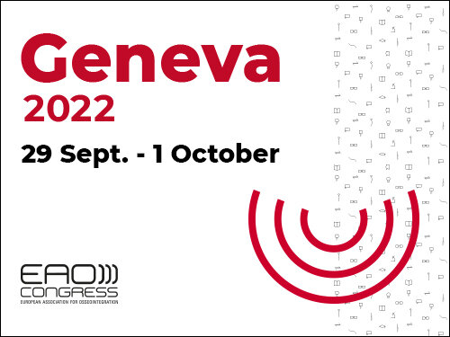
29th EAO annual scientific meeting
Uniting Nations through Innovations29. Sep 2022 — 1. Oct 2022Palexpo, Le Grand-Saconnex, Switzerland
Speakers: Thabo Beeler, Nitzan Bichacho, Rino Burkhardt, Luigi Canullo, Matteo Chiapasco, Luca De Stavola, Mirela Feraru, Alfonso L. Gil, Klaus Gotfredsen, Markus Gross, Ueli Grunder, Christoph Hämmerle, Björn Klinge, Ivo Krejci, Sebastian Kühl, Niklaus P. Lang, Sonia Leziy, Daniele Manfredini, Konrad H. Meyenberg, Francesco Mintrone, Ricardo Mitrani, Sven Mühlemann, José M. Navarro, Florian Probst, Pablo Ramírez, Christoph Andreas Ramseier, Mario roccuzzo, Fidel Ruggia, Mariano Sanz, Rubens Spin-Neto, Dennis P. Tarnow, Daniel Thoma, Istvan Urban, Hans-Peter Weber
European Association for Osseintegration (EAO)
The 14th International Symposium on Periodontics and Restorative Dentistry (ISPRD)
9. Jun 2022 — 12. Jun 2022Boston Marriott Copley Place, Boston, MA, United States of America
Speakers: Tara Aghaloo, Edward P. Allen, Evanthia Anadioti, Wael Att, Vinay Bhide, Markus B. Blatz, Scotty Bolding, Lorenzo Breschi, Jeff Brucia, Daniel Buser, Luigi Canullo, Daniele Cardaropoli, Stephen J. Chu, Donald Clem, Christian Coachman, Lyndon F. Cooper, Daniel Cullum, Lee Culp, José Carlos Martins da Rosa, Sergio De Paoli, Marco Degidi, Nicholas Dello Russo, Serge Dibart, Joseph P. Fiorellini, Mauro Fradeani, Stuart J. Froum, David Garber, Maria L. Geisinger, William Giannobile, Luca Gobbato, Ueli Grunder, Galip Gürel, Chad Gwaltney, Christoph Hämmerle, Robert A. Horowitz, Marc Hürzeler, David Kim, Gregg Kinzer, Christopher Köttgen, Ina Köttgen, Purnima S. Kumar, Burton Langer, Lydia Legg, Pascal Magne, Kenneth A. Malament, Jay Malmquist, George Mandelaris, Pamela K. McClain, Michael K. McGuire, Mauro Merli, Konrad H. Meyenberg, Craig M. Misch, Julie A. Mitchell, Marc L. Nevins, Myron Nevins, Michael G. Newman, Miguel A. Ortiz, Jacinthe M. Paquette, Stefano Parma-Benfenati, Michael A. Pikos, Giulio Rasperini, Pamela S. Ray, Christopher R. Richardson, Isabella Rocchietta, Marisa Roncati, Marco Ronda, Paul S. Rosen, Maria Emanuel Ryan, Irena Sailer, Maurice Salama, David M. Sarver, Takeshi Sasaki, Todd Scheyer, Massimo Simion, Michael Sonick, Sergio Spinato, Dennis P. Tarnow, Lorenzo Tavelli, Douglas A. Terry, Tiziano Testori, Carlo Tinti, Istvan Urban, Hom-Lay Wang, Robert Winter, Giovanni Zucchelli
Quintessence Publishing Co., Inc. USA
IRED Lugano Symposium 2022
DON'T PANIC! Complications in dental medicine6. May 2022 — 7. May 2022Palazzo dei Congressi Lugano, Lugano, Switzerland
Speakers: Goran Benic, Eriberto Bressan, Urs Brodbeck, Luca Cordaro, Petra Gierthmühlen, Christoph Hämmerle, Marc Hürzeler, Gaetano Paolone, Giulio Rasperini, Fidel Ruggia, Irena Sailer, Giovanni E. Salvi, Matthias Zehnder, Giovanni Zucchelli
Institute for Research and Education in Dental Medicine
Farewell Symposium Christoph Hämmerle
Contemporary prosthodontics: Where do we stand, where are we heading?22. Jan 2022, 9:00 — 17:30 (CET)University of Zurich Irchel Campus, Zürich, Switzerland
Speakers: Thomas Attin, Beatrice Beck Schimmer, Tord Berglundh, Rino Burkhardt, Lyndon F. Cooper, Christer Dahlin, Stefano Gracis, Christoph Hämmerle, Lisa J. A. Heitz-Mayfield, Marc Hürzeler, Ronald Jung, Nadja Nänni, Mutlu Özcan, Marc Quirynen, Irena Sailer, Christian S. Stohler, Daniel Thoma
Purpose: To assess the clinical concept of patient treatment with fixed tooth- and implant-supported restorations in a university-based undergraduate program after 13 to 15 years. Materials and Methods: In total, 30 patients (mean age 56 years) who had received multiple tooth- and implant-supported restorations were recalled after 13 to 15 years. The clinical assessment comprised biologic and technical parameters as well as patient satisfaction. Data were analyzed descriptively, and the 13- to 15-year survival rates for tooth- and implant-supported single crowns and fixed dental prostheses (FDPs) were calculated. Results: The survival rate of tooth-supported restorations amounted to 88.3% (single crowns) and 69.6% (FDPs); in implants, it reached 100% for all types of restorations. Overall, 92.4% of all restorations were free of technical complications. The most common technical complication was chipping of the veneering ceramic (tooth-supported restorations: 5.5%; implant-supported restorations: 13% to 15.9%) regardless of the material used. For tooth-supported restorations, increased probing depth ≥ 5 mm was the most frequent biologic complication (22.8%), followed by endodontic complications of root canal–treated teeth (14%) and loss of vitality at abutment teeth (8.2%). Peri-implantitis was diagnosed in 10.2% of implants. Conclusions: The results of this study indicate that the clinical concept implemented in the undergraduate program and performed by undergraduate students works well. The clinical outcomes are similar to those reported in the literature. In general, the majority of biologic complications occur in reconstructed teeth, whereas implant-supported restorations are more prone to technical complications.
Purpose: To assess oral health–related quality of life (OHRQoL) and patient satisfaction with a three-implant-retained mandibular overdenture. Materials and Methods: In this randomized crossover clinical trial, 20 edentulous patients received a new set of conventional complete dentures (CDs; baseline). Subsequently, three implants were placed in the anterior mandible: two were placed in the canine regions bilaterally and one in the midline. After successful osseointegration, CDs were attached to the implants using resilient attachments. The overdenture was retained either by three implants (test group) or two implants (control group). The sequence of treatment was randomized such that each patient experienced both treatment options for 6 months each. OHRQoL was assessed at baseline and after 6 months of function for each treatment option using the Oral Health Impact Profile (OHIP-14) and visual analog scale (VAS) scores. Statistical analyses were performed using Friedman and Wilcoxon signed rank tests. Results: CD resulted in significantly higher OHIP-14 and VAS scores (25.25 + 6.42, 8.55 + 1.73) compared to both the control group (11.15 + 5.39, 4 + 2; P < .001) and the test group (6.25 + 4.02, 2.06 + 1.48; P < .001). Similarly, significantly higher mean OHIP-14 and VAS scores were noted for the control group compared to the test group (P < .001). Conclusions: Overdentures retained by three implants resulted in better OHRQoL scores and higher patient satisfaction compared to overdentures retained by two implants and CDs. Int J Prosthodont 2023;36:554–56
Purpose: To assess marginal bone loss (MBL) and implant stability when implant site preparation is performed with conventional drilling and the osteotome technique in the posterior maxilla. Materials and Methods: In total, 30 patients (mean age: 46.97 + 7.48 years) receiving 60 implants were enrolled in this study. In each patient, implant site preparation was done using either conventional drilling (conventional group; n = 30) or the osteotome technique (osteotome group; n = 30). The implant sites were further divided into groups based on the implant length used (implant length < 10 mm, implant length ≥ 10 mm). Marginal bone levels and implant stability quotient (ISQ) values were evaluated at the time of crown insertion and 1 year later. Independent t test and paired t test were used for intergroup and intragroup comparison, respectively. Results: The osteotome group showed statistically significant higher initial ISQ (ISQi) and final ISQ (ISQf) values (ISQi: 61 ± 3.6; ISQf: 64.08 ± 3.7) compared to the conventional group (ISQi: 58.01 ± 4.6; ISQf: 61.32 ± 4.8). Statistically significant higher mean MBL was noted in the conventional group (–0.33 ± 0.12 mm) compared to the osteotome group (–0.26 ± 0.10 mm). Higher MBL was noted in the osteotome group (–0.32 ± 0.09 mm) compared to the conventional group (–0.30 ± 0.14 mm) for implants shorter than 10 mm. For implants ≥ 10 mm in length, significantly higher MBL was noted in the conventional group (–0.37 ± 0.09 mm) compared to the osteotome group (–0.19 ± .06 mm). Conclusions: Osteotome technique could be used as an alternative to conventional drilling, especially when implants longer than 10 mm are planned in the posterior maxilla.
International Journal of Computerized Dentistry, 3/2023
ScienceDOI: 10.3290/j.ijcd.b3781703, PubMed ID (PMID): 36632986Pages 237-245, Language: English, GermanGil, Alfonso / Eliades, George / Özcan, Mutlu / Jung, Ronald E. / Hämmerle, Christoph H. F. / Ioannidis, Alexis
Aim: To evaluate the fracture load and type of failure of two different monolithic restorative materials bonded to standardized titanium bases and fabricated by two different procedures regarding the bonding interface.
Materials and methods: All screw-retained implant crown specimens (n = 40), subjected to fatigue by thermomechanical loading, differed in the restorative material (lithium disilicate [LDS] or polymer-infiltrated ceramic network [PICN], referred to as ‘hybrid ceramic’ [HYC]) and the interface type between the restorative material and the titanium base abutment (prefabricated ex-factory or produced during a CAM-milling procedure). This resulted in the following groups (n = 10/group): 1) LDS-M: lithium disilicate crown with a CAM-milled interface; 2) LDS-P: lithium disilicate crown with a prefabricated interface; 3) HYC-M: PICN crown with a CAM-milled interface; and 4) HYC-P: PICN crown with a prefabricated interface. Aged specimens underwent static fracture load testing. The load (N) at which the initial crack occurred was denoted as Finitial, and the maximal load (N) at which the restorations fractured as Fmax. All specimens were examined under a stereomicroscope to determine the failure mode.
Results: The median Finitial values were 180 N for LDS-M, 343 N for LDS-P, 340 N for HYC-M, and 190 N for HYC-P. The median Fmax values were 1822 N for LDS-M, 2039 N for LDS-P, 1454 N for HYC-M, and 1581 N for HYC-P. The intergroup differences were significant for Finitial (KW: P = 0.0042) and for Fmax (KW: P = 0.0010). The failure types also showed differences between the restorative groups.
Conclusions: The choice of restorative material had a stronger influence on the fracture load than the abutment interface workflow. Lithium disilicate showed the highest load for initial crack appearance (Finitial) and for complete fracture of the restoration (Fmax).
Keywords: lithium disilicate, dental materials, polymer-infiltrated ceramic network, thermomechanical aging, fracture load, failure mode, prosthetic dentistry, restorative material, abutment interface
The International Journal of Prosthodontics, 5/2021
DOI: 10.11607/ijp.6999Pages 560-566d, Language: EnglishStucki, Lukas / Asgeirsson, Asgeir G / Jung, Ronald E / Sailer, Irena / Hämmerle, Christoph Hf / Thoma, Daniel S
Purpose: To assess the clinical, technical, and esthetic outcomes of directly veneered zirconia abutments cemented onto nonoriginal titanium bases over a 3-year follow-up.
Materials and Methods: A total of 24 healthy patients with a single missing tooth in the maxilla or mandible (incisors, canines, or premolars) received a two-piece implant with a screw-retained veneered zirconia restoration extraorally cemented onto a titanium base abutment. Baseline measurements and follow-up examinations were performed at 6 months, 1 year, and 3 years following loading. Radiographic, clinical, technical, and esthetic parameters were assessed. Wilcoxon signed rank test was used to analyze the data.
Results: Mean marginal bone levels measured 0.54 ± 0.39 mm (median: 0.47 mm, range: 0.07 to 1.75 mm) at baseline and 0.52 ± 0.39 mm (median: 0.39 mm, range: 0.06 to 1.33 mm) at 3 years. Mean probing depth around the implants increased from 3.0 ± 0.6 mm at baseline to 3.8 ± 0.8 mm at 3 years (P = .001). Bleeding on probing changed from 27.1% ± 20.7% at baseline to 51.5% ± 26.1% at 3 years (P = .001). The mean plaque control record amounted to 11.1% ± 21.2% at baseline and 14.4% ± 13.89% at 3 years (P = .261). Two implants were lost at 3.5 and 30 months postloading due to periimplantitis, resulting in a 91.7% implant survival rate. Patient satisfaction was high at 3 years.
Conclusion: Zirconia restorations cemented onto the tested nonoriginal titanium bases should not be recommended for daily clinical use, as they were associated with significant increases in BOP and PD values and varying marginal bone levels 3 years after placement.
Die Diagnostik ist entscheidend für den vorhersagbaren Erfolg einer restaurativen Therapie. Hierbei müssen sich Patient und Zahnarzt vor der Anfertigung der definitiven Restauration auf ein gemeinsames Behandlungsziel einigen, um später Enttäuschungen zu vermeiden. Allerdings kann es schwierig sein, die Patientenwünsche vollständig zu erfassen. Ein nützliches Hilfsmittel zur Lösung dieses Problems sind das diagnostische Wax-up und Mock-up. Mit diesem zeitaufwendigen Verfahren wird jedoch nur eine einzige Variante des möglichen Behandlungsergebnisses visualisiert. Die moderne Digitaltechnik bietet nützliche Funktionen, die bei diesem diagnostischen Schritt zu einer Entscheidungsfindung beitragen können. Im vorliegende Beitrag werden die Möglichkeiten der Digitaltechnik für die prothetische Diagnostik erörtert und die Verfahren anhand von Patientenfällen beschrieben.
Purpose: To measure the effect of simulated aging on stained resin-ceramic CAD/CAM materials regarding the durability of color and gloss.
Materials and Methods: Test specimens (n = 15 per material) were prepared out of CAD/CAM ingots from two resin nanoceramics (Lava Ultimate [LVU], Cerasmart [CER]) and a polymerinfiltrated ceramic (ENA, VITA Enamic) stained with the manufacturer’s recommended staining kit using photopolymerization. Control specimens were made of feldspathic ceramic (VITA Mark II [VM2]) and stained by means of ceramic firing. Negative control specimens (n = 15) (no staining) were prepared for each group. Color and gloss measurements were performed before and after each aging cycle by means of mechanical abrasion with a toothbrush. Groups were compared using Kruskal-Wallis test and paired post hoc Conover test. Changes within a group were calculated using Wilcoxon signed-rank test (α = .05).
Results: The color difference (ΔE) was statistically significant for all stained CAD/CAM materials after simulated aging: CER (P < .001, 95% CI: 2.96 to 3.69), LVU (P = .004, 95% CI: 1.09 to 1.46), ENA (P = .004, 95% CI: 0.20 to 0.42), and VM2 (P < .001, 95% CI: 0.29 to 1.08). Aging resulted in a statistically significant increase in gloss in the LVU group (P < 0.001, 95% CI: 13.78 to 17.29), whereas in the ENA (P < .001, 95% CI: 7.83 to 12.72), CER (P < .001, 95% CI: 2.69 to 8.44), and VM2 (P = .014, 95% CI: 0.22 to 1.87) groups, a significant decrease in gloss was noted.
Conclusion: Color and gloss of stained resin-ceramic CAD/CAM materials changed significantly after aging by means of toothbrush abrasion in vitro.