Wir verwenden Cookies ausschließlich zu dem Zweck, technisch notwendige Funktionen wie das Login oder einen Warenkorb zu ermöglichen, oder Ihre Bestätigung zu speichern. Mehr Informationen zur Datenerhebung und -verarbeitung finden Sie in unserer Datenschutzerklärung.
Dr. Lambert schloss ihr Studium der Parodontologie an der Universität Lüttich ab, wo sie im selben Fach promovierte. Sie absolvierte zudem ein Stipendienprogramm in oraler Implantologie an der renommierten Harvard School of Dental Medicine sowie an der University of Washington. Derzeit ist sie Leiterin der Klinik für Parodontologie und Oralchirurgie an der Universität Lüttich. Ihre klinische Arbeit konzentriert sich ausschließlich auf Implantologie, mucogingivale Chirurgie und Parodontologie. Ihre wissenschaftlichen Forschungsgebiete umfassen verschiedene Aspekte der Verwendung von Biomaterialien und Zahnimplantaten.
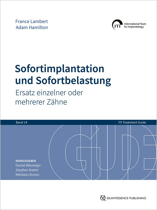
Reihe: ITI Treatment Guide Series, Band 14 1. Auflage 2024 Buch Hardcover, 21 x 28 cm, 288 Seiten, 856 Abbildungen Sprache: Deutsch Kategorie: Implantologie ISBN 978-3-86867-694-5 QP Deutschland
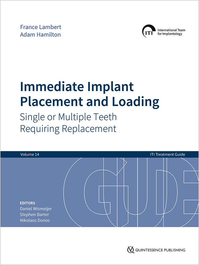
Reihe: ITI Treatment Guide Series, Band 14 1. Auflage 2023 Buch Hardcover, 21 x 28 cm, 288 Seiten, 856 Abbildungen Sprache: Englisch Kategorie: Implantologie Artikelnr.: BG164 ISBN 978-1-78698-127-1 QP Deutschland
Details make perfectionOktober 24, 2024 — Oktober 26, 2024MiCo - Milano Convention Centre, Milano, Italien
Referenten: Bilal Al-Nawas, Gil Alcoforado, Federico Hernández Alfaro, Sofia Aroca, Wael Att, Gustavo Avila-Ortiz, Kathrin Becker, Anne Benhamou, Juan Blanco Carrión, Dieter Bosshardt, Daniel Buser, Francesco Cairo, Paolo Casentini, Raffaele Cavalcanti, Tali Chackartchi, Renato Cocconi, Luca Cordaro, Luca De Stavola, Nuno Sousa Dias, Egon Euwe, Vincent Fehmer, Alberto Fonzar, Helena Francisco, Lukas Fürhauser, German O. Gallucci, Oscar Gonzalez-Martin, Dominik Groß, Robert Haas, Alexis Ioannidis, Simon Storgård Jensen, Ronald Jung, France Lambert, Luca Landi, Georg Mailath-Pokorny jun., Silvia Masiero, Iva Milinkovic, Carlo Monaco, Jose Nart, José M. Navarro, Katja Nelson, Manuel Nienkemper, David Nisand, Michael Payer, Sergio Piano, Bjarni E. Pjetursson, Sven Reich, Isabella Rocchietta, Giuseppe Romeo, Irena Sailer, Mariano Sanz, Ignacio Sanz Martín, Frank Schwarz, Shakeel Shahdad, Massimo Simion, Ralf Smeets, Benedikt Spies, Bogna Stawarczyk, Martina Stefanini, Hendrik Terheyden, Tiziano Testori, Daniel Thoma, Ana Torres Moneu, Piero Venezia, Lukas Waltenberger, Hom-Lay Wang, Stefan Wolfart, Giovanni Zucchelli, Otto Zuhr
European Association for Osseintegration (EAO)
International Esthetic Days
Digital excellence across disciplinesSeptember 19, 2024 — September 21, 2024Palau de Congressos de Palma, Palma de Mallorca, Spanien
Referenten: Eirik Aasland Salvesen, Wael Att, Andrea Bazzucchi, Andre Chen, Maja Chmielewska, Karim Dada, Mona Eide-Gast, Vincent Fehmer, Roshi Frafjord, Fernando Franch, Ophir Fromovich, Nuno Gil, Oscar Gonzalez-Martin, Sanaa Kader, France Lambert, Adriaens Laurence , Diego Lops, Mark Lowe, James Mah, Patrice Margossian, Alberto Monje, Léon Parienté, Lucrezia Paternò Holtzman, Alessandro Perucchi, Sonia Presencia Pascual, Algirdas Puišys, Cristian Scognamiglio, Prav Solanki, Gabor Tepper, Jasper Thoolen, Jochen Tunkel, Kay Vietor, Eglė Vindašiūtė-Narbutė, Sarah Weston
Straumann GmbH
35th EAED Spring Open Meeting
35 years of Esthetic Dentistry - The masters, the fundamentals, today's trendsMai 23, 2024 — Mai 25, 2024The Baron's Hall & Gallery, Vila Nova Gaia, Portugal
Referenten: Luís Azevedo, Urs C. Belser, Nitzan Bichacho, Markus Blatz, Jorge Cardoso, Nuno Sousa Dias, Petra Gierthmühlen, Aiste Gintaute, Oscar Gonzalez-Martin, Stefano Gracis, Ueli Grunder, Arndt Happe, Marc Hürzeler, France Lambert, Amélie Mainjot, Alexandros Manolakis, Konrad H. Meyenberg, Nazariy Mykhaylyuk, José M. Navarro, Gaetano Paolone, Stavros Pelekanos, Roberto Perasso, João Pitta, Pablo Ramírez, Andrea Ricci, Giano Ricci, Giuseppe Romeo, Irena Sailer, David Winkler, Yu Zhang, Giovanni Zucchelli
European Academy of Esthetic Dentistry
Zeitschriftenbeiträge dieses Autors
The International Journal of Prosthodontics, 3/2024
Online OnlyDOI: 10.11607/ijp.8328, PubMed-ID: 37988434Seiten: e136-e148, Sprache: EnglischRoth, Deborah / Compere, Philippe / Van Hede, Dorien / Lamy, Marc / Lambert, France
Purpose: To validate the effectiveness of a simplified ultrasonic cleaning protocol to clean customized abutments, and to investigate the percentage of pollutants on customized abutments provided by the implant company and the additional effect of dental laboratory manipulations. Materials and Methods: A total of 24 customized abutments were divided in two groups, 12 returning from the implant company and 12 others returning from the dental laboratory. In each group, there were 6 zirconia (Zr02) abutments and 6 .tanium (Ti) abutments. For each condition, half of the samples were cleaned with the experimental protocol, and the others were left as delivered by the company. The two-step cleaning protocol consisted of mechanical treatment with a sterile compress soaked in a detergent over the transgingival part of the abutment, followed by three successive ultrasound baths for two minutes/bath. The presence of pollutants was quantified using scanning electron microscopy. Results: The suggested cleaning method allowed to significantly decrease the quantity of pollutants (P = .0006). The abutments returning from the dental laboratory were significantly more polluted than those coming directly from the implant company (P = .0043). The cleaning effect was highly significant in both groups (P < .0001). The quantity of pollutants before cleaning were similar in the titanium and in the zirconia groups and the cleaning effect was highly significant in both groups (P = .0009). Conclusions: The cleaning protocol tested was successful on the customized abutments from each group.
Anwendung bei Zahnverschleiß und Schneidezahnnichtanlagen
In der restaurativen Zahnmedizin kann fehlender okklusaler Freiraum dazu führen, dass gesundes Gewebe geopfert werden muss, um Platz für das Restaurationsmaterial zu schaffen. Eine Versorgung ohne Präparation (No-Prep) lässt sich realisieren, indem Restaurationen in Supraokklusion eingesetzt werden und der dadurch hervorgerufene offene Biss durch einfache kieferorthopädische Extrusion (Simple Orthodontic Extrusion, SOE) der übrigen Zähne wieder geschlossen wird. Diese schnelle, partielle kieferorthopädische Maßnahme wird von den Patienten gut angenommen. Sie verwendet einfache Knöpfchen und benötigt nur wenige Wochen, um die Okklusionskontakte wiederherzustellen. Die SOE stellt eine Weiterentwicklung des Dahl-Prinzips dar und behält dessen Vorteile ohne die inhärenten Nachteile bei. In diesem Beitrag werden zwei mögliche Anwendungen der SOE-Technik gezeigt: die Restauration stark abgenutzter Frontzähne durch palatinale NoPrep-Veneers aus einer Hybridkeramik (duales Feldspatkeramik-Polymer-Netzwerk) und der Ersatz fehlender seitlicher Schneidezähne durch No-Prep-Adhäsivbrücken aus Zirkonoxid. Außerdem wird eine neuartige 3-D-gedruckte Kunststoffschiene zur korrekten Positionierung von Adhäsivbrücken und Erleichterung der Entfernung von überschüssigem Befestigungskomposit vorgestellt. Der Beitrag unterstreicht die Vorteile einer multidisziplinären Zusammenarbeit im Bereich der minimalinvasiven Zahnmedizin.
Schlagwörter: Adhäsivbrücken, Hybridkeramik, Kieferorthopädie, minimalinvasive Zahnmedizin, No-Prep, Okklusion, Zahnverschleiß, Zirkonoxid
Illustration in tooth wear and resin-bonded bridges
In restorative dentistry, the lack of occlusal space may lead to the mutilation of healthy tissue in order to provide sufficient space for the restorative material. Noprep dentistry can be achieved by placing high-bite restorations, followed by Simple Orthodontic Extrusion (SOE) of other teeth to close the created open bite. This rapid, partial orthodontic treatment is well accepted by patients as it can be easily performed using simple buttons, and it takes only a few weeks to reestablish occlusal contacts. The SOE technique is a further development of the Dahl concept. It has the advantages without the disadvantages. Two applications of this technique are presented in this article: the treatment of the severe wear of anterior teeth with no-prep palatal veneers made of Polymer-infiltrated Ceramic Network (PICN, “hybrid ceramic”) material and the realization of no-prep zirconia resin-bonded bridges (RBBs) to replace missing lateral incisors. An original 3D-printed resin guide for correctly positioning RBBs and facilitating the removal of excess composite cement is also presented. This work highlights the considerable advantages of multidisciplinary collaboration in the field of minimally invasive dentistry.
Schlagwörter: hybrid ceramic, minimally invasive dentistry, occlusion, orthodontics, PICN, resin-bonded bridges, tooth wear, zirconia
Purpose: To assess two types of abutment materials routinely used in daily practice—direct polymethyl methacrylate (PMMA) and a zirconia-on—Ti-base abutment—and their effects on peri-implant soft tissues and bone remodeling in a minipig model. Materials and Methods: A total of 40 implants were placed in five minipigs in a single-stage surgery. Four different types of abutment materials (n = 10 per group) were used: (1) titanium (control); (2) zirconia (control); (3) PMMA (test 1); and (4) Ti-base (zirconia bonded to a titanium framework; test 2). After 3 months of healing, the samples were collected and subjected to nondecalcified histology. The soft tissue dimensions (sulcus, junctional epithelium, and connective tissue attachment) were assessed on each abutment mesially and distally, and the distance from the implant margin to the first bone-to-implant contact (BIC) was measured. Results: No statistically significant differences were found among the four groups regarding soft tissue dimensions (P = .21), and a long junctional epithelium (mean: 4.1 mm) and a short connective tissue attachment (mean: 0.3 mm) were found in the majority of abutments. In some samples, the junctional epithelium extended all the way to the bone level. The measured peri-implant bone remodeling was similar in all four groups (P = .25). Conclusions: The present findings indicate that both direct PMMA and zirconia-on–Ti-base abutments seem to allow soft tissue integration similar to that of titanium and zirconia abutments. However, clinical studies are warranted to either confirm or refute the observed findings and to further investigate the influence of different materials on mucointegration.
Ziel: Ziel dieser Fallserie war die Auswertung der 4-Jahres-Ergebnisse von Implantaten in der ästhetischen Zone, die statisch geführt (static computer-assisted implant surgery, s-CAIS) eingesetzt und ohne Abutmentwechsel (One-Abutment-One-Time-Konzept, OAOT) mit vor der Implantatsetzung angefertigten individuellen Zirkonoxidabutments und zementierten Kronenprovisorien versorgt wurden.
Material und Methode: Zehn Implantate wurden sukzedan in der ästhetischen Zone eingesetzt. Nach digitaler Planung der Implantatchirurgie und prothetischen Versorgung wurden Implantatschablonen bestellt, die zunächst für die Herstellung von definitiven individuellen Zirkonoxidabutments und PMMA-Provisorien verwendet wurden. Anschließend wurden mithilfe der Schablonen die Implantate eingesetzt und sofort mit den prothetischen Komponenten versorgt. Die Implantatergebnisse wurden zum Zeitpunkt der Implantatsetzung sowie 4 Monate und 4 Jahre danach ausgewertet.
Ergebnisse: Alle Implantate konnten erfolgreich mithilfe der Implantatschablonen eingesetzt und mit den definitiven Abutments und Kronenprovisorien versorgt werden. Größere prothetische Komplikationen wurden nicht beobachtet. Nach 4 Jahren lag die Implantatüberlebensrate bei 100 % und es wurden kleinere periimplantäre Sondierungsblutungen, aber sehr stabile periimplantäre Knochenhöhen beobachtet. Der Pink-Esthetic-Score zeigt, dass alle prothetischen Komponenten gut integriert waren und eine stabile periimplantäre Weichgewebesituation vorlag.
Schlussfolgerung: Innerhalb der Grenzen dieser Fallserie zeigen die erzielten Ergebnisse, dass neue digitale Workflows die Anfertigung individueller definitiver Abutments und Provisorien vor der Implantatoperation ermöglichen. Der individualisierte OAOT-Ansatz kann zementbedingte Komplikationen verringern und das ästhetische Ergebnis verbessern, indem die Gewebeheilung durch die prothetischen Komponenten optimiert wird. Allerdings haben sich Protokolle dieser Art inzwischen zu volldigitalen Workflows entwickelt, die zunächst klinische Vergleichsstudien erfordern.
Schlagwörter: statisch geführte Implantatchirurgie, s-CAIS, digitaler Workflow, One Abutment One Time, individuelles Abutment, Knochenremodellierung, Knochen-Implantat-Kontakt, Pink Esthetic Score
Purpose: To study the practices of general dentists, periodontists and dental hygienists who are members of the European Federation of Periodontology, regarding oral hygiene education, plaque control assessment, recommended dental and interdental hygiene tools, and antimicrobial agents.
Materials and Methods: A web-based survey was sent to 13,622 members of the European Federation of Periodontology (EFP) through its 29 national member societies. It targeted general dentists (GD), specialists in periodontology (DSP) and dental hygienists (DH). Data were collected between 24 April and 17 May 2015. A data-driven statistical analysis was conducted and differences between professions were explored.
Results: A total of 2076 answers were collected. Only the 2009 answers originating from GD, DSP and DH were analysed (67 answers originated from other professions and were excluded). Among those 2009 respondents, 43.2% were DSP and 37.2% were GD. Overall, DH, DSP and GD reported spending 17.1 minutes for the initial teaching of OH, with differences between professions (p < 0.0001). DH, GD and DSP exhibited differences in the type of toothbrushes they recommend (p < 0.0001). DSP recommended electric and manual toothbrushes (TB) equally. DH predominantly recommended electric TB (56.8%). Overall, 95% of DH, DSP and GD recommended interdental brushes, with no statistically significant differences between professions. Low concentration chlorhexidine was considered the most relevant antimicrobial agent for daily oral care of periodontitis patients. Half of GD prescribed antimicrobial mouthrinses for long-term use in 70%–100% of their patients with periodontitis.
Conclusion: EFP-affiliated practitioners allocate a significant amount of time to educating patients on oral health. Their practices are mostly in line with the current scientific evidence. Some discrepancies were found between the different professions. Similar surveys could be conducted over time to monitor the evolution of practices.
Schlagwörter: dental hygienist, oral hygiene, patient education as topic, periodontal diseases, periodontists, surveys and questionnaires
Purpose: The purpose of this study was to evaluate the 2-year outcomes of the one-tooth onetime complete digital workflow, allowing the immediate loading of a single implant in the posterior region with a final CAD/CAM crown made of a polymer-infiltrated ceramic network.
Materials and methods: A series of 10 implants were placed, and an intraoral scan was taken after surgery. A final screw-retained polymer-infiltrated ceramic network crown was manufactured chairside and placed the same day in full occlusion. Marginal peri-implant bone changes and soft tissue health were evaluated, and restoration performance was assessed using FDI World Dental Federation criteria and pink and white aesthetic scores. Patient-reported outcome measures and data on the time required to perform the procedures were collected.
Results: After 2 years, the implant survival rate was 100%. The debonding of one crown from its titanium base led to prosthodontic survival rate of 90% and the remaining crowns were all considered successful. The mean marginal peri-implant bone changes yielded 0.87 mm (standard deviation 0.96 mm) and 0.55 mm (standard deviation 0.53 mm) after 1 and 2 years, respectively. Mild or no inflammation of peri-implant soft tissue was observed in most implants. The total treatment time was 175 minutes and patient-reported outcome measures displayed high patient satisfaction.
Conclusions: This study constitutes the first report examining immediate loading of a single implant in the posterior region with a final crown in occlusion. In this case series, the 2-year outcomes of the one-tooth one-time protocol seem rather promising and fulfilled patient expectations. However, these preliminary results need to be confirmed by randomised controlled trials, and patient selection is likely to be a key factor in the success of this procedure.
Schlagwörter: CAD/CAM, dental implant, digital workflow, immediate loading, intraoral scan (IOS), polymerinfiltrated ceramic network (PICN)
Amélie Mainjot cohabits with the founder of the company MaJEB, which contributes to the development of polymer-infiltrated ceramic network materials. Prof Lambert has collaboration agreement
The primary objective of this pilot study was to evaluate a new socket preservation technique involving the intentional expansion of the extraction socket buccal plate using a flapless internal corticotomy and biomaterials. A total of 11 patients requiring tooth extraction were enrolled in this study. The aim of this technique was to maintain or improve the hard and soft tissue contour of the ridge after tooth extraction. All surgical sites healed uneventfully. Significant alveolar bone dimension changes were observed in the coronal region of the ridge (−1.4 ± 0.9 mm); however, it was only slightly lower at the medium (−0.35 ± 0.7 mm) and apical levels (−0.3 ± 0.8 mm) (P > .05). The ridge dimensional changes were significantly higher in the buccal aspect than in the palatal aspect in all patients. Vertical bone resorption was not significant. Concerning the soft tissue contour, the horizontal distance between the preoperative and postoperative buccal profiles ranged from 0.94 to −2.88 mm. The proposed ridge preservation technique may help maintain the volume of the healed ridge but cannot completely prevent contour changes after tooth extraction.
Purpose: To evaluate the effectiveness of a bone substitute covered with a resorbable membrane versus open flap debridement for the treatment of periodontal infrabony defects.
Materials and methods: Ninety-seven patients with one infrabony defect, which was 3 mm or deeper and at least 2 mm wide were randomly allocated either to grafting with a bone substitute covered with a resorbable barrier (BG group) or open flap debridement (OFD group) according to a parallel group design in five European centres. Blinded outcome measures assessed tooth loss, complications, patient's satisfaction with treatment and aesthetics, changes in probing attachment levels (PAL), probing pocket depths (PPD), gingival recessions (REC), radiographic bone levels (RAD) on standardised periapical radiographs, plaque index (PI) and marginal bleeding index (MBI).
Results: 49 patients were randomly allocated to the BG group and 48 to the OFD group. At baseline there were more mobile teeth in the BG group (29 versus 15). One year after treatment two patients dropped out from the BG group and no teeth were lost. Three complications (minor postoperative wound dehiscence) occurred in the BG group versus none in the OFD group, where the difference was not statistically significant. The BG group obtained significantly greater statistical PAL gain (mean difference = -0.8 mm, 95%CI [-1.51; -0.03], P = 0.0428), PPD reduction (mean difference = -1.1 mm, 95%CI [-1.84; -0.19], P = 0.0165) and RAD gain (mean difference = -1.2 mm, 95%CI [-2.0; -0.4], P = 0.0058) compared to the OFD group. No statistically significant differences between the groups were observed for gingival recession, or the patient's satisfaction with the treatment and aesthetics. There were some statistically significant differences between the centres for PAL and PPD with the Italian centres reporting better outcomes.
Conclusions: The use of a bone substitute covered with a resorbable membrane yielded significantly better statistical clinical outcomes than open flap debridement in the treatment of periodontal infrabony defects deeper than 3 mm, with regard to PAL gain, PPD reduction and RAD gain.
Schlagwörter: bone substitutes, infrabony defect, periodontitis, randomised controlled trial
Purpose: Subsinus bone regeneration procedures are reliable and known to be effective with the use of biomaterials alone. Nevertheless, many types of biomaterials are available, and the efficacy of each in terms of bone formation and resorption rate has rarely been compared. This study aimed to compare bone formation, resorption rate, osteoconductivity, and three-dimensional volume changes of three biomaterials often used for alveolar ridge augmentation.
Materials and Methods: Rabbits underwent bilateral sinus elevation using three different types of space fillers: bovine hydroxyapatite (BHA), beta-tricalcium phosphate (ß-TCP), or biphasic calcium phosphate (BCP). Animals were sacrificed at 1 week, 5 weeks, and 6 months. Samples were subjected to microcomputed tomography and histologic examination. Qualitative analysis was performed on nondecalcified sections, and quantitative histomorphometric analysis was conducted using scanning electron microscopy. Volume differences in augmented bone were calculated at different time points.
Results: All three particulated biomaterials promoted osteogenesis in this particular animal model. At 6 months, biomaterial resorption rates were significantly different across the three groups. The highest resorption rate was found with ß-TCP, in which only 6.7% of the baseline particle surface remained. At 6 months, bone was in close contact with the BHA particles, constituting a composite network; in contrast, BCP particles were often surrounded by soft tissue. Within each group, no significant differences in volume were found at the different time points.
Conclusions: Despite the limitations of the study, the three studied biomaterials proved to be effective in promoting osteogenesis. High resorption rates and complete replacement of the biomaterials by bone seemed to withstand intrasinusal pressure. Further investigations in humans should consider longer follow-up periods.