Auf unserer Website kommen verschiedene Cookies zum Einsatz: Technisch notwendige Cookies verwenden wir zu dem Zweck, Funktionen wie das Login oder einen Warenkorb zu ermöglichen. Optionale Cookies verwenden wir zu Marketing- und Optimierungszwecken, insbesondere um für Sie relevante und interessante Anzeigen bei den Plattformen von Meta (Facebook, Instagram) zu schalten. Optionale Cookies können Sie ablehnen. Mehr Informationen zur Datenerhebung und -verarbeitung finden Sie in unserer Datenschutzerklärung.
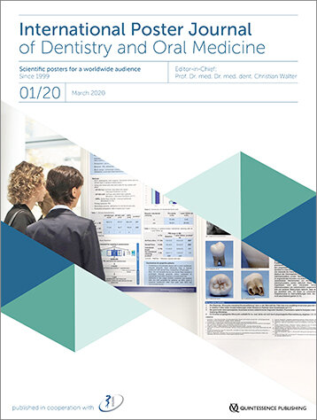
Das "International Poster Journal of Dentistry and Oral Medicine" wird seit März 1999 in Zusammenarbeit mit der Deutschen Gesellschaft für Zahn-, Mund- und Kieferheilkunde (DGZMK) und dem Quintessenz Verlag herausgegeben.
Die vierteljährlich erscheinende Publikation bietet ein Forum für die Veröffentlichung von preisgekrönten und begutachteten wissenschaftlichen Postern aus dem Bereich der Zahn-, Mund- und Kieferheilkunde, die auf internationalen Ausstellungen präsentiert werden.
Schriftleitung / Chefredaktion
Prof. Dr. Dr. Christian Walter MD DDS PhD
Deutschland, Mainz
1993-2004: Studium der Medizin (Universität Mainz), Technik in der Medizin (Technische Universität Kaiserslautern) und Zahnmedizin (Universität Mainz). 2003: Wissenschaftlicher Mitarbeiter an der Klinik für Mund-, Kiefer- und Gesichtschirurgie Mainz. 2009: Bestellung zum Oberarzt. 2010: Habilitation im Fach Mund-, Kiefer- und Gesichtschirurgie.
Koordinierende Redaktion
Dipl.-Ing. Karin Klewer
Deutschland, Berlin
Vorschau
Beschreibung
Dateiformat
Download
Autorenrichtlinien International Poster Journal of Dentistry and Oral Medicine ISSN 1612-7749
International Poster Journal of Dentistry and Oral Medicine, Supplement OMD/2021
Themenschwerpunkt: 30th Annual Meeting of the Portuguese Dental Association
In Forensic Odontology, the estimation of the stature often has a crucial role in the reconstructive phase of disjointed populations. The femur, being the longest bone in the human body, is usually the most reliable source in height estimation. This investigation aims to estimate the stature as well as to identify the minimum number of individuals (MNI) of the catastrophic population concerning the earthquake that occurred in 1755 in Lisbon, whose skeletal remains were found in the Cloister’s South Wing of Academia das Ciências de Lisboa in 2004. This study was conducted on eight complete femurs and twenty-one fragments, which were measured, using an absolute digimatic Mitutoyo® caliper or a vernier Mitutoyo® caliper, and weighted. Mildred Trotter and Goldine C. Gleser’s (1952) study was used to estimate the height using whole femurs. Based on the fragmented femurs, the total length of the femur was calculated using Steele and Mckern’s (1970) study. Afterwards, Mildred Trotter and Goldine C. Gleser’s (1952) study was applied in order to estimate each individuum’s height. It was possible to determine a minimum number of individuals (MNI) of 29, correspondent to 8 left whole femurs (from a total number of 14) and 21 left femur bodies (from a total number of 58). The results showed that, for the whole femur, the height varies between 148 cm and 169 cm and regarding the fragments, they vary between 151,67 cm and 181,59 cm. The results of the estimated heights are compatible with the expected for that period of time, therefore the carried-out investigation reveals that the applied methods seem to be viable.
Schlagwörter: Forensic odontology, femur, estimation of height, catastrophic population
In regards to Forensic Odontology, sex classification from unknown skeletal remains is vital in the reconstruction phase, and the most accurate methods to do it have been researched extensively on various bones of the human skeleton. The present work consists in the analysis of 129 coxal bones belonging to a population attributed to the 1755 Lisbon’s Earthquake, aiming to characterize the population whose skeletal remains were found in 2004 in the Cloister’s South Wing of Academia das Ciências de Lisboa. To achieve the final objective, six measurements were taken, in anatomic position, with a Mitutoyo Vernier Caliper and a Mitutoyo Absolute Digimatic Caliper®, and seven indices were calculated. Morphological characteristics were also observed through four different methods. From measurements and indices, there were 16 females and 5 males identified, but the overall results were not reliable, since there was no consensus between the different measurements, and so the majority of the coxal bones were classified as ambiguous. It was found that the morphological observations were more discriminating and, therefore, the Phenice’s Method showed the most precise results, because of the inclusion of three parameters simultaneously.
Schlagwörter: Forensic odontology, sex classification, coxal, linear measurements, morphology
Introduction: All preparation techniques and instruments are associated with apically extruded debris (AED). Different preparation techniques promote different amounts of AED justified by the instruments design, namely its cross section, conicity, tip design and also the number of files, kinematics and cutting efficiency, associated with the quantity, type and technique of irrigation.
Objective: Compare the amount of AED, created by two hybrid techniques and two single system techniques. The null hypothesis formulated stated that all groups present the same amount of AED.
Materials and Methods: Eighty single root canal teeth were randomly assigned into 4 groups (n=20), according to the instrumentation technique: hybrid technique TruNatomy®/ProTaper Gold® (TN/PTG), hybrid technique TruNatomy/ProTaper Next® (TNP/TN), system ProTaper Gold (PTG) and system ProTaper Next (PTN) (Dentsply Sirona, Ballaigues, Switzerland). During preparation, for each specimen, a total of 13 mL 3% sodium hypochlorite (NaOCl) was used. AED was collected in Eppendorf tubes (ET). After instrumentation, each tooth was removed from the ET and, after filling with 1.5 mL NaOCl, each ET was stored in an incubator at 70ºC for 5 days. Three ET, without AED, containing 1.5mL 3% NaOCl were used as control. After the incubation period, each ET was weighed three times to obtain an average weight. The amount of AED, in each ET, was calculated by subtracting the control-weighed ET to the post-preparation weight. After checking normality (Shapiro-Wilk test), the Kruskal-Wallis non-parametric test was performed followed by the Mann-Whitney test with Bonferroni correction to identify significant differences (α=0.05).
Results: Significant differences in the AED were detected for the various techniques (p<0.001). The hybrid techniques TN/PTG and TN/PTN groups produced significantly less extrusion compared to the PTG and PTN techniques (p=0.012 and p=0.046, respectively). The TN/PTN technique had a significantly lower AED than the other groups (p≤0.046) and the PTG technique significantly higher (p≤0.012).
Discussion: In this study, the superior performance of the PTN system can be justified by the type of movement, less taper than PTG files and the type of cross-section that these instruments have, as well as the fact that this system has a smaller number of files. The significantly smaller differences related to AED, between TN/PTG and TN/PTN hybrid techniques and PTG and PTN alone, respectively, may be related to the fact that TN instruments have a maximum cervical diameter of 0.8mm which promotes greater dentin preservation when compared to S1, S2 and X1 files which cut the pericervical dentin more aggressively, as they have 1.20 maximum cervical diameter. Consequently, performing Preflaring with smaller cervical diameter before Root Canal preparation can reduce the amount of AED.
Conclusion: Hybrid instrumentation techniques TN/PTN and TN/PTG produced significantly less AED than the PTN and PTG techniques.
Schlagwörter: Glide Path, debris extrusion, TruNatomy, ProTaper
Objectives: The Objective of the present study was to describe the effects observed after conservative treatment of TMJ osteoarthritis and osteoarthrosis. The procedure consisted of stabilization(1) using occlusal appliance and physiotherapy(2-4) for functional recovery and restoration of joint biomechanics. These interventions were associated with minimally invasive treatment injecting hyaluronic acid (5-9), (AH) into the upper and lower joint space and the results evaluated by cone beam computed tomography (CBCT).
Materials and methods: 60 year old man, diagnosed using DC/TMD with myofascial pain, osteoarthritis, osteoarthrosis, and presumption of diagnosis of disc displacement without reduction, of the two temporo-mandibular joints associated with primary or idiopathic(9-12) wake and sleep bruxism, was submitted to a sequential protocol of injection of medium molecular weight HA(13) (Osteonil plus-TRB pharma) in the posterosuperior compartment and low molecular weight HA (Hylart-Bagó pharma) interspersed with Osteonil plus-TRB pharma for the anteroinferior compartment of both TMJs, monthly for 4 months (13,14) . Evaluation with CBCT was performed before treatment and six months after the last infiltration. The patient underwent physiotherapy after each viscosupplementation session and the following week, home oriented exercises(2,15) and monthly clinical evaluation with measurement of oral opening amplitude (ROM) with therabite and pain using the visual analogue scale (VAS)(16). The patient was instructed to use applications for mobile phone to control bruxism while awake and occlusal split to control sleep bruxism (17,18).
Results: Significant structural gain seen in shape and volume of the mandible head in both TMJs, functional joint improvement, with increased amplitude of oral opening, The initial opening was 30 mm and after the end of treatment 44 mm, absence of pain at the end of treatment with obvious improvement in activities of daily living such as eating or speaking.
Conclusions: Sequential viscosupplementation with AH of the two joint compartments associated with conservative treatment and control of awake and sleep bruxism, were effective in the treatment of osteoarthritis and osteoarthrosis of patient TMJs.
Schlagwörter: viscosupplementation, hyaluronic acid, temporomandibular joint, osteoarthritis, osteoarthrosis
Introduction: Obstructive sleep apnea syndrome (OSAS) is a sleep-related breathing disorder. The objective of this paper is to present a clinical case with OSAS successfully treated through orthognathic surgery.
Material and Methods: Male patient, 43 years and 3 months old, with moderate OSAS and an apnea-hypopnea index of 22. In the cephalometric analysis, a skeletal Class I (< ANB 3º) with a setback in the position of the maxilla (< SNA 77º) and mandible (< SNB 74º) in relation to the skull base was observed. This skeletal pattern favors the narrowing or obstruction of the upper airway, which was confirmed by the radiographic examination. The proposed treatment plan consisted of: orthognathic surgery, using the Surgery First technique, with Le Fort I maxillary advancement of 7 mm and impaction of 2 mm and bilateral sagittal osteotomy with mandibular advancement of 7 mm.
Results: Two months after surgery, there were no post-surgical complications, the patient reported an improvement in symptoms associated with OSAS, and in the assessment of radiographic examinations was observed an increase in the volume and the minimum axial dimension of the upper airway.
Discussion: The bimaxillary advancement surgery allows the anterior pharyngeal wall to move forward, resulting in the enlargement of the pharyngeal airway and, consequently, a decrease in the apnea-hypopnea index.
Conclusions: The Surgery First technique allowed for immediate surgery and the consequent improvement of OSAS. Bimaxillary advancement movement increased the volume and minimum axial distance of the upper airway.
Schlagwörter: Sleep apnea, obstructive, upper airway, orthognathic surgery, surgery first, osteotomy, Le Fort, osteotomy, sagittal split ramus
Introduction: A skeletal class II patient may present a mandibular retrognathia. This condition can lead to an inefficient breathing pattern, suboptimal mastication due to the improper occlusion pattern as well as an unsatisfactory profile. Although mandibular hypoplasia is a rather common occurrence, the combination of orthodontic and orthognathic procedures can be the most appropriate treatment for moderate to severe discrepancies. The lengthening of the mandible ramus can be achieved through a split ramus osteotomy or, in more severe cases, through a distraction device. This intervention can be chosen in order to achieve better aesthetics and function therefore improving the patient’s quality of life. The aim of this poster is to present a surgical-orthodontic approach for effective treatment of a skeletal class II associated with severe mandibular retrognathia.
Material and Methods: A twenty-three-year-old patient with severe skeletal class II presented to the Institute of Orthodontics looking for malocclusion correction. The patient had undergone previous orthodontic treatment at another centre, however she presented both breathing and feeding difficulties as well as unappealing facial aesthetics. After careful consideration of the initial case tooth-borne distraction osteogenesis for mandibular lengthening and surgical orthodontic treatment were suggested as the best treatment option.
Results: Roth 0,018 prescription fixed appliances were placed to level the arches followed by the placement of the tooth-borne distraction osteogenesis device which was activated twice bilaterally every 12 hours. At the end of the distraction, 11 mm of mandibular lengthening were obtained. After this intervention the orthodontic treatment progressed in order to prepare the patient for a orthognathic surgery of maxillary impaction and mandibular repositioning. A significant improvement in the ANB angle was achieved from 16º to 4º. Despite the facial soft tissue improvement, the convex profile still did not meet the patient’s aesthetic expectations so the patient was subsequently submitted to a genioplasty.
Discussion and Conclusions: The patient had re-established a functional occlusion, a more suitable breathing pattern, a desirable profile and an overall pleasant facial aesthetic.
Schlagwörter: Orthodontics, orthognatic surgery, class II, mandibular retrognathia
Full mouth rehabilitation with immediate loading implants has become a predictable procedure whereas implant and prosthesis survival are concerned. However less information is known from the patient centered outcomes measures perspective. This was a practice based prospective single cohort study aiming at studying the Impact of immediate loading implant supported full mouth rehabilitation on the oral health related quality of life (OHRQoL). 43 patients were recruited in a Portuguese dental clinic. Inclusion criteria were indication of full mouth rehabilitation with immediate loading protocol, exclusion criteria were nonnative Portuguese language. A Portuguese version of the Oral Health Impact Profile 14 was administered at study baseline before the intervention (visit 1), day 10 (visit2), 1 month (visit 3) and 6 months follow up (visit 4). Predictive variables were age, gender, household owning, marital status, education, profession, dental needs, type of rehabilitation provided and visit number. Primary outcome was total OHIP 14 PT score and partial domains. Glass effect size was calculated. Mean differences were tested with paired student t test and significance was set at alpha 5 %. Total OHIP 14 scores at visits 1, 2, 3 and 4 were 18,00±11,20 SD; 11,47±7,95 SD; 5,84±4,07 SD; 5,02±3,04 respectively. There was an extremely significant improvement in OHRQoL along the study follow up (p<0,001 Paired Student t Test) except for comparisons between visit 3 and 4 for every OHIP 14 domain. Full mouth rehabilitation with immediate loading protocol significantly increases the OHRQoL, from baseline through six months follow up.
Schlagwörter: quality of life, immediate dental implant loading, mouth rehabilitation, implant-supported, prosthodontics, dentistry
Through current scientific evidence, the application of pit and fissure sealants in permanent dentition is a procedure consistently based on the prevention of caries, however, little evidence supports the performance of this technique in primary molars, and its application is reserved for high risk conditions. The objective of this systematic review was to answer the following question, formulated through the PICO strategy: Does the application of sealants contribute effectively to the prevention of caries in primary molars? Additionally, it was intended to understand the success rates inherent to the retention of sealants in this dentition. Bibliographic research of scientific articles published between 2005 and 2020 in the electronic databases PubMed, B-On, Cochrane Library and ScienceDirect, through research terms: pit and fissure sealants; primary teeth; primary molars; dental caries; prevention and retention, articulated through the boolean AND marker. Inclusion and exclusion criteria were established. For inclusion: clinical trials, randomized controlled trials, cohort and case-control studies, conducted in primary dentition and whose dental outcome was: the development of caries and/or, the rate of retention of pit and fissure sealants, comparing the intervention (sealant application) with the absence of application or with the implementation of other preventive measures. The methodological line followed for the selection of articles is presented in the PRISMA flow diagram. The methodological quality assessment of clinical trials was performed using a Cochrane tool and the Newcastle-Ottawa scale was used for the cohort study.
Regarding the efficacy of the application of sealants in the prevention of caries, three of the four studies included showed a positive association between the application of this technique and the lower incidence of caries. Although the studies of Joshi et al.; Hong et al. and Chadwick et al. share a similar methodology (application vs. non-application of sealant), in the latter, there was no significant association between the intervention and the outcome evaluated. Regarding the retention rate, it is verified that the authors consider, in a different way, different variables, namely, light curing cycles, acid conditioning techniques and different types of sealants, making it difficult to accurately evaluate them. However, it is verified that, overall, the application of this technique presents satisfactory retention rates in this dentition and can be applied effectively in primary molars. Given the effectiveness of the application of sealants in the prevention of caries in primary dentition, this measure should be included in a holistic preventive program, without being specifically reserved for children at high risk for this disease. Despite the anatomical characteristics less favorable to the retention of sealants, in this dentition, it is verified that due to their shorter period of permanence in the oral cavity, the retention rates are satisfactory. It is denoted, however, the need for future studies with stricter methodologies in order to obtain more robust results on this theme.
In conclusion, there is scientific evidence that demonstrates that the application of pit and fissure sealants in primary molars is an effective method to prevent dental caries.
Schlagwörter: Pit and fissure sealants, primary teeth, prevention, retention, pediatric dentistry, preventive dentistry
Epidermolysis Bullosa (EB) is a rare genetic disease characterized by a recurrent formation of blisters due to structural weakness of the skin and mucous membranes. It is caused by mutations in genes, which alter the cohesion between epithelial cells or between these cells and connective tissue. The disease can be classified into four types: EB simplex; junctional EB; dystrophic EB (DEB) and Kindler syndrome. There is no predominance of sex or race.
The most common clinical manifestations of this pathology are blisters that, when they rupture, leave painful ulcers. Changes in the shape and structure of teeth, ankyloglossia, susceptibility to tooth decay, limited mouth opening, among others, may also occur. In recessive dystrophic epidermolysis bullosa (RDEB), oral lesions appear with minimal trauma, requiring specific care in dental treatment. A 5-year-old caucasian male patient attended a dental appointment in 2016, having been diagnosed with RDEB since he was 23 months old. Clinically, the patient had bullous and hemorrhagic lesions on the oral mucosa, ulcers and skin scars. He had a high plaque index, tartar deposition, gingival inflammation, tooth decay, dental number changes such as gemination in a deciduous dentition tooth and left unilateral crossbite. The reason for the consultation was the presence of tooth decay and difficulty in oral hygiene. Initially, specific oral hygiene and dietary instructions were addressed. Scaling were performed, as well as the wear of the edges of the cusps that traumatized the mucous membranes and topical applications of fluoride. The treatment of caries and extraction of certain deciduous teeth that had not exfoliated began. Appointments were scheduled quarterly, although given the patient's little cooperation, they were limited to urgent treatments. DEB can be inherited in an autosomal dominant or recessive manner. In the present case, the mutation has maternal origin and atypical, given the benignity of the phenotype. The clinical and radiographic findings showed that we were facing a patient with a high risk of tooth decay. Brushing has always been very difficult, the patient is unable to use dental floss and doesn't tolerate manipulation of the oral cavity. As such, preventive measures were adopted. Treatments were sometimes postponed due to painful symptoms prior to the appointments. Currently, scaling is performed with Guided Biofilm Therapy®, which was well tolerated by the patient and didn’t form new bullous lesions. For a correct follow-up in the field of dentistry, both the dentist and caregivers must be aware of preventive measures and appropriate recommendations for these patients. Considering the fragility of the oral mucosa in patients with EB, preventive dentistry plays a fundamental role in reducing the need for dental treatments, and it is essential that the dentist knows the care to be taken.
Schlagwörter: epidermolysis bullosa, prevention, guidelines