Various cookies are used on our website: We use technically necessary cookies for the purpose of enabling functions such as login or a shopping cart. We use optional cookies for marketing and optimization purposes, in particular to place relevant and interesting ads for you on Meta's platforms (Facebook, Instagram). You can refuse optional cookies. More information on data collection and processing can be found in our privacy policy.
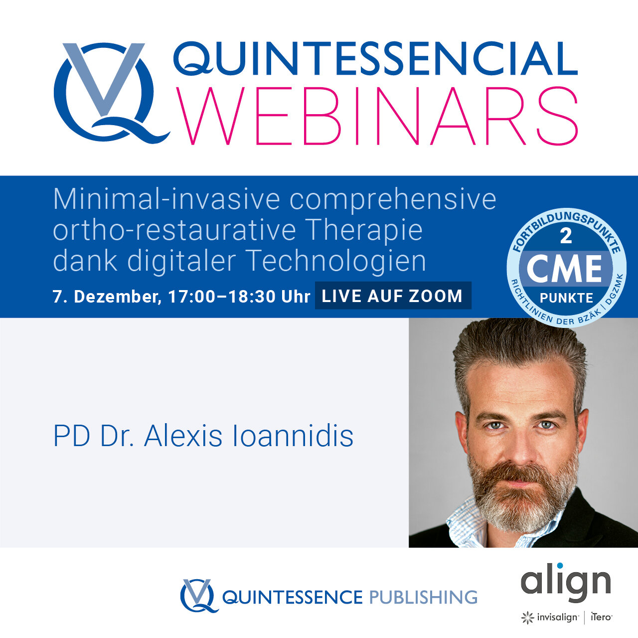
Alexis Ioannidis is a Senior Teaching and Research Assistant at the Clinic of Reconstructive Dentistry, University of Zurich, Switzerland. He graduated 2009 at the University of Zurich and received the title „Dr. med. dent.“ at the same University (2013). After completion of a 3-year post-graduate training at the Clinic of Reconstructive Dentistry at the University of Zurich in 2016, he received a stipend founded by the Clinical Research Foundation to spend 6 months as a research fellow at the Department of Biomaterials at the University of Athens, Greece. Since 2017 he serves as a full-time Senior Teaching and Research Assistant at the Clinic of Reconstructive Dentistry at the University of Zurich. His clinical focus is on the comprehensive treatment of complex, fully dentated or partially edentulous patients applying all available options of prosthetic dentistry including dental implants. He is a specialist for reconstructive dentistry (Swiss Society for Reconstructive Dentistry), an EPA Recognized Specialist in Prosthetic Dentistry (European Prosthodontic Association), has a Master of Advanced Studies in Periodontology and a Master of Advanced Studies in Oral Implantology (University of Zurich). His main scientific interest is related to minimally invasive prosthetic treatment concepts. He has published > 25 scientific and clinical articles, mostly in high-ranked peer-reviewed journals.
Events
31st EAO Annual Scientific Meeting
Details make perfection24. Oct 2024 — 26. Oct 2024MiCo - Milano Convention Centre, Milano, Italy
Speakers: Bilal Al-Nawas, Gil Alcoforado, Federico Hernández Alfaro, Sofia Aroca, Wael Att, Gustavo Avila-Ortiz, Kathrin Becker, Anne Benhamou, Juan Blanco Carrión, Dieter Bosshardt, Daniel Buser, Francesco Cairo, Paolo Casentini, Raffaele Cavalcanti, Tali Chackartchi, Renato Cocconi, Luca Cordaro, Luca De Stavola, Nuno Sousa Dias, Egon Euwe, Vincent Fehmer, Alberto Fonzar, Helena Francisco, Lukas Fürhauser, German O. Gallucci, Oscar Gonzalez-Martin, Dominik Groß, Robert Haas, Alexis Ioannidis, Simon Storgård Jensen, Ronald Jung, France Lambert, Luca Landi, Georg Mailath-Pokorny jun., Silvia Masiero, Iva Milinkovic, Carlo Monaco, Jose Nart, José M. Navarro, Katja Nelson, Manuel Nienkemper, David Nisand, Michael Payer, Sergio Piano, Bjarni E. Pjetursson, Sven Reich, Isabella Rocchietta, Giuseppe Romeo, Irena Sailer, Mariano Sanz, Ignacio Sanz Martín, Frank Schwarz, Shakeel Shahdad, Massimo Simion, Ralf Smeets, Benedikt Spies, Bogna Stawarczyk, Martina Stefanini, Hendrik Terheyden, Tiziano Testori, Daniel Thoma, Ana Torres Moneu, Piero Venezia, Lukas Waltenberger, Hom-Lay Wang, Stefan Wolfart, Giovanni Zucchelli, Otto Zuhr
Implantology: Beyond your expectations12. Oct 2021 — 14. Oct 2021online
Speakers: Enrico Agliardi, Alessandro Agnini, Andrea Mastrorosa Agnini, Mauricio Araujo, Goran Benic, Juan Blanco Carrión, Daniel Buser, Raffaele Cavalcanti, Tali Chackartchi, Luca Cordaro, Jan Cosyn, Holger Essig, Vincent Fehmer, Stefan Fickl, Alberto Fonzar, Helena Francisco, German O. Gallucci, Ramin Gomez-Meda, Oscar Gonzalez-Martin, Robert Haas, Arndt Happe, Alexis Ioannidis, Ronald Jung, Niklaus P. Lang, Tomas Linkevičius, Iva Milinkovic, Sven Mühlemann, Katja Nelson, Sergio Piano, Michael A. Pikos, Bjarni E. Pjetursson, Marc Quirynen, Franck Renouard, Isabella Rocchietta, Dennis Rohner, Irena Sailer, Henning Schliephake, Shakeel Shahdad, Massimo Simion, Ali Tahmaseb, Hendrik Terheyden, Jochen Tunkel, Stefan Vandeweghe, Piero Venezia, Stijn Vervaeke, Martin Wanendeya, Georg Watzek, Giovanni Zucchelli
European Association for Osseintegration (EAO)
This author's journal articles
International Journal of Esthetic Dentistry (EN), 4/2024
IJED EspressoPubMed ID (PMID): 39429066Pages 310-311, Language: EnglishPirc, Miha / Fischli, Tobias / Thoma, Daniel / Ioannidis, Alexis / Jung, Ronald / Bienz, Stefan
Einteilung nach verfahrenstechnischen und werkstoffkundlichen Aspekten
Durch die wachsende Nachfrage nach monolithischen Zirkonoxid-Restaurationen wächst der Anspruch an die Veredelungstechniken. Die stetige Weiterentwicklung des Gerüstmaterials schafft neue Möglichkeiten der Finalisierung. So lassen sich sowohl einige Vorteile als auch neue Herausforderungen durch das Mikrolayering-System beobachten. Die Datenlage ist derzeit noch begrenzt und ein Vergleich der verschiedenen Massen hat noch nicht stattgefunden. Eine Einteilung hinsichtlich verfahrenstechnischer und werkstoffkundlicher Aspekte verschafft einen Überblick.
Keywords: Mikrolayering, Minimalverblendung, Beschichtung, Veredelung, Zirkonoxid
International Journal of Computerized Dentistry, 3/2023
ScienceDOI: 10.3290/j.ijcd.b3781703, PubMed ID (PMID): 36632986Pages 237-245, Language: English, GermanGil, Alfonso / Eliades, George / Özcan, Mutlu / Jung, Ronald E. / Hämmerle, Christoph H. F. / Ioannidis, Alexis
Aim: To evaluate the fracture load and type of failure of two different monolithic restorative materials bonded to standardized titanium bases and fabricated by two different procedures regarding the bonding interface.
Materials and methods: All screw-retained implant crown specimens (n = 40), subjected to fatigue by thermomechanical loading, differed in the restorative material (lithium disilicate [LDS] or polymer-infiltrated ceramic network [PICN], referred to as ‘hybrid ceramic’ [HYC]) and the interface type between the restorative material and the titanium base abutment (prefabricated ex-factory or produced during a CAM-milling procedure). This resulted in the following groups (n = 10/group): 1) LDS-M: lithium disilicate crown with a CAM-milled interface; 2) LDS-P: lithium disilicate crown with a prefabricated interface; 3) HYC-M: PICN crown with a CAM-milled interface; and 4) HYC-P: PICN crown with a prefabricated interface. Aged specimens underwent static fracture load testing. The load (N) at which the initial crack occurred was denoted as Finitial, and the maximal load (N) at which the restorations fractured as Fmax. All specimens were examined under a stereomicroscope to determine the failure mode.
Results: The median Finitial values were 180 N for LDS-M, 343 N for LDS-P, 340 N for HYC-M, and 190 N for HYC-P. The median Fmax values were 1822 N for LDS-M, 2039 N for LDS-P, 1454 N for HYC-M, and 1581 N for HYC-P. The intergroup differences were significant for Finitial (KW: P = 0.0042) and for Fmax (KW: P = 0.0010). The failure types also showed differences between the restorative groups.
Conclusions: The choice of restorative material had a stronger influence on the fracture load than the abutment interface workflow. Lithium disilicate showed the highest load for initial crack appearance (Finitial) and for complete fracture of the restoration (Fmax).
Keywords: lithium disilicate, dental materials, polymer-infiltrated ceramic network, thermomechanical aging, fracture load, failure mode, prosthetic dentistry, restorative material, abutment interface
Purpose: To evaluate the minimum ceramic thickness needed to increase the lightness by one value by means of glass-ceramic restorations, as perceived by dental technicians, dentists, and laypersons.
Materials and Methods: A total of 15 assessment pairs (= reference and test sample) were formed using glass-ceramic blocks in four different colors. Each assessment pair was comprised of two underground blocks differing by one value of lightness. On top of the underground blocks, glass-ceramic platelets were cemented in 5 different thicknesses (0.1 to 0.5 mm) in the same color as the reference. Dental technicians, dentists, and laypersons (n = 41/group) were asked to determine the presence of a color difference between the two samples under standardized light conditions. The threshold ceramic thickness was defined as the thickness at which ≥ 50% of the evaluators were not able to perceive a difference within an assessment pair. The thresholds were analyzed, and groups were compared by applying chi-square test (P < .05).
Results: The majority of dentists and dental technicians (> 50%) detected a lightness difference between test and reference samples up to a ceramic thickness of 0.5 mm. The majority of laypersons (≥ 50%) did not perceive lightness differences with ceramic thicknesses of 0.5 mm. If separated by the different color changes, the threshold ceramic thickness started at 0.4 mm and varied within the groups of evaluators and the lightness of the assessed color.
Conclusions: A considerable number of evaluators perceived a lightness difference when minimally invasive ceramic restorations of 0.5-mm thickness were applied. The threshold ceramic thickness, however, was reduced when the lightness of the substrate was lower.
Ein Überblick über die wissenschaftliche Datenlage
Begleitend zu der stetig steigenden Nachfrage nach monolithischen Zirkonoxid-Restaurationen, drängt die Methode der minimalen Verblendung und Bemalung zunehmend in den Vordergrund. Das sogenannte Mikrolayering oder die sogenannten 3-D-Massen gelten als Schlüsselbegriffe dieser Konzepte. Die momentane wissenschaftliche Datenlage zu diesen Systemen, bestehend aus Mal-, Textur- und Glasurmassen, ist unzureichend. Zahlreiche Anwenderbeispiele spiegeln positive Bewertungen hinsichtlich Ästhetik, Reproduzierbarkeit und Effizienz wider.
Keywords: Mikrolayering, 3-D-Massen, Zirkonoxid, Pastenkeramik, Minimalschichtung
Aktueller Stand und Wünsche an die neue Technologie
Wenn bei Patientinnen und Patienten restaurativer Bedarf besteht, so macht es aus biologischer Sicht Sinn, möglichst substanzschonend zu arbeiten. Nebst konventionellen Herstellungsverfahren stehen heute auch subtraktive und additive Verfahren für die Herstellung von minimal-invasiven Restaurationen zur Verfügung. Besonders im Zusammenhang mit minimal-invasiven dünnen Restaurationen zeigen subtraktive Fertigungstechniken ihre Limitationen. Die additive Fertigung von Keramiken für minimal-invasive Restaurationen birgt deshalb großes Potenzial. Erste In-vitro-Versuche liefern hoffnungsvolle Ergebnisse. Diese müssen jedoch noch weiter in vitro und später klinisch bestätigt werden.
Keywords: additive Fertigung, minimal-invasive Restaurationen, Lithiumdisilikat, Passung, Belastbarkeit
Purpose: To measure the effect of simulated aging on stained resin-ceramic CAD/CAM materials regarding the durability of color and gloss.
Materials and Methods: Test specimens (n = 15 per material) were prepared out of CAD/CAM ingots from two resin nanoceramics (Lava Ultimate [LVU], Cerasmart [CER]) and a polymerinfiltrated ceramic (ENA, VITA Enamic) stained with the manufacturer’s recommended staining kit using photopolymerization. Control specimens were made of feldspathic ceramic (VITA Mark II [VM2]) and stained by means of ceramic firing. Negative control specimens (n = 15) (no staining) were prepared for each group. Color and gloss measurements were performed before and after each aging cycle by means of mechanical abrasion with a toothbrush. Groups were compared using Kruskal-Wallis test and paired post hoc Conover test. Changes within a group were calculated using Wilcoxon signed-rank test (α = .05).
Results: The color difference (ΔE) was statistically significant for all stained CAD/CAM materials after simulated aging: CER (P < .001, 95% CI: 2.96 to 3.69), LVU (P = .004, 95% CI: 1.09 to 1.46), ENA (P = .004, 95% CI: 0.20 to 0.42), and VM2 (P < .001, 95% CI: 0.29 to 1.08). Aging resulted in a statistically significant increase in gloss in the LVU group (P < 0.001, 95% CI: 13.78 to 17.29), whereas in the ENA (P < .001, 95% CI: 7.83 to 12.72), CER (P < .001, 95% CI: 2.69 to 8.44), and VM2 (P = .014, 95% CI: 0.22 to 1.87) groups, a significant decrease in gloss was noted.
Conclusion: Color and gloss of stained resin-ceramic CAD/CAM materials changed significantly after aging by means of toothbrush abrasion in vitro.
Purpose: To investigate the effect of aging on the morphology of the interface between monolithic implant crowns and standardized titanium base abutments.
Materials and Methods: Four groups of hybrid abutment crowns differing in restorative material (lithium disilicate [LD] or polymer-infiltrated ceramic network [PICN]) and in fabrication procedure of the interfacial zone for luting to a titanium abutment (milled during CAD/ CAM procedure [M] or prefabricated [P]) were formed: LDS-M, LDS-P, PICN-M, and PICN-P (n = 10 each). The morphology of the crown-abutment interface was examined before and after artificial aging using scanning electron microscopy. The total gap length per specimen was measured at both time points, and intergroup (Kruskal-Wallis [KW]) plus pairwise (Wilcoxon Mann-Whitney [WMW]) comparisons were performed (α = .05).
Results: Before aging, statistically significant differences in gap length were identified among groups (KW: P = .0369) for PICN-P > LDS-P (WMW: P = .0496) and LDS-M > LDS-P (WMW: P = .0060). The effect of aging among the groups, expressed as an increment of total gap length, was 50% in LDS-M, 30% in LDS-P, 20% in PICN-M, and 30% in PICN-P. After aging, the statistically significant differences in gap length identified among groups (KW: P = .0048) were for PICN-P > LDS-P (WMW: P = .0134); LDS-M > PICN-M (WMW: P = .0204); PICN-P > PICN-M (WMW: P = .0486); and LDS-M > LDS-P (P = .0022). However, comparison of the difference in gap length from before to after aging among the groups was not statistically significant (KW: P = .3549).
Conclusion: The cementation interfaces of CAD/CAM crowns on standardized titanium base abutments demonstrated a high percentage of gaps before and after thermomechanical loading. The composition of the restorative material and the nature of the interface influenced the interfacial gap dimension.
The International Journal of Oral & Maxillofacial Implants, 3/2018
Online OnlyDOI: 10.11607/jomi.6417, PubMed ID (PMID): 29763503Pages e45-e65, Language: EnglishAntonoglou, Georgios N. / Stavropoulos, Andreas / Samara, Maria D. / Ioannidis, Alexis / Benic, Goran I. / Papageorgiou, Spyridon N. / Sándor, George K.
Purpose: The purpose of this systematic review was to assess the survival of implants placed in augmented sinuses on a medium- to long-term basis, and identify factors affecting implant survival such as surgical technique, bone grafts, and timing of implant placement.
Materials and Methods: A literature search up to July 2016 was performed to identify prospective clinical studies on sinus floor augmentation in conjunction with implant placement with a minimum follow-up of 3 years. Meta-analytic methods were implemented to calculate implant survival rates and relative risks (RR) for failure and the effect of surgical technique, use of bone graft, graft type, use of membrane, mean residual bone height, and timing of implant insertion.
Results: A total of 17 clinical trials (1 randomized and 16 prospective nonrandomized) were included, which pertained to 637 patients (at least 48% male) and 1,610 implants placed after sinus floor augmentation with the osteotome (transalveolar) or lateral window approach. The pooled implant survival rate at 3 to 6 years of follow-up was 97.7% (17 studies; 95% CI = 94.4% to 99.7%) with high heterogeneity. Smoking was associated with significantly worse implant survival (2 studies; RR = 4.8; 95% CI = 1.2 to 19.4; P .05). However, evidence of influencing factors varied from very low to moderate after adopting the GRADE approach, due to risk of bias, imprecision, inconsistency, and small-study effects.
Conclusion: Current evidence suggests that implants in augmented sinuses have high survival rates, with smoking playing a potentially important negative role in their prognosis. Both indirect and direct maxillary sinus floor augmentation seem to have a low frequency of manageable complications.
Keywords: atrophic maxilla, bone graft, implant success, implant survival, peri-implantitis, sinus elevation
International Journal of Periodontics & Restorative Dentistry, 6/2016
DOI: 10.11607/prd.2937, PubMed ID (PMID): 27922642Pages 776-783, Language: EnglishThoma, Daniel S. / Ioannidis, Alexis / Fehmer, Vincent / Michelotti, Gioia / Jung, Ronald E. / Sailer, Irena
The aim of the present study was to assess the threshold values for color changes on the level of the tooth separately for laypeople, dentists, and dental technicians. Standardized presentations were made of five cases (maxilla, left to right lateral incisors) with the gingiva and teeth separated. The color parameters L (lightness), a, and b (chroma) (CIE L*a*b* parameters) of the tooth layers were adjusted to induce darker and lighter colors. In the presentations, the left part of the pictures (maxillary right central and lateral incisors) was unchanged, whereas the right part was modified. A group of 10 dentists, 10 dental technicians, and 10 laypeople evaluated the color differences between the pictures. The median threshold values were 1.8 (laypeople), 1.8 (dental technicians), and 1.9 (dentists). The overall ΔE amounted to 1.8.