Various cookies are used on our website: We use technically necessary cookies for the purpose of enabling functions such as login or a shopping cart. We use optional cookies for marketing and optimization purposes, in particular to place relevant and interesting ads for you on Meta's platforms (Facebook, Instagram). You can refuse optional cookies. More information on data collection and processing can be found in our privacy policy.
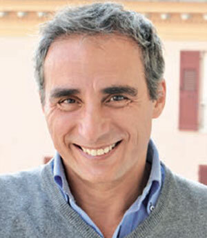
Marco Ronda, MD, DDS, annually attends the meetings organized by the American Academy of Implant Dentistry and several specialized courses on the techniques of horizontal and vertical bone regeneration. He also participates in annual continuing dental education at the New York University College of Dentistry. Dr Ronda maintains a private practice in Genoa, Italy, where he periodically provides practical training courses in implantology and bone regeneration techniques.
Events
The 14th International Symposium on Periodontics and Restorative Dentistry (ISPRD)
9. Jun 2022 — 12. Jun 2022Boston Marriott Copley Place, Boston, MA, United States of America
Speakers: Tara Aghaloo, Edward P. Allen, Evanthia Anadioti, Wael Att, Vinay Bhide, Markus B. Blatz, Scotty Bolding, Lorenzo Breschi, Jeff Brucia, Daniel Buser, Luigi Canullo, Daniele Cardaropoli, Stephen J. Chu, Donald Clem, Christian Coachman, Lyndon F. Cooper, Daniel Cullum, Lee Culp, José Carlos Martins da Rosa, Sergio De Paoli, Marco Degidi, Nicholas Dello Russo, Serge Dibart, Joseph P. Fiorellini, Mauro Fradeani, Stuart J. Froum, David Garber, Maria L. Geisinger, William Giannobile, Luca Gobbato, Ueli Grunder, Galip Gürel, Chad Gwaltney, Christoph Hämmerle, Robert A. Horowitz, Marc Hürzeler, David Kim, Gregg Kinzer, Christopher Köttgen, Ina Köttgen, Purnima S. Kumar, Burton Langer, Lydia Legg, Pascal Magne, Kenneth A. Malament, Jay Malmquist, George Mandelaris, Pamela K. McClain, Michael K. McGuire, Mauro Merli, Konrad H. Meyenberg, Craig M. Misch, Julie A. Mitchell, Marc L. Nevins, Myron Nevins, Michael G. Newman, Miguel A. Ortiz, Jacinthe M. Paquette, Stefano Parma-Benfenati, Michael A. Pikos, Giulio Rasperini, Pamela S. Ray, Christopher R. Richardson, Isabella Rocchietta, Marisa Roncati, Marco Ronda, Paul S. Rosen, Maria Emanuel Ryan, Irena Sailer, Maurice Salama, David M. Sarver, Takeshi Sasaki, Todd Scheyer, Massimo Simion, Michael Sonick, Sergio Spinato, Dennis P. Tarnow, Lorenzo Tavelli, Douglas A. Terry, Tiziano Testori, Carlo Tinti, Istvan Urban, Hom-Lay Wang, Robert Winter, Giovanni Zucchelli
Quintessence Publishing Co., Inc. USA
This author's journal articles
International Journal of Periodontics & Restorative Dentistry, Pre-Print
DOI: 10.11607/prd.7246, PubMed ID (PMID): 392703126. Sep 2024,Pages 1-25, Language: EnglishRonda, Marco / Desantis, Viviana / Bruno, Diego / Veneriano, Luca / Elli, Camilla / Pispero, Alberto
If a prosthetically guided implant rehabilitation is to be carried out, the patient must have adequate bone volumes; as a result of bone resorption in the alveolar process, the latter are not always present. An effective technique in regenerative surgery involves the use of titanium mesh to recreate the missing bone volume. New generation meshes have been used in this study: they present a new texture with micro holes and they can have a space-creating effect and a barrier effect. The primary objective of the study is to measure the height and width of the regenerated bone following the use of a new generation customized titanium mesh. This retrospective case series involved 18 patients, with a total of 23 bone atrophies who have undergone bone regeneration surgery with titanium mesh and subsequently implants have been placed. We achieved a vertical gain of 5.1 ± 2.9 mm and a medium horizontal gain is 12.3 ± 2.6 mm, with an average volume obtained during regeneration is 3.2 ± 1.3 cm3; we report one failure (4.3%) and in 3 cases (13%) it was necessary to perform a second minor surgery. The mean follow-up of implants after loading was 22.3 ± 15.6 months. This study showed the innovation of new generation titanium mesh, with a micro-perforated texture that offers both a spacemaking effect and a partial barrier effect. This has led to excellent results in terms of regeneration, with a significant improvement in vertical and horizontal defects treated and reduction in operating time. Future studies with longer follow-up will be necessary.
Purpose: To establish consensus-driven guidelines that could support the clinical decision-making process for implant-supported rehabilitation of the posterior atrophic maxilla and ultimately improve long-term treatment outcomes and patient satisfaction.
Materials and methods: A total of 33 participants were enrolled (18 active members of the Italian Academy of Osseointegration and 15 international experts). Based on the available evidence, the development group discussed and proposed an initial list of 20 statements, which were later evalu-ated by all participants. After the forms were completed, the responses were sent for blinded ana-lysis. In most cases, when a consensus was not reached, the statements were rephrased and sent to the participants for another round of evaluation. Three rounds were planned.
Results: After the first round of voting, participants came close to reaching a consensus on six statements, but no consensus was achieved for the other fourteen. Following this, nineteen statements were rephrased and sent to participants again for the second round of voting, after which a consensus was reached for six statements and almost reached for three statements, but no consensus was achieved for the other ten. All 13 statements upon which no consensus was reached were rephrased and included in the third round. After this round, a consensus was achieved for an additional nine statements and almost achieved for three statements, but no consensus was reached for the remaining statement.
Conclusion: This Delphi consensus highlights the importance of accurate preoperative planning, taking into consideration the maxillomandibular relationship to meet the functional and aesthetic requirements of the final restoration. Emphasis is placed on the role played by the sinus bony walls and floor in providing essential elements for bone formation, and on evaluation of bucco-palatal sinus width for choosing between lateral and transcrestal sinus floor elevation. Tilted and trans-sinus implants are considered viable options, whereas caution is advised when placing pterygoid implants. Zygomatic implants are seen as a potential option in specific cases, such as for completely edentulous elderly or oncological patients, for whom conventional alternatives are unsuitable.
Keywords: diagnostic procedure, implant dentistry, lateral window technique, pterygoid implants, sinus floor elevation, transcrestal sinus floor elevation, zygomatic implants
The authors report no conflicts of interest relating to this study.
An adequate flap release is necessary to perform a tension-free suture over an augmented area. This is a fundamental requisite to attain and maintain a reliable biological seal, protecting the graft from bacterial contamination during the healing period. In the posterior mandible, in particular, the use of conventional periosteal incisions is not always sufficient for a proper buccal flap passivation, as they are often limited by anatomical factors. This article reports a series of 76 consecutive cases of vertical guided bone regeneration in the posterior mandible introducing a novel surgical technique to enhance the coronal advancement of the buccal flap in a safe and predictable way.
One of the crucial factors in the success of guided bone regeneration procedures is the correct management of the soft tissues. This allows for stable primary wound closure without tension, which can result in premature exposure of the augmentation area, jeopardizing the final outcome. The use of vertical and periosteal incisions to passivate buccal and lingual flaps in the posterior mandible is often limited by anatomical factors. This paper reports on a series of 69 consecutive cases introducing a novel surgical technique to release and advance the lingual flap coronally in a safe and predictable manner.