Various cookies are used on our website: We use technically necessary cookies for the purpose of enabling functions such as login or a shopping cart. We use optional cookies for marketing and optimization purposes, in particular to place relevant and interesting ads for you on Meta's platforms (Facebook, Instagram). You can refuse optional cookies. More information on data collection and processing can be found in our privacy policy.
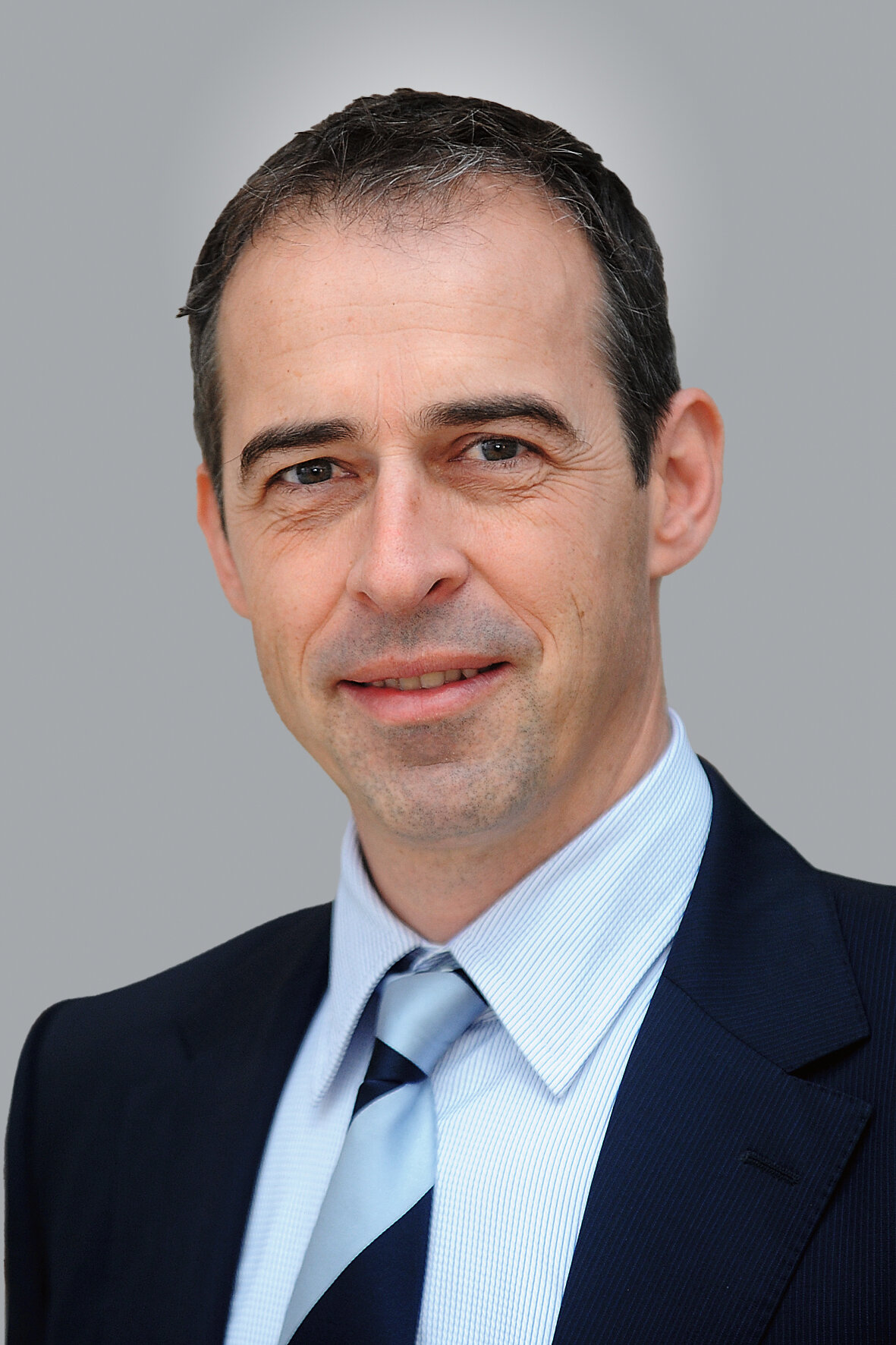
1987-1992 Studium der Zahnheilkunde an der Johann Wolfgang Goethe-Universität in Frankfurt am Main Seit 1993 Tätigkeit als Wissenschaftlicher Mitarbeiter an der Poliklinik für Zahnärztliche Prothetik am Zentrum der Zahn-, Mund- und Kieferheilkunde (Carolinum) der Johann Wolfgang Goethe-Universität, Frankfurt am Main 1994 Promotion zum Dr. med. dent., Frankfurt am Main 2001-2002 Forschungsaufenthalt an der Oregon Health & Science University in Portland, Oregon, USA Seit 2002 Ernennung zum Oberarzt an der Poliklinik für Zahnärztliche Prothetik am Zentrum der Zahn-, Mund- und Kieferheilkunde (Carolinum) der Johann Wolfgang Goethe-Universität, Frankfurt am Main Ernennung zum qualifiziert fortgebildeten Spezialisten der Deutschen Gesellschaft für Zahnärztliche Prothetik und Werkstoffkunde (DGPro) Zertifikat im Bereich der Implantologie der Deutschen Gesellschaft für Implantologie im Zahn-, Mund- und Kieferbereich (DGI) 2006 Habilitation im Fachbereich der Medizin der Johann Wolfgang Goethe-Universität Frankfurt am Main Seit 01.03.2012 Leiter des Lehrstuhls für Zahnärztliche Prothetik und Dentale Technologie, Universität Witten/Herdecke
Events
26. Prothetik Symposium
Touch the past to create the future2. Dec 2023Marriott Hotel Berlin, Berlin, Germany
Speakers: Shahab Esfarjani, Karl-Uwe Jülich, Erwin Klampfer, Friedhelm Klingenburg, Karl-Heinz Körholz, Andree Piwowarczyk, Daniel Reinke, Ralph Riquier, Stefan Sander, Eric Standop, Pawlos Stilos, Tonguç Sülün, Gerhard Zips, Richard Zips
Merz Dental GmbH
Deutscher Zahnärztetag 2018
MISSERFOLGE - erkennen, beherrschen, vermeiden9. Nov 2018 — 10. Nov 2018Congress Center Messe Frankfurt
Speakers: Karl-Ludwig Ackermann, Sarah Al-Maawi, Bilal Al-Nawas, Kurt Werner Alt, Anna Greta Barbe, Tobias Bauer, Daniel Bäumer, Marco Baz Bartels, Grietje Beck, Katrin Bekes, Christoph Benz, Dirk Bleiel, Johannes Boesch, Martin Boost, Wolfgang Buchalla, Oskar Bunz, Fabian Cieplik, Monika Daubländer, Sybille David-Hebgen, Andreas Dehler, Renate Deinzer, Sonja H. M. Derman, Konstanze Diekmeyer, Ingmar Dobberstein, Heike Dyrna, Thomas Eger, Guido Elsäßer, Anne Sophie Engel, Peter Engel, Norbert Enkling, Susanne Fath, Stefan Fickl, Michael Frank, Roland Frankenberger, Rene Franzen, Kerstin Galler, Carolina Ganß, Roland Garve, Christian Ralf Gernhardt, Werner Geurtsen, Shahram Ghanaati, Petra Gierthmühlen, Christiane Gleissner, Steffani Görl, Werner Götz, Susanne Grässel, Dominik Groß, Stefan Grümer, Claus Grundmann, Martin Guffart, Heinz-Michael Günther, Norbert Gutknecht, Peter Hahner, Elmar Hellwig, Christian Henrici, Katrin Hertrampf, Fabian Huettig, Michael Hülsmann, Bruno Imhoff, Holger Jentsch, A. Rainer Jordan, Ana Elisa Kauling, Moritz Kebschull, Christian Kirschneck, Joachim Klimek, Andrea Klink, Thomas Klinke, Birte Koch, Thomas Kocher, Eva Köllensperger, Heike Maria Korbmacher-Steiner, Bernd Kordaß, Hannah Kottmann, Pablo Krämer-Fernandez, Gabriel Krastl, Birgit Krause, Till Kreutzer, Conrad Kühnöl, Stefanie Kurzschenkel, Thorsten Kuypers, Günter Lauer, Hans-Christoph Lauer, Elfi Laurisch, Tina Lawall, Karl Martin Lehmann, Silke Lehmann-Binder M.Sc., Dirk Leisenberg, Ulrike Lübbert, Michael Lüpke, Thomas Malik, Jutta Margraf-Stiksrud, Lorenz Meinel, Gudrun Mentel, Wibke Merten, Louisa Mewes, Johanna Isabel Moosmüller, Martin U. Müller, Wolfgang Müller, Nicole Nicklisch, Ina Nitschke, Michael J. Noack, Marina Nörr-Müller, Karina Obreja, Dietmar Oesterreich, Puria Parvini, Ingrid Peroz, Waldemar Petker, Oksana Petruchin, Andree Piwowarczyk, Peter Pospiech, Peter Proff, Sven Reich, Katharina Reichenmiller, Katharina Reinecke, Bernd Reiss, Svenja Rink, Christiane Rinnen, Jerome Rotgans, Didem Sahin, Sonja Sälzer, Petra Santander, Heidrun Schaaf, Jürgen Schäffer, Elisabeth Schiffner, Ulrich Schiffner, Markus Schlee, Maximiliane Amelie Schlenz, Peter Schmidt, Andrea-Maria Schmidt-Westhausen, Claas Ole Schmitt, Sigmar Schnutenhaus, Jörg Schröder, Gerd Schröter, Andreas Schulte, Philipp Schwaab, Frank Schwarz, Falk Schwendicke, Clemens Schwerin, Sinan Sen, Önder Solakoglu, Hansmartin Spatzier, Christian H. Splieth, Norbert Staab, Bernd Stadlinger, Sabine Steding, Marcus Stoetzer, Giorgio Tabanella, Gisela Tascher, Hendrik Terheyden, Valentina A. Tesky, Jan Tetsch, Juliane von Hoyningen-Huene, Maximilian Voß, Michael Walter, Alexander Welk, Dietmar Weng, Hans-Jürgen Wenz, Jens Westemeier, Lotta Westphal, Annette Wiegand, Karl Frederick Wilms, Michael M. Wolf, Diana Wolff, Anne Wolowski, Johann-Dietrich Wörner, Sylvia Wuttig, Mohamed Younis, Stefan Zimmer, Lisa Zumpe
Methode: Die digitale Bilddaten von 343 Patienten, bei denen sowohl eine DVT- als auch eine PSA-Aufnahme vorlag, wurden auf apikale Läsionen um Unterkiefer untersucht. Für entdeckte Läsionen wurden die Abmessungen der Läsion und die Dicke des kortikalen Knochens in der betroffenen Region gemessen. Die Ergebnisse wurden einer statistisches Analyse unter Verwendung einer Statistiksoftware (IBM SPSS 25, Fa. IBM, Armonk, USA) unterzogen.
Ergebnisse: Mittels DVT wurden an 115 Zähnen apikale Läsionen entdeckt. Von diesen Läsionen waren 77 auch in der PSA sichtbar. Die Unterschiede zwischen den beiden bildgebenden Verfahren waren in der Prämolaren- und Molarenregion signifikant (McNemar-Test, p < 0,001), nicht jedoch im Frontzahnbereich (p = 0,063). Bezüglich der Läsionsgröße wurden keine signifikanten Unterschiede gefunden (Wilcoxon-Test, Frontzähne: p = 0,60, Prämolaren: p = 0,90, Molaren: p = 0,61). Der Mann-Whitney-U-Test ergab weder für die vestibuläre noch für die linguale Kortikalisdicke einen signifikanten Einfluss auf die Sichtbarkeit der Läsionen in DVT und PSA (vestibulär: p = 0,93, palatinal: p = 0,15).
Schlussfolgerung: Apikale Läsionen scheinen in digitalen Volumentomografien wesentlich besser sichtbar zu sein. Die Dicke des kortikalen Knochens und die Größe der Läsionen haben keinen signifikanten Einfluss auf die Darstellung apikaler Läsionen.
Keywords: apikale Läsion, DVT, digitale Volumentomografie, kortikale Knochendicke, Panoramaschichtaufnahme, PSA
Introduction: This study compares the accuracy of panoramic radiography and CBCT in detecting and diagnosing mandibular apical lesions, analysing the effect of the thickness of the cortical bone on the radiological visibility of apical lesions.
Methods: Digital images of 343 patients who had CBCT scans and panoramic radiographs were inspected for mandibular apical lesions. The dimensions of the lesion and the thickness of the cortical bone in the affected region were measured. Statistical analyses were made using statistical software (IBM SPSS 25, IBM, Armonk, NY, USA).
Results: CBCT detected apical lesions around 115 teeth; 77 of these were also visible on a panoramic radiography. The differences between the two methods were statistically significant in the premolar and molar regions (McNemar test; p 0.001) but not in the anterior region (p = 0.063). For the size of the lesions no significant differences were found (Wilcoxon test; anterior, p = 0.60; premolar, p = 0.90; molar, p = 0.61). In the Mann-Whitney U test, buccal and palatal cortical thickness did not significantly influence the visibility of lesions in CBCT and panoramic measurements (buccal, p = 0.93; palatal, p = 0.15).
Conclusion: Apical lesions seem to be much more readily visible on CBCT scans. The thickness of the cortical bone and the size of the lesions do not significantly influence the representation of apical lesions.
Keywords: apical lesions, CBCT, cone-beam computed tomography, cortical bone thickness, panoramic radiography
Introduction: Since the loss of one's own teeth decreases with age, the various effects of various wear and tear on tooth structure and their therapy have become increasingly important. Erosive and abrasive/attritive processes may overlap. Attritive processes can also progress more rapidly in erosively damaged teeth. If the erosive loss of hard tooth substance particularly affects the palatal and occlusal surfaces of the maxillary teeth, an endogenous aetiology in the sense of chronic gastrointestinal disturbances or regular vomiting with bulimia nervosa can be assumed. However, buccal defects in the lower jaw due to the nocturnal lying position allow reflux diseases to be distinguished from chronic vomiting. Bulimia nervosa has a gender ratio of 1:20 (m:f) and a prevalence of 0.5-5% in western industrial countries. Frequently, affected patients practice intensified mechanical oral hygiene, which leads to increased loss of tooth substance. If erosive and mechanical wear occur simultaneously, a temporally linear correlation can no longer be assumed.
Case report: In the case presented, the patient describes a pronounced nocturnal bruxism (abrasion/attrition) and long-term bulimia nervosa. The functional symptoms of the patient (severe head- and earaches) support the dental findings regarding bruxism. Bulimia patients usually show erosions in the area of the oral and occlusal tooth surfaces, especially the incisors. At the time of initial presentation, the patient had already suffered for years from aesthetic, functional, and phonetic limitations. Due to restorations with crowns and bridges in the posterior region, the vertical dimension was largely retained. Teeth 12-22 showed a clinical coronal residual height of approx. 3 mm. Since all 4 anterior teeth still showed vitality, it had to be considered whether they should be devitalised for the forthcoming prosthetic restoration. Since root-canal-treated teeth are more susceptible to fractures than vital teeth, this should be avoided wherever possible. The front of the upper jaw was clearly visible, especially when laughing (gummy smile). Although there was no increased loss of vertical dimension, there was not enough intermaxillary space in the anterior region to reconstruct the anterior teeth. To avoid devitalisation and to harmonise the gingival margin, a crown extension was performed. The required slight bite elevation was achieved by crowning all posterior maxillary teeth that had previously been treated with insufficient restorations. A functional wax-up was created. A Michigan splint with a modelled maxillary front served as a therapeutic denture for 6 months. Then a surgical crown extension was performed with special consideration for the harmonisation of the gingival margin. A mock-up was used as a template and later as a temporary. The patient showed no symptoms after 6 months and was fitted with full zircon crowns in the maxilla.
Discussion: Since the loss of hard tooth substance was already at an advanced stage, extensive restoration work had to be carried out. The patient was fitted with a Michigan splint to protect the restorations from attritive secondary damage. In the therapy or prevention of non-carious tooth substance defects, the patient's personal responsibility for success must be made clear.
Keywords: erosion, bulimia nervosa, prosthetic rehabilitation
Dental restorations without tooth preparation are among today's more advanced treatment options. This article presents the case of a young man who was treated with a non-prep laminate veneer that provided 3.5 mm of incisal elongation. A Photoshop assessment and functional evaluation, as well as a wax-up and mock-up, were used during pre-prosthetic planning. A combination of pressable and feldspathic ceramics was used as a restorative material, which was applied to the tooth surface without any preparation of dental hard tissue. An ideal functional and esthetic restoration was achieved thanks to close collaboration between the patient, the dentist, and the technician.
Keywords: Creation CP, esthetics, mock-up, non-preparation laminate veneer, non-prep veneer, pressable ceramics, "tenuia" veneer
Introduction: The purpose of this study was to examine whether a unilateral occlusion block, using a 1 or 2 mm thick silicone panel, can significantly change the upper part of the body posture.
Materials and methods: This study was carried out with 38 adults without remarkable discomfort in the temporomandibular system or body movement apparatus. The measurement of the upper part of the body posture was performed with a three-dimensional back scanner. Every test person placed the 1 or 2 mm thick silicone panel systematically between the left/right premolars. The statistical comparison of the measuring data was carried out between the reference measurement and the provoked lower jaw position.
Results: If the occlusion was changed dextrally (to the right) or sinistrally (to the left) by the silicone panel, the rotation in the shoulder area improved (right 1 or 2 mm: P = 0.00; left 1 mm: P = 0.00) in comparison to the habitual starting position. With a dextral occlusion disruption by 1 mm of silicone, the pelvic rotation also changed (P = 0.05).
Conclusion: A unilateral occlusion block in the premolar region has an influence on the upper body rotation in the standing position, particularly in the shoulder area. Thus the results demonstrate functional relationships between the chewing and the movement system, which may explain complaints in one of the subsystems influenced by therapeutic measures in an otherwise unaffected system.
Keywords: asymmetrical occlusion blocking, three-dimensional back scan, upper body posture
The International Journal of Prosthodontics, 1/2009
PubMed ID (PMID): 19260423Pages 20-32, Language: EnglishLandes, Constantin Alexander / Paffrath, Christian / Koehler, Christian / Thai, Van Dung / Stübinger, Stefan / Sader, Robert / Lauer, Hans-Christoph / Piwowarczyk, Andree
Purpose: This study presents successful maxillofacial prosthetic rehabilitation using telescopic and crowns on zygoma implants as abutments.
Materials and Methods: Fifteen patients received 36 zygomatic and 24 dental implants and were followed-up for an average of 65 months (range: 13 to 102 months). Machined zygoma implants were positioned classically in the maxillary molar region. In larger defects, premolar and canine implants were also used. Follow-up included implant and prosthetic success parameters as well as the completion of the Oral Health Impact Profile (OHIP14G).
Results: Seventy-three percent of patients during the study period did not encounter notable complications after prosthetic rehabilitation. There was an 89% cumulative 8-year zygoma implant survival rate and a 100% survival rate for the dental implants. Three losses occurred due to overloading and persistent infection; each was immediately replaced. Five successfully osseointegrated implants had to be removed in two patients due to recurrences of disease; one patient died. Peri-implant bleeding and plaque index scores decreased. After prosthetic treatment with electroplated gold or galvanotelescopes, all patients who had participated in the follow-up declared function (ie, retention, speech, and mastication) and esthetics as having improved. Other positive aspects mentioned were good hygiene, comfortable usage, and a decrease in sore spots. OHIP scores were 25 ± 12 on a scale of 0 (no impairment) to 56 (maximum impairment).
Conclusion: Within the limitations of this study (a variable evaluation period), it was observed that zygomatic implants are reliable retention for maxillofacial prostheses. Losses were diagnosed as occuring primarily from chronic infection and overloading. A trapezoid prosthesis design support is recommended with a sufficient number of implants.
Objective: This study determined the long-term bond strength of various luting cements to high-gold-content alloy.Materials and Methods: High-gold-content alloy (Portadur P4; Wieland, Germany) specimens (Ø = 10 mm, h = 1 mm) were fabricated. Bonding substrates were placed into plastic rings and embedded with slow-polymerizing epoxy resin. Material surfaces were ground sequentially to 600-grit using SiC sandpaper. The surface of all specimens were 100 microns Al2O3 abraded at 2.8 bar, 10 s at 10 mm distance. Following luting cements were tested: PermaCem (DMG), RelyX ARC (3M ESPE), Panavia F (Kuraray), Nexus 2 (Kerr), Calibra (Dentsply DeTrey) and RelyX Unicem (3M ESPE). Eight specimens per group were fabricated: Group (A) was aged and tested after 150 day 37°C water storage; Group (B) 150 day at 37°C water storage, then thermocycled (37.500X, 5-55°C). Shear loading tests were performed using a universal testing machine at a constant crosshead speed of 0.5 mm/min. Two-way ANOVA of variance was used to analyze the data (α=0.05).
Results: Significant differences were found between all main effects (all p
Keywords: luting cement, bond durability, artificial aging
Der Anwendungsbereich des LAVA-Systems ermöglicht bisher die Herstellung von vollkeramischen Kronen und dreigliedrigen Brücken im Front- und Seitenzahnbereich. Bei der Gerüstkeramik handelt es sich um mit ca. 3 mol-% Yttriumoxid teilstabilisiertes tetragonales polykristallines Zirkonoxid. Mittels CAD/CAM-Technologie werden Zirkonoxid-Rohlinge im ungesinterten Zustand gefräst und ggf. vor dem Sintern manuell zur Formkorrektur und Oberflächenglättung nachbearbeitet. Die nach dem Sinterungsprozess bei einer Temperatur von etwa 1.500 °C hergestellten LAVA-Gerüste zeichnen sich durch sehr gute mechanische Eigenschaften aus. Eine speziell auf den Wärmeausdehnungskoeffizient der Gerüste abgestimmte Verblendkeramik bietet exzellente ästhetische Möglichkeiten, ein günstiges Abrasionsverhalten und eine für die entzündungsfreie Anlagerung des Parodontiums ideale Oberflächenstruktur. Die vorliegende Arbeit gibt einen Überblick über bisher erfolgte wissenschaftliche Untersuchungen und beschreibt anhand von zwei Falldokumentationen das klinische Vorgehen.
Keywords: CAD/CAM-Technologie, Zirkonoxid, Y-TZP, vollkeramische Kronen, vollkeramische Front- und Seitenzahnbrücken
Objective: The objective of this study was to determine quantitatively the wear resistance of a newly developed light-curing resin for two different types of occlusal splints over a period in situ of six months.
Materials and Methods: The study included two samples of ten patients each that had been provided with either stabilization or distraction splints. These occlusal splints were repositioned on remount casts and the occlusal aspect was surveyed prior to insertion and at the four-week, three-month, and six-month recalls. The test setup consisted of an anti-vibration table framework, three translation stages, an inductive displacement transducer, a stereomicroscope, and a digital measurement unit for data acquisition and analysis. The coordinates for the occlusal contacts were stored at the baseline examinations and reproducibly sought out during the recall examinations. Each examination included ten repeat measurements each in the regions of teeth 13, 23, 16, and 26.
Results: After six months, the stabilization splints showed a median vertical gain of 1 µm in the region of tooth 13 and a median vertical loss of 24 µm in the region of tooth 23. Regions 16 and 26 exhibited median vertical losses of 37 µm and 33 µm, respectively. In the second sample (distraction splint), the corresponding vertical loss values were 1 µm (tooth 13), 23 µm (tooth 23), 33 µm (tooth 16), and 64 µm (tooth 26). Statistical analysis (Mann-Whitney U-test, p Conclusions: The present study clinically confirms the good wear resistance results of the new resin splint material obtained in a previous in-vitro study [Ottl et al., Dtsch Zahnärztl Z 52, 342 (1997)]. This parameter is of great importance for maintaining the therapeutic mandibular position during the treatment period.
Keywords: craniomandibular disorders, temporomandibular joint, clinical follow-up study, occlusal splint therapy, resin, wear resistance
Purpose: This study evaluated the dimensional accuracy of various impression materials for monophase elastic impression making. To isolate this parameter, a direct measurement of the impressions was made without taking the model material into consideration.
Materials and Methods: A total of eight materials were tested; six impression materials were addition-curing silicones, and two were polyether impression materials. All materials were processed according to the manufacturers' instructions. A specially developed precision mold made of stainless steel served as a basis for measuring the elastomeric impression materials. Using a stereomicroscope at a temperature of 23.0 ± 1.0°C and with a precision linear adjustment and lathe, sights were set on the marking points of customized posts. The measurement was performed after the earliest time possible for fabricating the model according to the manufacturer (time 1) and after 90 minutes (time 2). In a one-way analysis of variance, multiple average comparisons of dimensional accuracy were made (P = .05) between the impression materials under investigation.
Results: Under the conditions of this study, the impression materials tested demonstrated a very high dimensional accuracy. The arithmetic means of the dimensional changes ranged from -11 to 19 µm for both measuring times.
Conclusion: Since as a rule, no significant dimensional changes occurred for the different impression materials between time 1 and time 2, this time interval for fabricating a model can be recommended.