Various cookies are used on our website: We use technically necessary cookies for the purpose of enabling functions such as login or a shopping cart. We use optional cookies for marketing and optimization purposes, in particular to place relevant and interesting ads for you on Meta's platforms (Facebook, Instagram). You can refuse optional cookies. More information on data collection and processing can be found in our privacy policy.
Born on 10th of August 1974 in Alessandria, Italy. Graduated in Dental School at Genoa University as DDS in 1999. ADHESTHETICS founder. EAED (European Academy of Esthetic Dentistry) Active Member since 2006. AIC (Italian Academy of Conservative) Active Member since 2007. Vice-president from 2016 to 2019. President elect for the biennium 2022-2023. SCAD (Society for Color and Appearance in Dentistry) Regional Councilor for Europe of for the biennium 2013/14 and 2015/16. Secretary for biennium 2017-2018. President elect for the biennium 2021-2022. Member of the Editorial Board of IJED (International Journal of Esthetic Dentistry) since 2010. AARD (American Academy of Restorative Dentistry) Member since 2014. DSD (Digital Smile Design) Master since 2013. International speaker at dental congresses and courses in more than thirty Countries. Other informations: National President of AISO (Italian Association of Dental Students) in 1997-99. Trainer at the Dr. Massironi Prosthodontics Annual Master Course from 2004 to 2018. Co-author of several Italian and International scientific publications including the book on Prosthodontics “Precision in Dental Esthetics” by D. Massironi, R. Pascetta, and G. Romeo published in 2004. Active and Founder Member of MSC (Massironi Study Club) from 2007 to 2018. Active and Founder Member of GICC (Interdisciplinar Gymnasium CAD CAM) from 2007 to 2016. Visiting Professor in Esthetic and Restorative Dentistry in different Universities: Genoa University (IT), University of Pisa (IT), Insubria University of Varese (IT), Alma Mater University of Bologna (IT), Dental School of Torino (IT), CEU University San Pablo of Madrid (ES), University of Almeria (ES), Universidad Europea de Madrid (ES), New York University (USA), University of Washington in Seattle (USA). Adjunct Assistant Professor in the Department of Preventive and Restorative Sciences in the Associated Faculty of the School of Dental Medicine of the Upenn University in Philadelphia since January 2020 until June 2022. IAED (Italian Academy of Esthetic Dentistry) Active Member since 2011. IAAD (International Academy for Adhesive Dentistry) Member since 2013. Dental practice in Alessandria, focusing in Operative Dentistry and Prosthodontics.
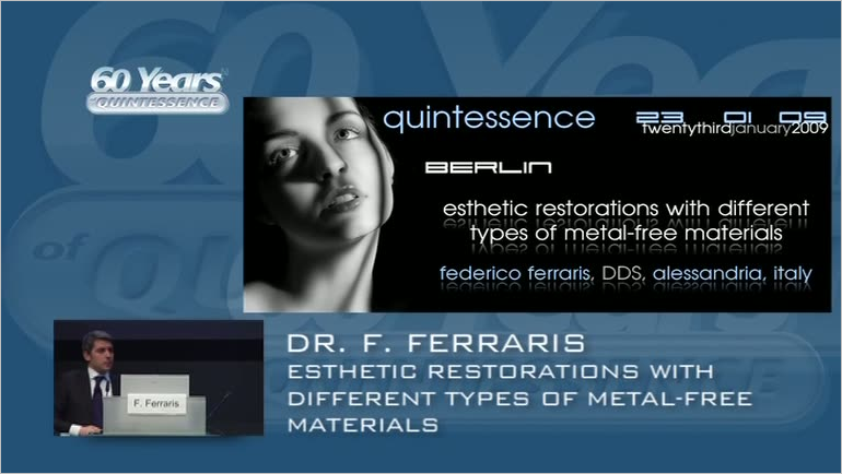
Length: 23 minutes Production year: 2009 Language: English Source: 60 Jahre Quintessenz Categories: Dental Technology, Esthetic Dentistry available since: 8. Feb 2010
Events
25th Annual Congress Italian Academy of Conservative Dentistry (AIC)
The Conservative Approach and Digital Workflows: The State of the Art according to AIC9. May 2024 — 11. May 2024Palazzo dei Congressi Bologna, Bologna, Italy
Speakers: Riccardo Ammannato, Lorenzo Breschi, Victor Clavijo, Walter Devoto, Mirela Feraru, Federico Ferraris, Iñaki Gamborena, Francesco Mintrone, Vincenzo Musella, Gaetano Paolone, Nikolaos Perakis, Bruno Pereira, Andrea Ricci, Daniele Rondoni, Giovanni Sammarco, Nicola Scotti
Accademia Italiana di Odontoiatra Conservativa e Restaurativa (AIC)
7 Decades of Quintessence Publishing
10. Jan 2019 — 12. Jan 2019Estrel Convention Center
Speakers: Jiro Abe, Michèle Aerden, Wael Att, Stavros Avgerinos, Avijit Banerjee, Vesna Barac Furtinger, Klaus-Dieter Bastendorf, Lars Bergmans, Ashwini Bhalerao, Jaroslav Bláha, Sebastian Bürklein, Daniel Buser, Josette Camilleri, Sevim Canlar, Sandra Chmieleck, Bun San Chong, Victor Clavijo, Carsten Czerny, Bettina Dannewitz, Alessandro Devigus, Didier Dietschi, Irina Dragan, Daniel H.-J. Edelhoff, Peter Eickholz, Karim Elhennawy, Peter Engel, Wolfgang Eßer, Marco Esposito, Susanne Fath, Vincent Fehmer, Federico Ferraris, Stefan Fickl, Mauro Fradeani, Roland Frankenberger, Eiji Funakoshi, Petra Gierthmühlen, Christiane Gleissner, Florian Göttfert, Dennis Grosse, Galip Gürel, Christian Haase, Horst-Wolfgang Haase, Manuela Hackenberg, Jörg Haist, Anke Handrock, Arndt Happe, Karsten Heegewaldt, Rüdiger Henrici, Michael Hülsmann, Hajime Igarashi, Tomohiro Ishikawa, Hideaki Katsuyama, Kathryn Kell, Matthias Kern, Fouad Khoury, Marko Knauf, Ralf J. Kohal, Stefen Koubi, Fabian Langenbach, Henriette Terezia Lerner, Thomas Malik, Siegfried Marquardt, Henrike März, Kathleen Menzel, Helen Möhrke, Kotaro Nakata, Marc L. Nevins, Masayuki Okawa, Rebecca Otto, Mark Stephen Pace, Shanon Patel, Karin Probst, Domenico Ricucci, Katrin Rinke, Irena Sailer, Edgar Schäfer, Ralf Schäfermeier, Jan Schellenberger, Tom Schloss, Gottfried Schmalz, Devorah Schwartz-Arad, Frank Schwarz, Thomas A. Schwenk, Anton Sculean, Bernd Stadlinger, Athanasios Stamos, Ana Stevanovic, Masana Suzuki, Senichi Suzuki, Hiroyuki Takino, Sameh Talaat, Mitsuhiro Tsukiboshi, Hideaki Ueda, Istvan Urban, Luc W. M. van der Sluis, Eric Van Dooren, Bart Van Meerbeek, Paula Vassallo, Juliane von Hoyningen-Huene, Michael Walter, Siegbert Witkowski, Stefan Wolfart, Sylvia Wuttig, Masao Yamazaki, Maciej Zarow, Matthias Zehnder, Raquel Zita Gomes, Giovanni Zucchelli, Otto Zuhr, Bettina Zydatiß
Quintessenz Verlags-GmbH
This author's journal articles
International Journal of Esthetic Dentistry (DE), 2/2022
Kariesläsionen mit tiefen Rändern sind in der zahnärztlichen Praxis erhebliche Herausforderungen. Folgende Schlüsselaspekte dieses Problems werden im vorliegenden Beitrag diskutiert: Wie ist unter restaurativem Gesichtspunkt mit einem tiefen Kavitätenrand umzugehen? Wann und wie sollte chirurgisch interveniert werden? Welcher ist der beste Zeitpunkt für die definitive restaurative Versorgung nach einer chirurgischen Intervention? Die mittlerweile verfügbaren Restaurationsmaterialien und Adhäsivtechniken ermöglichen minimalinvasive Behandlungsansätze und damit einen umfangreichen Substanzerhalt und helfen dabei, unerwünschte Reaktionen der parodontalen Gewebe zu vermeiden. Je nach klinischer Ausgangslage kommen bei subgingivalen Kavitätenrändern drei Behandlungsoptionen in Betracht. Liegt der Rand maximal 1,5 mm unter dem Gingivarand, ist eine Anhebung des Kavitätenrands unter Kofferdam möglich, was eine optimale Restauration und Weichgewebeintegration erlaubt. Liegt der Kavitätenrand mehr als 1,5 mm unter dem Gingivarand, ist vor der Restauration eine chirurgische Intervention erforderlich. Liegt der Rand noch mindestens 2 mm über dem Knochen, wird ein ästhetisches suprakrestales Gewebemanagement (Supracrestal Tissue Esthetic Management, STEM) durchgeführt. Das heißt, es genügt eine Osteoplastik, um das suprakrestale Attachment neu zu etablieren und die restaurative Behandlung durchführen zu können. Eine Ostektomie ist nicht nötig. Liegt der Kavitätenrand weniger als 2 mm koronal zum Knochenkamm, ist eine Kronenverlängerung mit einer minimalen Ostektomie und anschließenden Osteoplastik indiziert.
Carious lesions with deep margins represent a challenge in daily clinical practice. The following key points are discussed in this article: how to manage a deep margin from a restorative point of view; when and how to perform a surgical procedure; and when it is favorable to carry out definitive restoration work after surgery. The restorative materials and adhesive procedures available today allow minimally invasive techniques to be used on dental tissue with a high preservation of tooth structure. These materials and techniques help to avoid adverse periodontal tissue reactions. Depending on the clinical situation, three treatment options are available when dealing with a subgingival margin. If the depth of the cavity margin is at a maximum distance of 1.5 mm below the gingival margin, isolation with rubber dam allows the performance of interproximal margin relocation, thereby facilitating optimal restoration and periodontal tissue integration. If the margin is located deeper than 1.5 mm below the gingival margin, surgery is necessary before any restorative work can take place. When the margin is within 2 mm above the bone crest, a supracrestal tissue esthetic management (STEM) procedure is undertaken, which means that no ostectomy is required and only osteoplasty is necessary to reshape the preexisting supracrestal attachment, thereby allowing the restorative work to proceed. When the margin is less than 2 mm above the bone crest, crown lengthening with minimal ostectomy and subsequent osteoplasty becomes necessary.
Aim: To investigate whether different restoration designs, overlay types, and full crowns in posterior teeth have similarly acceptable marginal sealing and quality.
Materials and methods: For Part 1 of the present study (investigation of fracture resistance), 70 extracted molars were divided into five groups (N = 14), prepared with four different posterior indirect adhesive restoration (PIAR) overlay design types, according to the adhesthetics classification. The groups were: 1. Butt Joint; 2. Full Bevel; 3. Shoulder; 4. Full Crown; 5. Sound Tooth. For Part 2 of the study (present article; marginal quality), there was no group 5, and only 56 of the 70 extracted molars were used. Seven expert dentists performed all the preparation and cementation phases with codified protocols. A CAD/CAM workflow was used to realize the 56 monolithic lithium disilicate restorations. The samples were tested with thermomechanical loading (TML) and the marginal quality evaluated. The data relating to fracture resistance are presented in Part 1 of this study (Int J Esthet Dent 2021;16:2–17).
Results and conclusions: In terms of marginal quality after TML, within the limitations of the present study, in molar teeth (without endodontic treatments) restored with different monolithic ceramic lithium disilicate PIAR designs, it is possible to present the following
conclusions:
Ziel: Untersuchung dazu, ob verschiedene Restaurationsformen, Overlaytypen und Vollkronen im Seitenzahnbereich untereinander vergleichbare, akzeptable Randdichtigkeiten und -güten erreichen.
Material und Methoden: Für Teil 1 dieser Studie (Untersuchung der Bruchfestigkeit), waren 70 extrahierte Molaren in fünf Gruppen (jeweils n = 14) eingeordnet worden, von denen vier für indirekte adhäsive Seitenzahnrestaurationen (IASR) mit verschiedenen Präparationsdesigns gemäß Adhesthetics-Klassifikation präpariert wurden. Die fünf Gruppen waren: 1: Butt Joint, 2: Abschrägung, 3: Schulter, 4: Vollkrone, 5: gesunder Zahn (Kontrolle). Teil 2 der Untersuchung ist der vorliegende Artikel und betrifft die Randqualität. Hier entfiel die Gruppe 5 und es wurden nur 56 der 70 extrahierten Molaren betrachtet. Sieben erfahrene Zahnärzte führten alle Präparations- und Befestigungsschritte nach festen Protokollen durch. Die 56 monolithischen Lithiumdisilikat-Restaurationen wurden in einem CAD/CAM-Workflow hergestellt. Die Proben wurden thermomechanisch ermüdet und anschließend bezüglich ihrer Randqualität analysiert. (Die Daten zur Bruchfestigkeit sind in Teil 1 nachzulesen: Int J Esthet Dent 2021;16:158–181).
Ergebnisse und Schlussfolgerungen: Bezüglich der Randqualität nach thermomechanischer Ermüdung ergeben sich für monolithische Lithiumdisilikat-Restaurationen mit verschiedenen IASR-Designs auf (endodontisch unbehandelten) Molaren innerhalb der Grenzen dieser Untersuchung diese Schlussfolgerungen: Alle untersuchten IASR-Designs zeigten nach thermomechanischer Ermüdung entsprechend etwa fünf Jahren klinischer Funktion eine sehr gute Randadaptation (durchschnittlich 98,7 % kontinuierlicher Rand).Die Restaurationen der Gruppen 4 (Vollkrone; 99,7 %), 2 (Abschrägung; 99,4 %) und 3 (Schulter; 98,8 %) erreichten eine bessere Randadaptation (in %) als die der Gruppe 1 (Butt-Joint; 97,1 %).
Ziel: In dieser Studie sollte untersucht werden ob (1) im Rahmen der Adhäsivtechnik eine Vollkronenrestauration auf einem Molaren eine bessere Festigkeit aufweist als eine Restauration vom Overlay-Typ und (2) eine indirekte adhäsive Seitenzahnrestauration (IASR) unter mechanischen Gesichtspunkten mit einem gesunden Zahn vergleichbar ist.
Material und Methode: 70 extrahierte Molaren wurden in fünf Gruppen (n = 14) aufgeteilt (1: Butt Joint, 2: Abschrägung, 3: Schulter, 4: Vollkrone, 5: gesunder Zahn [Kontrolle]), von denen vier (1 bis 4) für verschiedene IASR-Designs (gemäß Adhesthetics-Klassifikation) präpariert wurden. Sieben erfahrene Zahnärzte führten alle Präparations- und Befestigungsschritte nach einem festen Protokoll durch. Die 56 monolithischen Lithiumdisilikat-Restaurationen wurden in einem CAD/CAM-Workflow hergestellt. Die Proben wurden thermomechanisch ermüdet, durchliefen anschließend einen Bruchlastversuch und wurden analysiert (Daten zur Randgüte folgen in Teil 2 dieser Artikelreihe). Ergebnisse und Schlussfolgerungen: Bei der Bruchfestigkeit in einer Überlastungssituation lässt sich innerhalb der Grenzen dieser Studie festhalten, dass die Gruppe 2 (Abschrägung) besser abschnitt als alle übrigen Gruppen. Alle IASR zeigten eine vergleichbare oder bessere Bewährung als die natürlichen Kontrollzähne der Gruppe 5 (gesunder Zahn). Die Bruchfestigkeit der Gruppe 4 (Vollkrone) war nicht besser als die der IASR vom Overlay-Typ. Die Frakturen betrafen bei 50 % der Proben oder mehr ausschließlich die Krone, während bei den übrigen der zervikale Anteil der Wurzel mitbetroffen war. Das Präparationsdesign mit der geringsten Wurzelbeteiligung war das der Gruppe 4 (14 %).
Aim: To investigate whether: 1) in the adhesive era, a full-crown restoration in a molar tooth is more resistant compared with an overlay-type restoration; b) a posterior indirect adhesive restoration (PIAR) is similar to a sound tooth from a mechanical point of view.
Materials and methods: Seventy extracted molars were divided into five groups (1. Butt Joint; 2. Full Bevel; 3. Shoulder; 4. Full Crown; 5. Sound Tooth (control); N = 14) and prepared with four different PIAR overlay design types (according to an adhesthetics classification). Seven expert dentists performed all the preparation and cementation phases with codified protocols. A CAD/CAM workflow was used to realize the 56 monolithic lithium disilicate restorations. The samples were tested with thermomechanical aging (margin quality data will be given in Part 2 of this article series), and the resistance to fracture was then tested and analyzed.
Results and conclusions: In terms of fracture resistance in a situation of overload and within the limitations of the present study, it is possible to conclude that the Full Bevel group showed higher fracture strength than all the other groups. All PIAR restorations performed equally or better than the natural control tooth in the Sound Tooth group. The Full Crown group did not perform better than partial overlay PIAR. The fracture types were limited to the crown in 50% or more of the samples; the rest involved the cervical part of the root. The preparation design that involved the root the least was the Full Crown group (14%).
Following the guidelines of the 'index technique' that were published in this journal in 2015, this article presents the 'digital index technique,' an updated no-prep restorative approach to the management of worn dentition. Patients with minimal, moderate, and severe hard tissue wear can be treated based on the application of minimally invasive or noninvasive adhesive composite restorations on posterior and anterior worn dentition. The technique allows for a purely additive treatment without sacrificing healthy hard tooth tissue. It follows the principles of biodentistry (maximum conservation of healthy tissue), and the reinforcing of residual dental structure. Depending on the severity of the enamel and dentin wear, the number of caries, and the size of the existing restorations, different treatment options can be applied to each tooth: direct and indirect partial restorations or full crowns. It is essential to diagnose and treat tooth surface loss in order to properly restore biomechanics, function, and esthetics by means of adhesive restorations. This article proposes that the update of the index technique through the digital workflow is a fast and conservative approach for the planning and management of a full-mouth adhesive restoration in all cases of light, moderate, and severe worn dentition. The technique is based on a 'copy-and-paste' guided approach, stamping composite resin directly onto the tooth surface by means of a transparent index created from a full-mouth digital restorative wax-up, following an initially planned increase in occlusal vertical dimension (OVD) through an esthetic and functional analysis.
Aufbauend auf den Prinzipien der "Indextechnik", die die Autoren 2015 in dieser Zeitschrift publiziert haben, stellt der vorliegende Artikel die "digitale Indextechnik" vor: einen aktualisierten, restaurativen Non-prep-Ansatz für die Behandlung im Abnutzungsgebiss. Patienten mit minimaler, moderater oder schwerer Abnutzung der Zahnhartsubstanzen können mit minimal- oder noninvasiven Kompositrestaurationen auf abgenutzten Front- und Seitenzähnen rehabilitiert werden. Die Technik ist rein additiv, sodass keine gesunde Hartsubstanz geopfert werden muss. Sie folgt den Prinzipien der Bioökonomie (maximaler Erhalt gesunder Substanz) und der Verstärkung der Restsubstanz. Abhängig vom Schweregrad der Schmelz- und Dentinabnutzung, der Anzahl kariöser Läsionen und der Größe der vorhandenen Restaurationen kommen für jeden Zahn unterschiedliche Restaurationsformen infrage: direkte oder indirekte Teilrestaurationen oder Vollkronen. Dabei ist es wichtig, den Substanzverlust korrekt zu diagnostizieren und zu behandeln, um mithilfe adhäsiver Restaurationen die Biomechanik, Funktion und Ästhetik adäquat wiederherstellen zu können. Dieser Beitrag präsentiert einen zeitsparenden, konservativen Ansatz für die Planung und Durchführung adhäsiver Gesamtrehabilitation in allen Fällen von leicht, moderat oder stark abgenutzten Zähnen als Aktualisierung der Indextechnik durch Digitalisierung des Arbeitsablaufs. Die Technik funktioniert nach dem "Copy-and-paste"-Prinzip: Komposit wird mithilfe eines transparenten Schlüssels direkt auf die Zähne aufgeformt. Dieser Schlüssel wird von einem digitalen Full-mouth-Wax-up abgenommen. Dieses berücksichtigt bereits eine Erhöhung der okklusalen Vertikaldimension, die zu Beginn auf Grundlage einer Ästhetik- und Funktionsanalyse geplant wurde.