Various cookies are used on our website: We use technically necessary cookies for the purpose of enabling functions such as login or a shopping cart. We use optional cookies for marketing and optimization purposes, in particular to place relevant and interesting ads for you on Meta's platforms (Facebook, Instagram). You can refuse optional cookies. More information on data collection and processing can be found in our privacy policy.
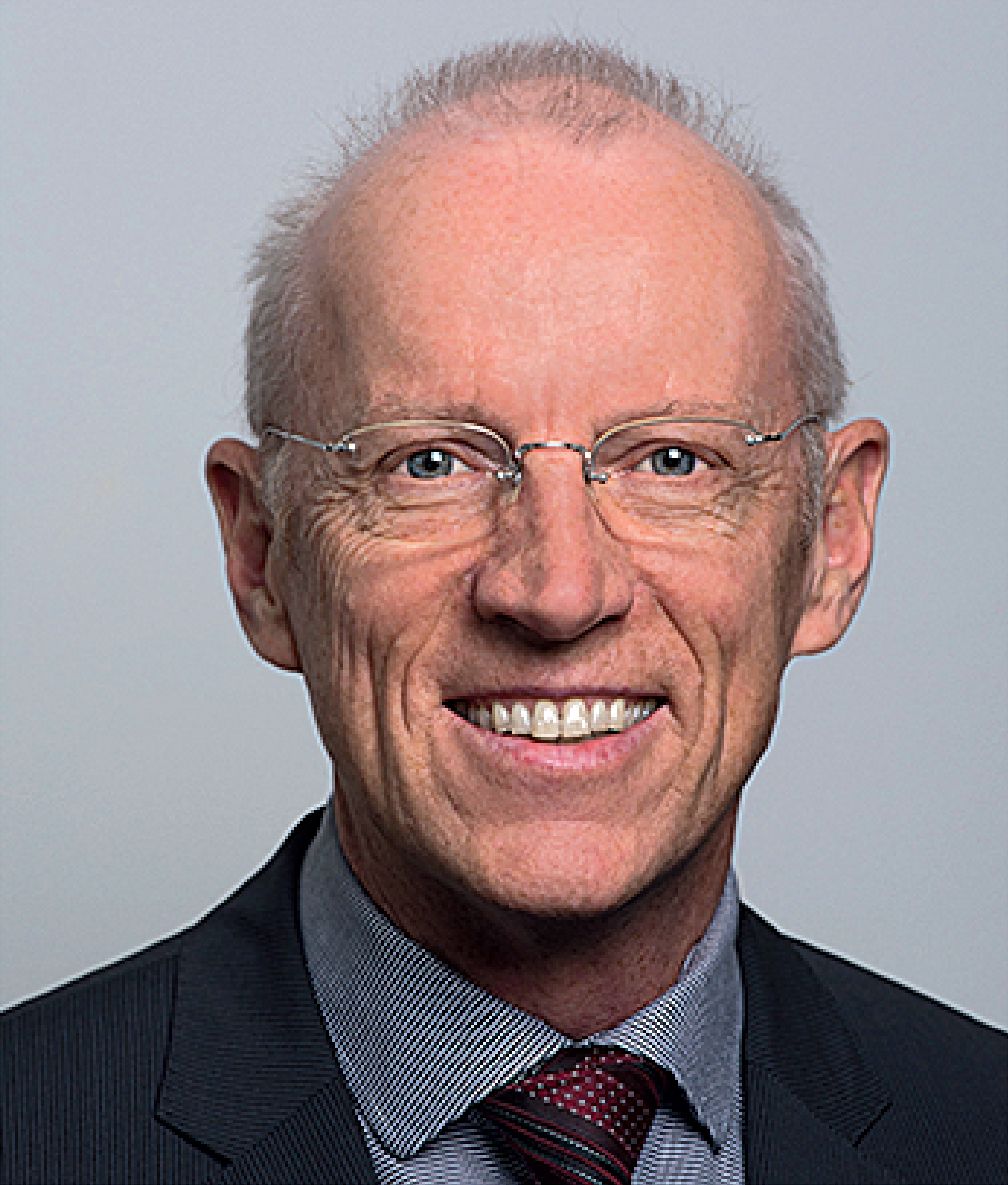
Prof. Dr. Daniel Thoma is the Head of Implantology and Regeneration at the Clinic of Reconstructive Dentistry, University of Zurich, Switzerland. He was trained in implant dentistry and prosthodontics at the Clinic of Reconstructive Dentistry, University of Zurich, Switzerland. Dr. Thoma was the recipient of an ITI scholarship and spent 1 year at the Department of Periodontics, University of Texas, Health Science Center, San Antonio, USA. In 2016, Dr. Thoma was a Visiting Associate Professor at the Department of Periodontology, Yonsei University, Seoul, South Korea. Since 2020, Dr. Thoma is an Adjunct Professor and serves as the Head of Implantology and Regeneration.
Details make perfection24. Oct 2024 — 26. Oct 2024MiCo - Milano Convention Centre, Milano, Italy
Speakers: Bilal Al-Nawas, Gil Alcoforado, Federico Hernández Alfaro, Sofia Aroca, Wael Att, Gustavo Avila-Ortiz, Kathrin Becker, Anne Benhamou, Juan Blanco Carrión, Dieter Bosshardt, Daniel Buser, Francesco Cairo, Paolo Casentini, Raffaele Cavalcanti, Tali Chackartchi, Renato Cocconi, Luca Cordaro, Luca De Stavola, Nuno Sousa Dias, Egon Euwe, Vincent Fehmer, Alberto Fonzar, Helena Francisco, Lukas Fürhauser, German O. Gallucci, Oscar Gonzalez-Martin, Dominik Groß, Robert Haas, Alexis Ioannidis, Simon Storgård Jensen, Ronald Jung, France Lambert, Luca Landi, Georg Mailath-Pokorny jun., Silvia Masiero, Iva Milinkovic, Carlo Monaco, Jose Nart, José M. Navarro, Katja Nelson, Manuel Nienkemper, David Nisand, Michael Payer, Sergio Piano, Bjarni E. Pjetursson, Sven Reich, Isabella Rocchietta, Giuseppe Romeo, Irena Sailer, Mariano Sanz, Ignacio Sanz Martín, Frank Schwarz, Shakeel Shahdad, Massimo Simion, Ralf Smeets, Benedikt Spies, Bogna Stawarczyk, Martina Stefanini, Hendrik Terheyden, Tiziano Testori, Daniel Thoma, Ana Torres Moneu, Piero Venezia, Lukas Waltenberger, Hom-Lay Wang, Stefan Wolfart, Giovanni Zucchelli, Otto Zuhr
European Association for Osseintegration (EAO)
29th EAO annual scientific meeting
Uniting Nations through Innovations29. Sep 2022 — 1. Oct 2022Palexpo, Le Grand-Saconnex, Switzerland
Speakers: Thabo Beeler, Nitzan Bichacho, Rino Burkhardt, Luigi Canullo, Matteo Chiapasco, Luca De Stavola, Mirela Feraru, Alfonso L. Gil, Klaus Gotfredsen, Markus Gross, Ueli Grunder, Christoph Hämmerle, Björn Klinge, Ivo Krejci, Sebastian Kühl, Niklaus P. Lang, Sonia Leziy, Daniele Manfredini, Konrad H. Meyenberg, Francesco Mintrone, Ricardo Mitrani, Sven Mühlemann, José M. Navarro, Florian Probst, Pablo Ramírez, Christoph Andreas Ramseier, Mario roccuzzo, Fidel Ruggia, Mariano Sanz, Rubens Spin-Neto, Dennis P. Tarnow, Daniel Thoma, Istvan Urban, Hans-Peter Weber
European Association for Osseintegration (EAO)
Farewell Symposium Christoph Hämmerle
Contemporary prosthodontics: Where do we stand, where are we heading?22. Jan 2022, 9:00 — 17:30 (CET)University of Zurich Irchel Campus, Zürich, Switzerland
Speakers: Thomas Attin, Beatrice Beck Schimmer, Tord Berglundh, Rino Burkhardt, Lyndon F. Cooper, Christer Dahlin, Stefano Gracis, Christoph Hämmerle, Lisa J. A. Heitz-Mayfield, Marc Hürzeler, Ronald Jung, Nadja Nänni, Mutlu Özcan, Marc Quirynen, Irena Sailer, Christian S. Stohler, Daniel Thoma
Disconnection and reconnection of abutments multiple times have known to affect the mucosal barrier around implants leading to marginal bone loss. This clinical report describes a novel technique that amalgamates the benefits of digital technologies encompassing the fabrication of surgical guides for implant placement, customized hybrid zirconia abutments and all ceramic lithium disilicate crowns prior to implant placement. A correct 3-dimensional implant positioning along with immediate placement of the definitive hybrid customized abutment and a lithium disilicate crown has the potential to reduce treatment time, visits and costs while delivering optimal esthetic outcomes.
The aim of the present clinical report is to introduce a novel surgical procedure, the “Apical Tooth Replantation with Surgical Intrusion Technique” (ATR-SIT) for managing teeth with hopeless prognosis compromised with a severe endodontal-periodontal lesion, pathologic tooth migration, and gingival recession. Two cases are presented managing teeth diagnosed with a hopeless prognosis. ATR-SIT involves tooth extraction, extra-oral root debridement, root surface conditioning, apicectomy, retrograde filling and the application of enamel matrix derivatives prior to reimplantation. Following reimplantation, the teeth are covered with a combination of autogenous bone chips and bone substitute materials, covered with resorbable membranes. Following ATR-SIT, the patients received either orthodontic treatment or tooth-supported fixed dental prostheses. The described ATR-SIT effectively improved the initially hopeless prognosis of the teeth and maintained periodontal health over time, evidenced by favourable clinical and radiographic outcomes. ATR-SIT might be a potential alternative to tooth extraction of hopeless teeth in patients with stage IV periodontitis.
Keywords: Endo-periodontal lesion, Stage Ⅳ periodontitis, Periodontal regeneration, Tooth replantation, Gingival recession, Hopeless tooth, Pathologic tooth migration
This article defines immunophenotypes of stromal inflammatory and endothelial cells and fibroblasts 3 months after augmentation of the peri-implant soft tissue using a porcine cross-linked collagen matrix (VCMX). Peri-implant soft tissue samples were obtained from 12 patients at the lining mucosa (LM)–masticatory mucosa (MM) junction before (1) and 3 months after (2) augmentation. Immunohistochemical stains were performed to identify inflammatory cells (T [CD3] and B [CD20] lymphocytes, plasma cells [CD138]), macrophages (CD68-proinflammatory, CD163-anti-inflammatory/ reparative), endothelial cells (CD31, CD34), and fibroblasts (CD90, TE-7). Differences in the mean positively stained cells pre- and postaugmentation were analyzed by Wilcoxon signed-rank test. CD31+ endothelial cells showed increased mean numbers in MM2 compared to MM1 (P = .025) and in LM2 compared to LM1 (P = .047). CD163+ anti-inflammatory macrophages showed higher mean numbers in MM2 than in MM1 (P = .021) and in LM2 than in LM1 (P = .012). All other cell phenotypes showed nonsignificant changes between pre- and postaugmentation. This molecular study provides novel insight on the frequency of stromal cell phenotypes in the wound healing process at 3 months postaugmentation with VCMX, with anti-inflammatory CD163+ macrophages being predominant. This should be further investigated to help find novel therapeutic approaches to modulate and promote the VCMX-related healing process.
Keywords: collagen fibers, collagen matrix, connective tissue, mucosal thickness, soft tissue augmentation
Purpose: To assess the clinical concept of patient treatment with fixed tooth- and implant-supported restorations in a university-based undergraduate program after 13 to 15 years. Materials and Methods: In total, 30 patients (mean age 56 years) who had received multiple tooth- and implant-supported restorations were recalled after 13 to 15 years. The clinical assessment comprised biologic and technical parameters as well as patient satisfaction. Data were analyzed descriptively, and the 13- to 15-year survival rates for tooth- and implant-supported single crowns and fixed dental prostheses (FDPs) were calculated. Results: The survival rate of tooth-supported restorations amounted to 88.3% (single crowns) and 69.6% (FDPs); in implants, it reached 100% for all types of restorations. Overall, 92.4% of all restorations were free of technical complications. The most common technical complication was chipping of the veneering ceramic (tooth-supported restorations: 5.5%; implant-supported restorations: 13% to 15.9%) regardless of the material used. For tooth-supported restorations, increased probing depth ≥ 5 mm was the most frequent biologic complication (22.8%), followed by endodontic complications of root canal–treated teeth (14%) and loss of vitality at abutment teeth (8.2%). Peri-implantitis was diagnosed in 10.2% of implants. Conclusions: The results of this study indicate that the clinical concept implemented in the undergraduate program and performed by undergraduate students works well. The clinical outcomes are similar to those reported in the literature. In general, the majority of biologic complications occur in reconstructed teeth, whereas implant-supported restorations are more prone to technical complications.
Die wissenschaftliche Evidenz für Zirkonoxidimplantate ist in den letzten Jahren erheblich gestiegen. Mittlerweile liegen Daten aus klinischen Studien vor, die eine Beobachtungszeit von 5−7 Jahren umfassen. Dennoch fehlen weiterhin randomisierte klinische Studien, die den Einsatz von Zirkonoxidimplantaten in verschiedenen klinischen Indikationen vergleichen. Es gibt bislang keine Studie, die den direkten Vergleich zwischen kurzen und längeren Zirkonoxidimplantaten untersucht. Ebenso fehlen klinische Daten über den Einsatz von kurzen Zirkonoxidimplantaten. Als kurze Implantate sind Implantate mit einer Länge von 6 mm oder weniger definiert. 8 mm „kurze“ Zirkonoxidimplantate können jedoch erfolgreich verwendet werden. Zudem zeigen Ergebnisse aus Kohortenstudien, dass die Implantatlänge keinen Einfluss auf das marginale Knochenniveau hat. Kurze Zirkonoxidimplantate könnten im Kieferkamm mit einer reduzierten vertikalen Höhe eine kostengünstige Behandlungsoption mit geringerer Morbidität darstellen, wenn dadurch eine aufwendige Knochenaugmentation verhindert werden kann. In Situationen, in denen ausreichend vertikale Höhe vorhanden ist, scheint die Verwendung von Implantaten mit Standardlänge die sicherere Option zu sein.
Manuskripteingang: 08.07.2023, Annahme: 18.08.2023
Keywords: Zirkonoxidimplantate, vollkeramische Restaurationen, metallfreie Restaurationen, kurze Implantate, wissenschaftliche Evidenz
The objective of this study was to assess volumetric and linear changes of buccal mucosal thickness at implant sites following soft tissue augmentation with a volume-stable collagen matrix (VCMX). Soft tissue augmentation using a VCMX was performed in 12 patients at the time of implant placement. Hydrocolloid impressions were taken prior to surgery and at 1 and 6 months postsurgery. Stone cast models were scanned, and stereolithography (STL) files from the three time points were uploaded to an image-analysis software. At all time points, linear and volumetric measurements of the contour changes up to 3 mm apical to the mucosal margin were performed and were analyzed statistically. At 1 mm apical to the mucosal margin, the change in soft tissue thickness between presurgery (T1) and 1 month (T2) amounted to 0.21 ± 1.22 mm, and the change between T1 and 6 months (T3) was 0.08 ± 1.47 mm. At 3 mm apical to the mucosal margin, the change in soft tissue thickness was 1.92 ± 1.70 mm between T1 and T2 and 0.31 ± 1.26 mm between T1 and T3. Contour (volumetric) changes revealed an increase of 0.58 ± 0.73 mm between T1 and T2 and an overall gain of 0.55 ± 0.73 mm between T1 and T3. Soft tissue augmentation with VCMX increased the ridge profile. The increase in ridge width was greater at 3 mm below the ridge crest than at 1 mm below the ridge crest. Remodeling processes during healing showed a decrease in the ridge contour between 1 and 6 months.
Festsitzender Zahnersatz, der von oralen Zahnimplantaten getragen wird, ist in der täglichen Praxis zu einer zuverlässigen Behandlungsoption geworden. Dennoch stellt der Ersatz von zwei benachbarten fehlenden Zähnen insbesondere bei ungünstigen anatomischen Verhältnissen eine klinische Herausforderung dar. Um diese Herausforderung zu meistern, hat die Verwendung von implantatgetragenem festsitzendem Zahnersatz mit einem Anhänger an Popularität gewonnen. Dies ist vor allem darauf zurückzuführen, dass keine oder weniger chirurgische Primäreingriffe erforderlich sind, die Kosten geringer ausfallen und somit die Morbidität des Patienten reduziert wird. Ziel der vorliegenden Übersichtsarbeit ist es, den aktuellen Stand der Erkenntnisse über die Verwendung von implantatgetragenem festsitzendem Teilzahnersatz mit Freiendverlängerung zusammenzufassen. Der Artikel befasst sich sowohl mit dem Seitenzahn- als auch mit dem Frontzahnbereich, wobei die Vor- und Nachteile der einzelnen Behandlungen hervorgehoben und die verfügbaren mittel- bis langfristigen Ergebnisse dargestellt werden.
Manuskripteingang: 13.05.2022, Manuskriptannahme: 27.07.2022
Keywords: Zahnimplantate, Freiendverlängerung, klinische Studie
The International Journal of Prosthodontics, 5/2021
DOI: 10.11607/ijp.6999Pages 560-566d, Language: EnglishStucki, Lukas / Asgeirsson, Asgeir G / Jung, Ronald E / Sailer, Irena / Hämmerle, Christoph Hf / Thoma, Daniel S
Purpose: To assess the clinical, technical, and esthetic outcomes of directly veneered zirconia abutments cemented onto nonoriginal titanium bases over a 3-year follow-up.
Materials and Methods: A total of 24 healthy patients with a single missing tooth in the maxilla or mandible (incisors, canines, or premolars) received a two-piece implant with a screw-retained veneered zirconia restoration extraorally cemented onto a titanium base abutment. Baseline measurements and follow-up examinations were performed at 6 months, 1 year, and 3 years following loading. Radiographic, clinical, technical, and esthetic parameters were assessed. Wilcoxon signed rank test was used to analyze the data.
Results: Mean marginal bone levels measured 0.54 ± 0.39 mm (median: 0.47 mm, range: 0.07 to 1.75 mm) at baseline and 0.52 ± 0.39 mm (median: 0.39 mm, range: 0.06 to 1.33 mm) at 3 years. Mean probing depth around the implants increased from 3.0 ± 0.6 mm at baseline to 3.8 ± 0.8 mm at 3 years (P = .001). Bleeding on probing changed from 27.1% ± 20.7% at baseline to 51.5% ± 26.1% at 3 years (P = .001). The mean plaque control record amounted to 11.1% ± 21.2% at baseline and 14.4% ± 13.89% at 3 years (P = .261). Two implants were lost at 3.5 and 30 months postloading due to periimplantitis, resulting in a 91.7% implant survival rate. Patient satisfaction was high at 3 years.
Conclusion: Zirconia restorations cemented onto the tested nonoriginal titanium bases should not be recommended for daily clinical use, as they were associated with significant increases in BOP and PD values and varying marginal bone levels 3 years after placement.
The International Journal of Prosthodontics, 5/2020
DOI: 10.11607/ijp.6470, PubMed ID (PMID): 32956429Pages 487-492, Language: EnglishWaller, Tobias / Evci, Erkan / Hämmerle, Christoph H.F. / Hüsler, Jürg / Jung, Ronald E. / Thoma, Daniel S.
Purpose: The aim of the present study was to assess the perceptibility and acceptability threshold values for color differentiation at the restoration and mucosa levels.
Materials and Methods: One restored single-tooth implant and the contralateral reference tooth were spectrophotometrically assessed in 20 patients. Perceptibility and acceptability were evaluated by dentists, dental technicians, and laypeople.
Results: Dental technicians had the highest sensitivity in the perception of tooth color differences (ΔE = 2.7), followed by dentists (ΔE = 3.3) and laypeople (ΔE = 4.4). Acceptability threshold values were generally higher than perceptibility threshold in all groups. Dental technicians exhibited the highest sensitivity in the perception of mucosa color differences (50% perceptibility at ΔE = 2.65), followed by dentists (ΔE > 3.7) and laypeople (ΔE > 6).
Conclusion: Color differences were tolerated with varying degrees among the three groups. Laypeople accepted higher color differences at the mucosa level.