Various cookies are used on our website: We use technically necessary cookies for the purpose of enabling functions such as login or a shopping cart. We use optional cookies for marketing and optimization purposes, in particular to place relevant and interesting ads for you on Meta's platforms (Facebook, Instagram). You can refuse optional cookies. More information on data collection and processing can be found in our privacy policy.
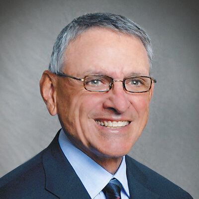
Myron Nevins, DDS, is an associate clinical professor of periodontology at the Harvard School of Dental Medicine and a clinical professor of periodontics at the University of Michigan School of Dentistry and Temple University Kornberg School of Dentistry. He is a past president of the American Academy of Periodontology and a former director and chairman of the American Board of Periodontology. He was also the coeditor-in-chief of The International Journal of Periodontics & Restorative Dentistry. Dr Nevins maintains a private practice limited to periodontics and implantology in Swampscott, Massachusetts, and is the founder and president of Perio Imp Research, Inc.
1st Edition 2020 DVD 17-Volume Set; NTSC/PAL; Runtime: 44 h 14 min Language: English Category: Implantology Stock No.: 9090 ISBN 978-1-78698-102-8 QP Deutschland
2nd Edition 2019 Book Hardcover, 22,2 x 30,3, 544 pages, 1782 illus Language: English Category: Implantology Stock No.: 21971 ISBN 978-0-86715-798-7 QP USA
1st Edition 2011 Book Hardcover, 256 pages, 235 illus colored Language: English Categories: Oral/Maxillofacial Surgery, Oral Surgery Stock No.: 18721 ISBN 978-1-85097-211-2 QP Deutschland
Length: 34 minutes Production year: 2009 Language: English Source: 60 Jahre Quintessenz available since: 8. Feb 2010
Events
The 14th International Symposium on Periodontics and Restorative Dentistry (ISPRD)
9. Jun 2022 — 12. Jun 2022Boston Marriott Copley Place, Boston, MA, United States of America
Speakers: Tara Aghaloo, Edward P. Allen, Evanthia Anadioti, Wael Att, Vinay Bhide, Markus B. Blatz, Scotty Bolding, Lorenzo Breschi, Jeff Brucia, Daniel Buser, Luigi Canullo, Daniele Cardaropoli, Stephen J. Chu, Donald Clem, Christian Coachman, Lyndon F. Cooper, Daniel Cullum, Lee Culp, José Carlos Martins da Rosa, Sergio De Paoli, Marco Degidi, Nicholas Dello Russo, Serge Dibart, Joseph P. Fiorellini, Mauro Fradeani, Stuart J. Froum, David Garber, Maria L. Geisinger, William Giannobile, Luca Gobbato, Ueli Grunder, Galip Gürel, Chad Gwaltney, Christoph Hämmerle, Robert A. Horowitz, Marc Hürzeler, David Kim, Gregg Kinzer, Christopher Köttgen, Ina Köttgen, Purnima S. Kumar, Burton Langer, Lydia Legg, Pascal Magne, Kenneth A. Malament, Jay Malmquist, George Mandelaris, Pamela K. McClain, Michael K. McGuire, Mauro Merli, Konrad H. Meyenberg, Craig M. Misch, Julie A. Mitchell, Marc L. Nevins, Myron Nevins, Michael G. Newman, Miguel A. Ortiz, Jacinthe M. Paquette, Stefano Parma-Benfenati, Michael A. Pikos, Giulio Rasperini, Pamela S. Ray, Christopher R. Richardson, Isabella Rocchietta, Marisa Roncati, Marco Ronda, Paul S. Rosen, Maria Emanuel Ryan, Irena Sailer, Maurice Salama, David M. Sarver, Takeshi Sasaki, Todd Scheyer, Massimo Simion, Michael Sonick, Sergio Spinato, Dennis P. Tarnow, Lorenzo Tavelli, Douglas A. Terry, Tiziano Testori, Carlo Tinti, Istvan Urban, Hom-Lay Wang, Robert Winter, Giovanni Zucchelli
Quintessence Publishing Co., Inc. USA
This author's journal articles
International Journal of Periodontics & Restorative Dentistry, Pre-Print
Background: The necessity of a minimum volume of keratinized tissue around implants to maintain tissue stability remains a matter of debate. The aim of this case series is to showcase the outcome and long-term maintenance of dental implants that received soft tissue augmentation. Case Presentation: Three cases of second stage following dental implant placement were evaluated for amount of keratinized tissue. Based on the Ono-Nevins classification described here, treatment was rendered including keratinized tissue grafting. The cases were followed for 10, 18 and 28 years. Each case demonstrated maintenance of soft and hard tissue profile around the dental implants with no incidence of peri-implantitis as evidenced on clinical examination and use of peri-apical radiographs. Conclusion: Soft tissue management around dental implants may play a key role in long-term maintenance of peri-implant health and prevention of peri-implantitis.
Keywords: periodontal disease, dental implant, peri-implantitis, soft tissue graft, case series
An advantage of treated implant surfaces is their increased degree of hydrophilicity and wettability compared with untreated, machined, smooth surfaces that are hydrophobic. The present preclinical in vivo study aimed to compare the two implant surface types, namely SLActive (Straumann, Basel, Switzerland) and nanohydroxyapatite (Hiossen, Englewood Cliffs, NJ, USA), in achieving early osseointegration. The authors hypothesised that the nanohydroxyapatite surface is comparable to SLActive for early bone–implant contact. Six male mixed foxhounds underwent mandibular premolar and first molar extraction, and the sockets healed for 42 days. The mandibles were randomised to receive implants with either SLActive (control group) or nanohydroxyapatite surfaces (test group). A total of 36 implants were placed in 6 animals, and they were sacrificed at 2 weeks (2 animals), 4 weeks (2 animals) and 6 weeks (2 animals) after implant surgery. When radiographic analysis was performed, the difference in bone level between the two groups was statistically significant at 4 weeks (P = 0.024) and 6 weeks (P = 0.008), indicating that the crestal bone level was better maintained for the test group versus the control group. The bone–implant contact was also higher for the test group at 2 (P = 0.012) and 4 weeks (P = 0.011), indicating early osseointegration. In conclusion, this study underscored the potential of implants with nanohydroxyapatite surfaces to achieve early osseointegration.
Keywords: acid-etched surface, bone–implant contact, dental implant, histology, histomorphometric analysis, nanohydroxyapatite, osseointegration, radiographs, SLActive, tooth extractions
This research was funded by Hiossen, Englewood Cliffs, NJ, USA. The funders played no role in the collection, analysis or interpretation of data, in the writing of the manuscript or in the decision to publish the results. The authors declare there are no conflicts of interest relating to this study.
This assessment evaluated the clinical feasibility of using narrow-diameter implants to support a mandibular overdenture. Twelve patients presenting with an edentulous mandible were recruited from nine dental offices. Each patient received two to four implants in the mandible. The implants were placed without additional bone augmentation, and a total of 36 implants were placed. All sites achieved satisfactory crestal bone stability and soft tissue maintenance 1 year after the final prosthetic restoration. This investigation was supported by the Osseointegration Foundation, working in conjunction with the Zest Anchors implant company. There are three basic valuable activities that emerge from a professional foundation, which were reflected in this case study. They include disseminating information to practitioners, persuading young clinicians to become research investigators, and reaching out to patients in need of treatment who cannot afford it without a charitable opportunity.
A 12-month prospective consecutive case series study was conducted to evaluate the effectiveness of a novel porcine collagen matrix to correct moderate to severe buccogingival recession defects. A total of 10 healthy patients (8 women and 2 men; age range: 30 to 68 years) with 26 maxillary and mandibular recession gingival defects > 4 mm deep were included. Healthy maturation of gingival tissues with natural color and texture matching the adjacent soft tissue areas were observed at all reevaluation visits. Complete root coverage was not achieved in all cases, possibly due to severe buccal bone loss in most of the selected cases, which negatively impacted the results. Nevertheless, when a novel porcine collagen matrix was used, a mean root coverage of 63.15% was achieved, and gains in clinical attachment level and keratinized tissue height were seen. Future randomized clinical trials are needed to further explore the potential of porcine collagen matrix for treatment of localized gingival recession defects.
This investigation was designed to evaluate crestal bone stability and soft tissue maintenance to Laser-Lok tapered tissue-level implants. Twelve patients presenting with an edentulous site adequate for the placement of two implants were recruited from four dental offices (2 to 4 patients per office). Each patient received two Laser-Lok tissue-level implants placed with a 3-mm interimplant distance according to a surgical stent. The implants were placed so that the Laser-Lok zone sat at the junction between hard and soft tissues. A total of 24 implants were placed, and all achieved satisfactory crestal bone stability and soft tissue maintenance 1 year after receiving the final prosthetic restoration.
Making treatment decisions in dental implantology has evolved over the last five decades. These decisions and the clinical management of sites thereafter are selected based on recent changes, including the achievement of osseointegration, reestablishment of biologic width bone remodeling, the peri-implant soft tissue phenotype, the way peri-implantitis is defined, and advancements in digital technology. This article discusses these key aspects and their effects and influence on implant therapy.
This ex vivo study evaluates the incidence of sinus membrane perforation during implant site osteotomy with two different types of drills and drilling techniques. Fifty goat heads with 50 sinus pairs (100 sinus sides) were assigned to two groups (osseodensification bur [OB] group and inverse conical shape bur [ICSB] group) to simulate transcrestal sinus elevation (50 sinus sides per group). An osteotomy was performed to pass through the lateral sinus wall no more than 3 mm. The integrity of the sinus membranes was examined and confirmed under a microscope. Of the 50 sinuses per group, the OB group presented with 14 (28%) perforated sinuses, while the ICSB group presented with 2 (4%) perforated sinuses. Of the 14 perforations from the OB group, 6 (42.9%) showed a pinpoint perforation pattern, 4 (28.5%) of which were not visible until direct air pressure was applied. Overall, the ICSB drill group demonstrated a lower sinus perforation rate than the OB group.
The present study aimed to evaluate the osseoconduction ability of an airborne particle-abraded and etched (SAE) titanium alloy surface when placed in humans with poor bone quality. Four patients scheduled to receive an implant-supported full-arch prosthesis received two additional reduced-diameter implants to be harvested after 6 months of submerged healing. Undecalcified vestibulopalatal/vestibulolingual histologic sections were prepared after the micro-computerized tomography (μCT) examination. Six implant sides from four biopsied implants displayed a type IV bone environment and were included in the present study. Bone-to-implant contact (BIC) was first measured on each implant side. The estimated initial BIC (E-iBIC) was evaluated by superimposing the implant profile 0.25 mm away from its actual position. The μCT provided information about the local and adjacent bony architecture. The mean BIC was 62.5% ± 10.6%, while the mean E-iBIC was 33.1% ± 4.4%. The E-iBIC/BIC ratio was 1.81 ± 0.38. The 3D μCT sections showed the thin bone trabeculae covering the implant surface; although they seemed to be separated from the rest of the bony scaffold, they were much more interconnected than what appeared to be on the 2D histologic preparations. This limited number of human histologic samples document, for the first time, that the SAE titanium alloy implant surface is apparently osseoconductive when placed in poor human bone quality. The average BIC was 1.81 times higher than the E-iBIC. This high osseoconductivity may explain the predictable clinical behavior of implants with this type of SAE textured surface in type IV bone.
The aim of this report is to present a technique for buccal soft tissue contour augmentation with the use of a porcine volume-stable collagen matrix (VSCM). Augmentation of buccal soft tissue at the time of implant placement is often a necessity but is mostly performed using autogenous tissue. The technique using a VSCM can be done at the time of implant placement or, in the case of a two-stage procedure, at the time of implant uncovering. Here, clinical outcomes are reported in two cases when using VSCM concurrently with implant placement at sites in need of buccal contour augmentation to achieve a functional, esthetic result. The use of a xenograft poses several advantages over autogenous tissue while providing similar gains in soft tissue thickness. By eliminating the need to harvest a soft tissue graft from the palate, patient morbidity is reduced, and the reliance on palatal tissue thickness, to determine the amount of achievable augmentation, is eliminated.
Certain bone morphologies and soft tissue thickness (ie, phenotype) are considered to be risk factors for the development of gingival recessions following orthodontic tooth movement. Preoperative evaluation of the periodontal phenotype, in the frame of orthodontic treatment plan, identify teeth at high risk for mucogingival complications related to orthodontic therapy. The new surgical technique is illustrated in a clinical case. A patient with a thin phenotype without visible gingival recession had bone dehiscences in the anterior mandible. Prior to orthodontic treatment, simultaneous bone and soft tissue augmentation was performed using the combination of a highly cross-linked ribose porcine type I collagen membrane and a subepithelial palatal connective tissue graft. Two years after augmentation surgery and initiation of orthodontic treatment, a thick buccal tissue with a wide band of attached gingiva was observed without any clinical signs of root prominences, indicating a substantial change in periodontal phenotype. The clinical findings were corroborated by the 3D analysis, demonstrating substantial bone apposition on the buccal aspect of all roots in the treated area. The described surgical technique offers a valuable approach for regenerating hard and soft tissues in deficient areas prior to orthodontic therapy, thus preventing the development of gingival recessions.