Various cookies are used on our website: We use technically necessary cookies for the purpose of enabling functions such as login or a shopping cart. We use optional cookies for marketing and optimization purposes, in particular to place relevant and interesting ads for you on Meta's platforms (Facebook, Instagram). You can refuse optional cookies. More information on data collection and processing can be found in our privacy policy.
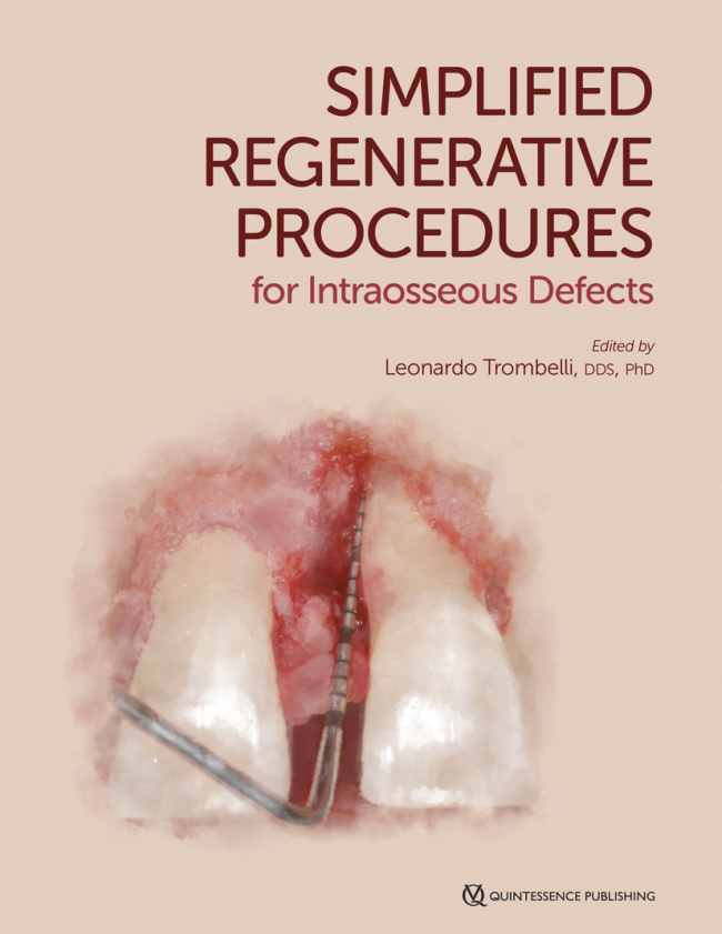
Leonardo Trombelli is a full professor and chair of the Department of Periodontology, School of Dentistry at the University of Ferrara. He has served as the director of the Research Center for the Study of Periodontal and Peri-implant Diseases at the University of Ferrara and the director of the Dental Clinic, University Hospital, Ferrara. He has been the dean of the Dental School at the University of Ferrara since 2013. He also served as the president of the Medical School at the University of Ferrara from 2014 to 2016. Past president of the SIO from 2007 to 2009. Dr Trombelli is an active member of SIdP and an editorial board member for the Journal of Clinical Periodontology.
1st Edition 2019 Book 2 Volumes, 21 x 29,7 cm, Hardcover in a slip case, 1356 pages, 4900 illus Language: English Categories: Implantology, Periodontics Stock No.: 21521 ISBN 978-0-86715-886-1 QP USA
390,00 €158,00 €
Events
SEPA 24 Bilbao
Clinical evidence based on scientific evidence29. May 2024 — 1. Jun 2024Bilbao, Spain
Speakers: Eduardo Anitua, Sofia Aroca, Serhat Aslan, Gustavo Avila-Ortiz, Juan Blanco Carrión, Gonzalo Blasi, Nagihan Bostanci, Iain L. C. Chapple, Jan Cosyn, Glécio Vaz de Campos, Luca De Stavola, Jan Derks, Vincent Fehmer, Elena Figuero, Sergio García, Alfonso L. Gil, Oscar Gonzalez-Martin, Adrian Guerrero, Sérgio Kahn, Alejandro Lanis, Antonio Liñares, Ferrán Llansana, Francesco Mangano, Dino Calzavara mantovani, Mauro Merli, Juan Mesquida, Alberto Monje, Eduardo Montero, Stefano Parma-Benfenati, Bjarni E. Pjetursson, Pablo Ramírez, Mariano Sanz, Ignacio Sanz Sànchez, Beatriz Solano Mendoza, Jacobo Somoza, Martina Stefanini, Maurizio S. Tonetti, Leonardo Trombelli, Ion Zabalegui
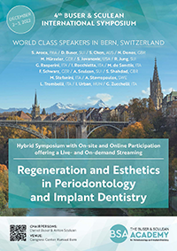
4th Buser & Sculean International Symposium
Regeneration and Esthetics in Periodontology and Implant Dentistry2. Dec 2022 — 3. Dec 2022Congress Center, Kursaal Bern, Bern, Switzerland
Speakers: Sofia Aroca, Daniel Buser, Stephen Chen, Nikolaos Donos, Marc Hürzeler, Sascha Jovanovic, Ronald Jung, Giulio Rasperini, Isabella Rocchietta, Frank Schwarz, Anton Sculean, Shakeel Shahdad, Andreas Stavropoulos, Martina Stefanini, Leonardo Trombelli, Istvan Urban, Giovanni Zucchelli
Buser & Sculean Academy
EuroPerio10
15. Jun 2022 — 18. Jun 2022Bella Center Copenhagen, Copenhagen, Denmark
Speakers: Mario Aimetti, Zvi Artzi, Serhat Aslan, Georgios Belibasakis, Florian Beuer, Juan Blanco Carrión, Michael M. Bornstein, Nagihan Bostanci, Philippe Bouchard, Darko Božić, Olivier Carcuac, Maria Clotilde Carra, Nelson Carranza, Iain L. C. Chapple, Pierpaolo Cortellini, Jan Cosyn, Mike Curtis, Francesco D'Aiuto, Bettina Dannewitz, Luca De Stavola, Jan Derks, Nikolaos Donos, Peter Eickholz, Bahar Eren Kuru, Ricardo Faria Almeida, Roberto Farina, Magda Feres, Elena Figuero, Dagmar Fosså Bunæs, Rok Gašperšič, William Giannobile, Cecilie Gjerde Gjengedal, Moshe Goldstein, Marjolaine Gosset, Klaus Gotfredsen, Filippo Graziani, Adrian Guerrero, George Hajishengallis, Hady Haririan, Lisa J. A. Heitz-Mayfield, Palle Holmstrup, Marc Hürzeler, Mark Ide, Søren Jepsen, Ronald Jung, Sérgio Kahn, Anhgela R. Kamer, Alpdogan Kantarci, Moritz Kebschull, Björn Klinge, Thomas Kocher, Odd Carsten Koldsland, Kenneth Kornman, Marja Laine, Markus Laky, Isabelle Laleman, Evanthia Lalla, France Lambert, Luca Landi, Niklaus P. Lang, Antonio Liñares, Tomas Linkevičius, Bruno Loos, Rodrigo Lopez, Eli Machtei, Aslan Mammadov, Mauro Merli, Andrea Mombelli, Eduardo Montero, Niki Moutsopoulos, Jose Nart, Gustavo G. Nascimento, Ian Needleman, Tiernan O'Brien, William Papaioannou, Panos N. Papapanou, Michael A. Pikos, Pawel Plakwicz, Constanza Pontarolo, Philip M. Preshaw, Marc Quirynen, Mia Rakic, Christoph Andreas Ramseier, Hélène Rangé, Papageorgiou Spyridon, Maurizio S. Tonetti, Leonardo Trombelli, Istvan Urban, Fridus van der Weijden, Fabio Vignoletti, Charalambos Vlachopoulos, Nicola West, Asaf Wilensky, Ion Zabalegui, Egija Zaura, Nicola Zitzmann, Giovanni Zucchelli, Otto Zuhr, Fardal Øystein
European Federation of Periodontology (EFP)
This author's journal articles
International Journal of Periodontics & Restorative Dentistry, 1/2024
The purpose of the present study was to describe a novel protocol for a minimally invasive pocket elimination surgery (MI-PES) in the posterior maxilla and mandible, which consists of the combined use of (1) an access flap based on an internally beveled gingivectomy with minimal to no papilla mobilization at the buccal aspect, and (2) a resective procedure with an apically positioned flap on the lingual aspect. The interproximal bone defects were accessed with a single (lingual) flap, and the bone architecture was modified by the adoption of piezoelectric inserts for controlled bone recontouring associated with fiber retention. Mean probing depth (PD) was 5.5 ± 0.8 mm before surgery and 2.7 ± 0.6 mm at the 6-month reevaluation. All treated pockets showed a postsurgical PD < 4 mm. Gingival recession (REC) was 0.3 ± 0.5 mm at baseline and increased to 1.6 ± 0.8 mm at 6 months. When buccal and lingual pockets were analyzed separately, a trend toward a similar PD reduction, less REC increase, and greater clinical attachment level gain was recorded for buccal pockets. These preliminary observations seem to support the use of MI-PES as a valuable option for pocket elimination, at least when residual pockets are associated with a shallow interproximal osseous crater in the posterior maxilla or mandible.
Purpose: To comparatively evaluate transcrestal sinus floor elevation (tSFE) and lateral sinus floor elevation (lSFE) at sites with different residual bone heights (RBHs). Materials and Methods: A re-analysis of data from a parallel-arm, randomized trial comparatively evaluating tSFE and lSFE was performed. Within each RBH interval (< 4 mm or ≥ 4 mm), tSFE and lSFE groups were compared for chair time, surgery-related costs, morbidity, and radiographic parameters (including the proportion of the implant surface in direct contact with the radiopaque area [totCON%]). Results: The intention-to-treat (ITT) population consisted of 29 and 28 patients in the tSFE and lSFE groups, respectively. Irrespective of RBH, both tSFE and lSFE lead to a median totCON% of 100%. At sites with RBH < 4 mm, pain severity was significantly higher at days 0 and 1 in the tSFE group, with no intergroup difference in the dose of analgesics. LSFE was associated with a significantly higher frequency of bruising and greater cost. At sites with RBH ≥ 4 mm, a significantly lower frequency of postoperative signs/symptoms, less chair time, and lower costs were observed in the tSFE group. Conclusions: The selection of tSFE or lSFE within the investigated RBH intervals seems to be supported by differences in chair time, costs, and morbidity between the two techniques. At sites with RBH < 4 mm, clinicians preferring tSFE should encourage the administration of analgesics according to a predefined plan in the early postoperative phase. At sites with RBH ≥ 4 mm, tSFE should be preferred to lSFE due to reduced chair time, costs, and morbidity.
Keywords: alveolar process, bone resorption, maxillary sinus, bone regeneration, minimally invasive, dental implants
International Journal of Oral Implantology, 4/2021
PubMed ID (PMID): 34726848Pages 385-399, Language: EnglishMonje, Alberto / Amerio, Ettore / Farina, Roberto / Nart, José / Ramanauskaite, Ausra / Renvert, Stefan / Roccuzzo, Andrea / Salvi, Giovanni E / Schwarz, Frank / Trombelli, Leonardo / Wang, Hom-Lay
Peri-implant diseases at implant sites represent the most considerable concern for many dental clinicians nowadays due to their detrimental effect on implant longevity. Preventive measures include patient education and motivation, supportive peri-implant therapy and routine assessment of the hard and soft tissues. Nevertheless, the reliability of clinical parameters to monitor peri-implant conditions is subject to debate. As such, the primary purpose of the present review was to gain further insight into the diagnostic accuracy of probing as a clinical tool to monitor dental implants and assist clinicians in preventing peri-implant diseases. Studies have recommended periodic probing to monitor the condition of the peri-implant tissues. Increased probing pocket depth, profuse bleeding on probing and suppuration at implants are clinical signs that have been associated with peri-implantitis; thus, if these clinical parameters are present, radiographic assessment is encouraged to make a definitive diagnosis considering potential inaccuracies related to local and/or systemic factors identified in the present review.
Keywords: complications, diagnostic procedure, peri-implantitis, surgical
Conflict-of-interest statement: The authors declare there are no conflicts of interest relating to this study.
Purpose: This case series illustrates a simplified soft tissue management, namely, the subperiosteal periimplant augmented layer (SPAL), to increase hard and soft tissue dimensions at the most coronal portion of an implant.
Materials and Methods: Twenty-seven implants in 16 patients presenting either a buccal bone dehiscence or a thin ( 1 mm) buccal cortical bone plate (BCBP) were consecutively treated. Briefly, a split-thickness flap (namely, the mucosal layer) was raised on the buccal aspect. Then, the periosteal layer was elevated from the bone crest. A full-thickness flap was elevated on the oral aspect. After implant site preparation, a xenograft was used to fill the space between the periosteal layer and the BCBP and/or exposed implant surface and, if present, to completely correct the bone dehiscence. The periosteal layer was sutured to the oral flap. The mucosal layer was coronally advanced and sutured to submerge both the graft and the implants. At 3 to 6 months, a re-entry procedure for implant exposure was performed.
Results: Healing was uneventful, with no signs of infection in all cases. A wound dehiscence was observed in three implants in two patients at 2 weeks postsurgery. Out of 15 implants showing an initial bone dehiscence, 12 implants (80%) showed a complete resolution, with a subperiosteal tissue thickness (SPTT) at the time of re-entry of 3.1 ± 1.0 mm. Three implants presented a residual dehiscence of 1 mm (two implants) or 2 mm (one implant), with a SPTT of at least 2 mm. Out of 12 implants showing a thin BCBP at implant placement, 10 implants (90%) revealed a SPTT ≥ 2 at the time of re-entry. Two implants revealed a SPTT of 1 mm.
Conclusion: The SPAL technique represents a valuable simplified surgical approach associated with a low rate of complications in the treatment of peri-implant bone dehiscence and in the horizontal augmentation of peri-implant tissue thickness.
Keywords: alveolar bone loss, bone dehiscence, implant dentistry, reconstructive surgical procedures, surgical flaps, wound healing
The present study was performed to evaluate the impact of smoking status on 6-month clinical outcomes of a regenerative procedure based on enamel matrix derivative and deproteinized bovine bone mineral in the treatment of intraosseous defects accessed with buccal single flap approach. A total of 22 defects were selected in smoking (n = 11) and nonsmoking (n = 11) patients. Regenerative treatment resulted in similar clinical attachment level (CAL) gain in both smokers and nonsmokers. A trend toward lower 6-month CAL gain and probing depth reduction was observed in patients smoking 11 to 20 cigarettes per day compared to those smoking 1 to 10 cigarettes per day.
Purpose: Implant-supported mandibular overdentures (OVDs) have been proposed as the gold standard for the treatment of edentulous mandibles. There is limited evidence on the clinical outcomes of immediate loading of two unsplinted implants supporting a mandibular OVD. The purpose of this randomized controlled trial was to evaluate the performance of two unsplinted implants supporting a Locator-retained mandibular OVD over 12 months loaded immediately or after a delay.
Materials and Methods: Each patient received two implants 4.0 mm in diameter and 8 to 15 mm long. Locator-retained mandibular OVDs were connected to the implants either immediately (IL) or 3 months postsurgery (DL). The primary response variable was radiographic bone loss (RBL) at 6 and 12 months postsurgery. Implant length, insertion torque, implant failure, prevalence of maintenance visits, and prosthetic complications were also recorded.
Results: Thirty participants (15 in the IL and 15 in the DL groups) were evaluated at 12 months. The implant cumulative survival rates were 100% and 93% for DL and IL, respectively. The mean RBL from baseline to 1 year was 0.54 (± 0.5) mm and 0.25 (± 0.5) mm for DL and IL, respectively. A statistically significant difference was observed at 12 months, with less RBL in the IL group. Insertion torque and implant length were not correlated with RBL. Also, no difference in frequency of maintenance visits and prosthetic complications was reported between the groups.
Conclusion: Immediate loading of two unsplinted implants supporting a Locator-retained mandibular OVD seems to be a suitable treatment option. Significantly less RBL was observed after 1 year of loading around IL implants than around DL implants. Furthermore, neither implant length nor insertion torque seemed to affect RBL 1 year after surgical placement.
Keywords: immediate loading, overdenture
Twenty-four periodontal intraosseous defects were accessed with a buccal single-flap approach (SFA) and treated with enamel matrix derivative (EMD) or EMD + deproteinized bovine bone mineral (DBBM) according to the surgeon's discretion. EMD with or without DBBM was clinically effective in the treatment of periodontal intraosseous defects accessed with a buccal SFA. The adjunctive use of DBBM in predominantly onewall defects seemed to compensate for, at least in part, the unfavorable osseous characteristics in terms of the outcomes of the procedure.
Kohortenstudie an einer Spezialklinik für Parodontologie
Zielsetzung: Untersuchung der Prävalenz und Verteilung von Sondierungsbluten (BOP = bleeding on probing) bei Patienten mit Parodontopathien. Untersuchung des Einflusses von patientenbezogenen Parametern auf die individuelle Variabilität von BOP. Beschreibung von zwei Teilgruppen von Patienten mit signifikant unterschiedlichem BOP-Wert. Material und Methode: 601 Patienten wurden in die retrospektive Untersuchung einbezogen. Der Prozentanteil der Messpunkte mit BOP wurde für das Gesamtgebiss (%BOPtotal), wie für verschiedene Unterbereiche des Gebisses berechnet. Außerdem wurde der mögliche Einfluss von patientenbezogenen Faktoren auf die interindividuelle Variabilität von %BOPtotal bewertet. Es wurden zwei Teilgruppen von Patienten mit geringem beziehungsweise starkem Sondierungsbluten, also niedrigem beziehungsweise hohem Median für %BOPtotal, definiert (unteres und oberes Quartil). Ergebnisse: Der Median für %BOPtotal lag bei 26,4 %. 99 % der Studienpopulation wiesen mindestens eine Messstelle mit BOP auf. Für %BOPtotal gab es positive Korrelationen mit dem Alter der Patienten und mit einer Sondierungstiefe (ST) >= 5 mm; BOP trat signifikant häufiger bei Messpunkten im Unter- als im Oberkiefer, im Seiten- als im Frontzahnbereich, an interproximalen als an oralen/bukkalen Messstellen sowie an Messstellen mit ST >= 5 mm gegenüber solchen mit ST = 4 mm auf. Patienten mit geringem und mit starkem Sondierungsbluten unterschieden sich signifikant hinsichtlich %BOPtotal und BOP bei Messpunkten mit ST = 4 und ST >= 5 mm, jedoch nicht hinsichtlich Geschlecht, Nikotinstatus, Zigarettenkonsum, Diabetes oder Messpunkten mit ST >= 5 mm. Schlussfolgerungen: Die Prävalenz und Variabilität von BOP war von Patient zu Patient und von Kieferbereich zu Kieferbereich hoch. Patienten- und ortsspezifische Faktoren (Alter, Zahl der Parodontaltaschen, Sondierungstiefe, Zahntyp, relative Position der Messpunkte) scheinen für die Variabilität von BOP teilweise mitverantwortlich zu sein. Patienten mit geringem und mit starkem BOP unterschieden sich nicht signifikant hinsichtlich der untersuchten Parameter mit potenziellem Vorhersagewert.
Keywords: Sondierungsbluten, Gingivitis, Parodontopathie
Objectives: To assess the prevalence and distribution of bleeding on probing (BoP) in a cohort of periodontally diseased patients, evaluate the influence of patient-related predictors on BoP variability, and characterize two subgroups of patients with a significantly different BoP score.
Method and Materials: Six hundred and one patients were retrospectively included. The percentage of BoP-positive sites was calculated for the entire dentition (%BoPtotal) and within different areas of the dentition. The influence of patient-related predictors on intersubject variability in %BoPtotal was evaluated. Two subgroups of patients, belonging to the lowest (LB, low bleeding) and highest (high bleeding, HB) quartile of patient distribution according to %BoPtotal, were identified.
Results: Median %BoPtotal was 26.4%, with 99% of the population showing at least one BoP-positive site. %BoPtotal was positively correlated with patient age and number of sites with pocket probing depth (PPD) >= 5 mm, and significantly more prevalent in mandibular vs maxillary sites, posterior vs anterior sites, interproximal vs oral/buccal sites, and sites with PPD >= 5 mm vs PPD = 4 mm. LB and HB patients were significantly different for %BoPtotal, as well as in sites with PPD = 4 mm and PPD >= 5 mm, but not for sex, age, smoking status, daily cigarette consumption, diabetic status, and number of sites with PPD >= 5 mm.
Conclusions: BoP was highly prevalent and variable in both patients and different areas of the dentition; patient-related factors and site-specific characteristics (such as age, number of periodontal pockets, probing depth, tooth type, and aspects) seem to be partly responsible for the individual variation in BoP; and HB and LB were not significantly different with respect to the investigated predictors.
Keywords: bleeding on probing, gingival inflammation, periodontal disease
The present report describes a minimally invasive technique for transcrestal sinus floor elevation, namely the Smart Lift technique, which is based on the controlled use of drills and osteotomes. The rationale of this technique is to provide predictable vertical bone augmentation into the sinus cavity by (1) using the existing residual bone as a viable graft and (2) limiting the incidence of membrane perforation and postsurgery morbidity. Surgical steps of the procedure are thoroughly illustrated by means of a paradigmatic case. The Smart Lift procedure was compatible with sinus floor elevation and implant stability for up to 1 year following surgery. No complications occurred during or after surgery. On periapical radiographs, a radiopaque area corresponding to the augmented sinus floor was present above the apical portion of the implant.
Keywords: maxillary sinus, minimally invasive surgery, sinus lift, transcrestal approach