Quintessence International, 3/2014
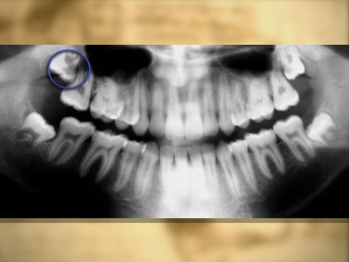
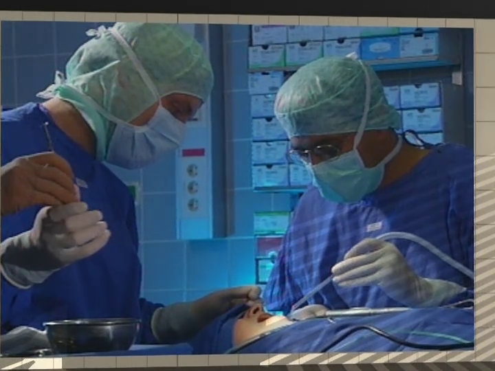
DOI: 10.3290/j.qi.a31204, PubMed ID (PMID): 24570991Pages 239-243, Language: EnglishPeisker, Andre / Kentouche, Karim / Raschke, Gregor Franziskus / Schultze-Mosgau, StefanPatients with hemophilia are at high risk of bleeding following oral surgery. As an X-linked recessive chromosomal bleeding disorder it is very rare in female patients. This is the first described case of management of third molar removal in a female patient suffering from severe hemophilia B. Excellent hemostasis was achieved by following a protocol using defined pre- and postoperative doses of factor IX and local hemostatic measures of collagen fleece, fibrin glue, primary suture, and tranexamic acid solution. Following defined protocols is essential in the management of oral surgery in patients with hemophilia and helps to prevent postoperative hemorrhages.
Keywords: bleeding disorder, factor replacement, female patient, hemophilia B, local measures, third molar removal
Quintessenz Zahnmedizin, 11/2013
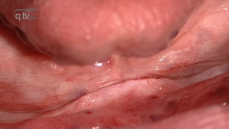
OralchirurgiePages 1395-1398, Language: GermanRoshanghias, Korosh/Peisker, Andre/Raschke, Gregor/Schultze-Mosgau, StefanEin FallberichtExostosen wie der Torus mandibularis werden bei zahnärztlichen Konsultationen häufig als Zufallsbefund diagnostiziert. Der Torus mandibularis stellt eine benigne Knochenhyperplasie dar, die mono-, aber auch gehäuft bilateral zwischen den Prämolaren im Unterkiefer auf der lingualen Seite in Erscheinung tritt. Die Ätiologie des Torus mandibularis ist sowohl genetischer als auch umweltbedingter Natur. Einige ethnische Bevölkerungsgruppen sind in Abhängigkeit von ihrer genetischen Konstitution und ihren Essgewohnheiten stärker betroffen. Klinisch symptomlose Befunde benötigen keine Therapie. Hingegen wird im Rahmen der prothetischen Rehabilitation von betroffenen Patienten oft eine operative Entfernung notwendig. Der Torus mandibularis ist als alternative Donorregion zur Gewinnung von autogenem Knochenmaterial für Augmentationen bei Sinusbodenelevationen im Oberkiefer beschrieben worden. Fallberichte zu Tori mandibulae sind in der Literatur rar. Der vorliegende Fallbericht zeigt eine Therapieoption bei einem symptomatischen Torus mandibularis.
Keywords: Torus mandibularis, Exostose, benigne Knochenhyperplasie, präprothetische Chirurgie
The International Journal of Oral & Maxillofacial Implants, 4/2011
PubMed ID (PMID): 21841985Pages 760-767, Language: EnglishMueller, Cornelia Katharina / Thorwarth, Michael / Schultze-Mosgau, StefanPurpose: The structure of peri-implant soft tissue that is regenerated after flapless and flap surgery has been shown to differ. However, its underlying mechanisms are relatively unknown. The present study sought to identify differences in the inflammatory cell infiltration and expression of gene transcripts during transmucosal healing between the two approaches with two different implant designs.
Materials and Methods: All mandibular premolars were removed from 12 minipigs. One month later, four implants (two NobelReplace Tapered Groovy and two NobelPerfect Groovy, Nobel Biocare) were placed in each quadrant. One quadrant was randomized to flapless insertion, while the other was chosen for flap surgery in each animal. Following 1, 2, 4, and 12 weeks of transmucosal implant healing, biopsy specimens were retrieved from the peri-implant soft tissue according to a standardized procedure to avoid crossover effects. Samples were subjected to a leukocyte count and a gene expression analysis.
Results: When the flapless placement technique was used, leukocyte influx in the peri-implant soft tissue was significantly smaller compared to open surgery for both implant designs. Gene expression analysis revealed significant overexpression of molecules associated with detoxification and reepithelialization in the flapless group. In contrast, myofibroblast-associated gene transcripts were significantly enriched in the flap surgery group.
Conclusions: The present data indicate perpetuation of inflammatory reactions as well as increased fibrotic scar tissue deposition in the peri-implant area following implant placement by the flap approach. Flapless implant insertion results in less inflammation and early reepithelialization, providing the potential for the formation of a fully functioning as well as esthetically preferable peri-implant soft tissue collar.
Keywords: flapless surgery, soft tissue-implant interaction, wound healing
Journal of Craniomandibular Function, 2/2011
Pages 115-126, Language: English, GermanMueller, Cornelia Katharina / Mueller, Andrea / Schultze-Mosgau, StefanObjective: A pathophysiologic link between malocclusions and temporomandibular disorders (TMD) is still being debated. This study correlates pain, joint sounds, and limited opening with a subset of orthodontic findings in different age groups.
Patients and methods: A case-control study employing 697 patients was conducted. Patients were subdivided into three age groups: 1 (n = 297): 7-12 years; 2 (n = 302): 13-18 years; 3 (n = 98): > 18 years. Orthodontic and TMD diagnostics were performed. A multivariate logistic regression approach was used to correlate malocclusions with TMD.
Results: A correlation between mandibular skeletal midline deviation and joint/muscle pain was found in group 1 (P 0.001), 2 (P 0.001) and 3 (P = 0.008). Furthermore, mandibular skeletal midline deviation was an independent predictor of joint sounds (P = 0.009) and limited opening (P = 0.014) in group 3.
Conclusion: Further longitudinal studies employing a group of patients with orthodontic treatment and a control group without treatment are needed to verify the potential of TMD prevention by early treatment for lateral force bite.
Keywords: malocclusion, multivariate analysis, temporomandibular disorders
The International Journal of Oral & Maxillofacial Implants, 2/2006
PubMed ID (PMID): 16634492Pages 225-231, Language: EnglishPetrovic, Ljubinko / Schlegel, Andreas K. / Schultze-Mosgau, Stefan / Wiltfang, JörgPurpose: To find the optimal scaffold for tissue-engineered bone, one approach is to test existing biomaterials on their suitability as scaffolds. In this study, the suitability of different alloplastic and xenogenic biomaterials as scaffolds for ex vivo osteoblast cultivation was investigated. materials and methods: Normal human osteoblast cells were cultured on the surface of bovine collagenous materials, bovine hydroxyapatite, porcine gelatin, synthetic polymer, and collagen-containing bovine hydroxyapatite, and the investigation of proliferation was performed after 24, 72, and 120 hours. Measurement of the differentiation marker alkaline phosphatase and osteocalcin was made after 20 days of incubation.
Results: The obtained data showed significantly higher proliferation and differentiation rates in cells cultivated on collagen-rich biomaterials in comparison to noncollagenous or collagen-poor biomaterials (P .05).
Discussion: In tissue engineering the scaffold should be biocompatible and serve as a proper matrix for the cells to produce the new structural environment of extracellular matrix ex vivo. Collagen supports initial cell attachment and cell proliferation, allowing immature osteogenic cells to differentiate into mature osteoblasts, but collagen may not be the only dominating factor for cell-matrix interaction during ex vivo bone formation.
Conclusion: These data suggest that a 3-dimensional collagen matrix can provide a more favorable environment for the attachment, proliferation, and differentiation of in vitro osteoblastlike cells, at least until the initial stage of differentiation, than noncollagenous biomaterials. (Basic Science)
Keywords: collagen, osteoblasts, tissue engineering
Implantologie, 1/2006
Pages 65-77, Language: GermanSchleier, Peter / Bierfreund, Gerrit / Rabe, Ute / Vollandt, Rüdiger / Moldenhauer, Franziska / Küpper, Harald / Schultze-Mosgau, StefanVerlaufsbeobachtung über einen Zeitraum von zwei Jahren nach Beginn der prothetischen BelastungDie Sinusbodenelevation ist ein etabliertes Verfahren zum Aufbau des atrophierten Oberkieferseitenzahngebiets. Das minimalinvasive Vorgehen nach Summers (interne Sinusbodenelevation) bietet Vorteile beim Patientenmanagement. Die Kombination des Verfahrens mit der endoskopischen Visualisierung wird in der vorliegenden klinischen Untersuchung evaluiert. Bei 30 Patienten (14 Männer, 16 Frauen) wurden insgesamt 52 Implantate mit Hilfe der endoskopisch kontrollierten internen Sinusbodenelevation inseriert. Die durchschnittliche präoperative Höhe des Oberkieferalveolarfortsatzes betrug am Implantationsort im Prämolarenbereich 8 mm und im Molarenbereich 7 mm. Die durchschnittliche Elevationshöhe im Bereich der Prämolaren betrug 3,5 mm und im Molarenbereich 4,5 mm. Es wurde klinisch und mit Hilfe der Resonanzfrequenzanalyse (4 und 12 Wochen postoperativ) eine ausreichende Stabilität (ISQ > 60) der Implantate festgestellt. In der Einheilungsphase gingen insgesamt drei Implantate verloren, was einer Erfolgsquote von 94 % entspricht. Bei Freilegung sowie in der darauf folgenden funktionellen Belastungsphase (Beobachtungszeitraum: zwei Jahre) trat kein weiteres Ereignis ein, das heißt, zum abschließenden Untersuchungszeitpunkt betrug die Erfolgsrate 100 %.
Keywords: Interne Sinusbodenelevation, atrophierter Oberkieferseitenzahnbereich, endoskopische Visualisierung, Resonanzfrequenzanalyse
Implantologie, 4/2005
Pages 379-394, Language: GermanHolst, Stefan / Blatz, Markus B. / Bergler, Michael / Schultze-Mosgau, Stefan / Wichmann, ManfredUmfangreiche In-vitro- und In-vivo-Untersuchungen haben zu zahlreichen Erkenntnissen und Weiterentwicklungen im Bereich der Implantologie geführt. Optimierte Implantatmaterialien, verbesserte chirurgische Techniken und die Berücksichtigung biomechanischer Gesichtspunkte ermöglichen neue Behandlungskonzepte. Während das konventionelle zweizeitige Verfahren eine etablierte Behandlungsmöglichkeit mit sehr guten Erfolgsraten darstellt, gewinnt auch die Sofortversorgung/-belastung von Implantaten verstärkt an Bedeutung. Der Artikel diskutiert wichtige chirurgische und prothetische Aspekte bei der Sofortversorgung zahnloser Patienten. Dabei wird herausgestellt, dass eine exakte Planung und die labortechnische Vorbereitung unabdingbar sind, um eine sowohl funktionell als auch phonetisch und ästhetisch adäquate Versorgung zu gewährleisten.
Keywords: Sofortversorgung, Sofortbelastung, Primärstabilität, strukturierte Implantatoberflächen, biomechanische Gesichtspunkte
Implantologie, 2/2005
Pages 133-144, Language: GermanWichmann, Manfred / Schultze-Mosgau, Stefan / Hamel, Jörg / Bergler, MichaelIm geringgradig atrophierten zahnlosen Kiefer wird der Ersatz der Zahnkronen mittels festsitzender implantatgetragener Suprakonstruktionen angestrebt. Mit zunehmender Länge der klinischen Kronen erreichen derartige Restaurationen jedoch ihre Grenzen. Demgegenüber ist im stark atrophierten zahnlosen Kiefer stets mit dem Ersatz sowohl der Zähne als auch des fehlenden Hart- und Weichgewebes zu rechnen, sodass die Suprakonstruktionen neben den künstlichen Zahnkronen einen zahnfleischfarbenen Anteil aufweisen. Auf Wunsch des Patienten kann der Ersatz von Zähnen sowie von Hart- und Weichgeweben mit festsitzenden (bedingt abnehmbaren) Suprakonstruktionen erfolgen. Diese Suprakonstruktionen werden in der Regel geteilt, um das gewünschte ästhetische Ergebnis zu erzielen. Die aus prothetischer Sicht günstigste Versorgungsform mit dem größten ästhetischen und funktionellen Potenzial ist beim stark atrophierten zahnlosen Kiefer der implantatgetragene, abnehmbare Zahnersatz. Aufgrund der technischen Gestaltungsmöglichkeiten kann nahezu jedes gewünschte ästhetische Ergebnis erreicht werden. Von einer "jugendlichen" Gestaltung bis hin zu einem bewusst individualisierten, altersgemäßen Aussehen sind alle Spielarten möglich. Damit stehen auch für Patienten mit stark atrophiertem zahnlosen Kiefer ästhetische Versorgungsmöglichkeiten zur Verfügung, die von Außenstehenden nicht von den eigenen Zähnen unterschieden werden können. Akzeptiert der Patient eine abnehmbare Suprakonstruktion, können selbst höchste ästhetische Ansprüche befriedigt werden.
Keywords: Atrophierter zahnloser Kiefer, festsitzende implantatgetragene Suprakonstruktion, implantatgetragener, abnehmbarer Zahnersatz, ästhetische Versorgungsmöglichkeiten
Quintessence International, 10/2005
Pages 759-769, Language: EnglishSchultze-Mosgau, Stefan/Blatz, Markus B./Wehrhan, Falk/Schlegel, Karl Andreas/Thorwart, Michael/Holst, StefanThe long-term clinical and esthetic success of implant-supported restorations is determined by osseointegration and optimal remodeling of peri-implant soft tissues. Complications of soft-tissue management are often caused by fibrotic regeneration of oral mucosa after multiple surgical procedures. Knowledge of the proliferative processes in wound healing is necessary to attain adequate soft-tissue conditions. Successful reconstruction of peri-implant soft tissues is feasible even in fibrotic conditions when appropriate surgical techniques are selected. The pleiotropic proliferative cytokine TGF-b is involved in the regulation of all phases of wound healing and tissue remodeling. The isoform TGF-b1 is a cytokine associated with the development of fibrotic tissue. Overexpression of TGF-b1 causes scarring and fibrosis, and results in limited clinical success of intraoral soft-tissue management. Experimental therapeutic approaches with neutralizing antibodies to block TGF-b1 resulted in less scarring and a reduction of fibrosis. Further molecular biologic research of cell-matrix-cytokine interactions in wound healing will provide highly specific antifibrotic therapeutic approaches in the future.
Keywords: fibrosis, implants, soft tissue, transforming growth factor-beta, wound healing
The International Journal of Oral & Maxillofacial Implants, 5/2004
Pages 716-720, Language: EnglishSchlegel, Karl Andreas / Schultze-Mosgau, Stefan / Eitner, Stephan / Wiltfang, Joerg / Rupprecht, StephanKarl Andreas Schlegel, MD, DDS/Stefan Schultze-Mosgau, PhD, MD, DDS/Stefan Eitner, DDS/Joerg Wiltfang, PhD, MD, DDS/Stephan Rupprecht, MD, DDSPurpose: Epithetic solutions in the maxillofacial region are indicated if plastic surgery reconstruction is not a valid option for an extensive defect. The purpose of this study was to examine whether the extraoral implants used provided sufficient retention to be used as anchoring aids.
Materials and Methods: Between November 1999 and September 2002, 33 identical modified Ankylos implants for extraoral anchorage were placed in 10 patients for the fixation of various epitheses in the midfacial (eye, nose) and ear regions in the course of a clinical trial.
Results: Over a follow-up period of 2 to 34 months, all implants remained osseointegrated (as confirmed radiographically), and the implants and epithetic restorations were clinically stable.
Discussion: The results demonstrated that the lasting retention of maxillofacial epitheses provided by implants assures patients that their epitheses are securely fixed.
Conclusion: The demonstrated extraoral implant system not only achieved sufficient osseointegration but also showed good clinical handling and easy fixation possibilities for epithetic anchorage.


