Various cookies are used on our website: We use technically necessary cookies for the purpose of enabling functions such as login or a shopping cart. We use optional cookies for marketing and optimization purposes, in particular to place relevant and interesting ads for you on Meta's platforms (Facebook, Instagram). You can refuse optional cookies. More information on data collection and processing can be found in our privacy policy.
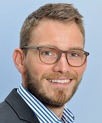
2008-2013: Studium der Zahnheilkunde an der Justus-Liebig-Universität Giessen. 2013: Staatsexamen und Approbation als Zahnarzt. 2013-2014: Wissenschaftlicher Mitarbeiter - Doktorand, Poliklinik für Zahnärztliche Prothetik, Universitätsklinikum der Justus-Liebig-Universität Giessen. 2015: Promotion. 2015-2016: Vorbereitungsassistent bei einem niedergelassenen Zahnarzt im Münsterland. seit 2016: Wissenschaftlicher Mitarbeiter - Zahnarzt, Poliklinik für Zahnärztliche Prothetik, Universitätsklinikum der Justus-Liebig-Universität Giessen. 2018: Spezialist für Prothetik der DGPro. 2017-2019:
Masterstudiengang Zahnmedizinische Prothetik an der Universität Greifswald
Events
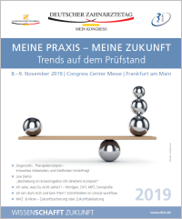
Deutscher Zahnärztetag 2019
8. Nov 2019 — 9. Nov 2019Congress Center Messe Frankfurt
Speakers: Karl-Ludwig Ackermann, Sarah Al-Maawi, Kurt Werner Alt, Jassin Arnold, Thomas Attin, Mustafa Ayna, Anna Greta Barbe, Ingo Baresel, Jens Baresel, Tobias Bauer, Ursula Becker, Wilfried Beckmann, Christoph Benz, Lisa Bitterich, Dirk Bleiel, Uwe Blunck, Martin Boost, Andreas Braun, Anne Bredel-Geissler, Wolfgang Buchalla, Egon Burian, Sebastian Bürklein, Iain L. C. Chapple, Wolfgang Christian, Fabian Cieplik, Bettina Dannewitz, Monika Daubländer, Sybille David-Hebgen, Isabel Deckwer, James Deschner, Annika Döding, Christof Dörfer, Heike Dyrna, Norbert Engel, Peter Engel, Susanne Fath, Michael Frank, Roland Frankenberger, Rene Franzen, Cornelia Frese, Tobias Fretwurst, Michael Gahlert, Roland Garve, Werner Geurtsen, Shahram Ghanaati, Christiane Gleissner, Ulrike Gonder, Werner Götz, Dominik Groß, Knut A. Grötz, Martin Guffart, Norbert Gutknecht, Cornelius Haffner, Thorsten Halling, Frederic Hermann, Carlos Herrera-Vizcaino, Tim Hilgenfeld, Jürgen Hoffmann, Martin Hoffmann, Fabian Huettig, Alfons Hugger, Christine Hutschenreuter, Bruno Imhoff, Silke Jacker-Guhr, Søren Jepsen, A. Rainer Jordan, Alexander Jürchott, Bärbel Kahl-Nieke, Peer W. Kämmerer, Philipp Kanzow, Nele Kettler, Christian Kirschneck, Lydia Kogler, Bernd Kordaß, Franz-Josef Kramer, Norbert Krämer, Felix Krause, Matthis Krischel, Joachim Krois, Christina Kühne, Conrad Kühnöl, Bernd Lapatki, Silke Lehmann-Binder M.Sc., Christian Leonhardt, Ivona Leventic, Daniel Lindel, Jörg Alexander Lisson, Ulrike Lübbert, Elmar Ludwig, Anne-Katrin Lührs, Michael Lüpke, Frank Georg Mathers, Wibke Merten, Georg Meyer, Wolfram Misselwitz, Karin Mölling, Mhd Said Mourad, Dietmar Friedrich Müller, Moritz Mutschler, Katja Nickel, Nicole Nicklisch, Ina Nitschke, Olaf Oberhofer, Karina Obreja, Dietmar Oesterreich, Rebecca Otto, Simon Peroz, Peter Pospiech, Florian Probst, Monika Probst, Michael Rädel, Sven Reich, Katharina Reichenmiller, Katharina Reinecke, Daniel R. Reißmann, Bernd Reiss, Stefan Ries, Christiane Rinnen, Katharina Röher, Jerome Rotgans, Uwe Rudol, Michael Rumpf, Heidrun Schaaf, Claudia Schaller, Karina Schick, Ulrich Schiffner, Maximiliane Amelie Schlenz, Alexander Schmidt, Mathias Schmidt, Andrea-Maria Schmidt-Westhausen, Julian Schmoeckel, Wolfgang Schneider, Sigmar Schnutenhaus, Holger Schön, Andreas Schulte, Nelly Schulz-Weidner, Karola Schulze, Ralf Schulze, Falk Schwendicke, Thomas A. Schwenk, Andreas Simka, Ralf Smeets, Önder Solakoglu, David Sonntag, Hansmartin Spatzier, Benedikt Spies, Norbert Staab, Sabine Steding, Angela Stillhart, Marcus Stoetzer, Hendrik Terheyden, Andrea Thumeyer, Marin Vodanovic, Kai Voß, Maximilian Voß, Wolfgang Wahlster, Michael Walter, Sandra Weber, Almut Johanna Weigel, Paul Weigl, Michael Weiss, Hans-Jürgen Wenz, Johannes-Simon Wenzler, Christian Wesemann, Jens Westemeier, Lotta Westphal, Matthias Widbiller, Annette Wiegand, Horst Willeweit, Karl Frederick Wilms, Sandra Windecker, Michael M. Wolf, Anne Wolowski, Bernd Wöstmann, Sylvia Wuttig
Quintessenz Verlags-GmbH
This author's journal articles
The International Journal of Prosthodontics, Pre-Print
DOI: 10.11607/ijp.8843, PubMed ID (PMID): 3853614822. Mar 2024,Pages 1-19, Language: EnglishSchmidt, Alexander / Berschin, Cara / Wöstmann, Bernd / Schlenz, Maximiliane Amelie
Purpose: To update data on the transfer accuracy of digital implant impressions by using a
coordinate-based analysis, latest intraoral scanners (IOSs) were investigated in an established
clinical close model set-up. Materials and Methods: An implant master model (IMM) of the
maxilla with four implants in the posterior area (#14/#24 and #16/#26) and a reference cube
was scanned with four different IOS (i700 (Medit), Primescan (Dentsply Sirona), Trios 4 and
Trios 5 (3Shape) ten times each. Datasets were compared with a reference dataset of IMM
that was generated with x-ray computed tomography in advance. 3D deviations for the
implant-abutment-interface points (IAIPs) were calculated. Statistical analysis was performed
by multifactorial ANOVA (p < .05). Results: Overall deviations for trueness (mean) ±
precision (SD) of the IAIPs ranged from 88±47 μm for the Primescan, followed by 112±57
μm for the i700, 121±42 μm for the Trios 4 and 124±43 μm for the Trios 5 with decreasing
accuracy along the scan path. For trueness, one significant difference between the Primescan
and the T4 was detected for one implant position. For precision, no significant differences
were noticed. Conclusions: Although the latest IOS showed a significant improvement in
transfer accuracy, the accumulating deviation along the scan path is not yet resolved.
Considering the Trios system, the innovation seems to be limited as no improvement could be
detected between Trios 4 and 5.
Eine randomisierte kontrollierte klinisch-volumetrische Studie
Die Bindegewebetransplantation zählt heute zu den Standardverfahren zur Kompensation von Volumendefiziten bei Sofortimplantation. Neue Biomaterialien wie azelluläre Matrices könnten die Gewinnung autogenen Gewebes auf ein absolut notwendiges Minimum reduzieren und damit die Häufigkeit und das Ausmaß postoperativer Beschwerden verringern. Die vorliegende randomisierte Studie verglich die klinischen Therapieergebnisse von Sofortimplantationen in der Oberkieferfront, bei denen sowohl eine knöcherne Augmentation als auch eine Weichgewebeverdickung durchgeführt wurde. Neben anorganischem bovinem Knochenmaterial (ABBM) kam entweder ein Bindegewebeersatz aus porciner Dermis, eine azelluläre dermale Matrix (ADM) oder ein autogenes Bindegewebetransplantat (BGT) zum Einsatz. An der Studie nahmen 20 Patienten (11 Männer, 9 Frauen) mit einem Durchschnittsalter von 48,9 Jahren (21−72 Jahre) teil. Die Zuordnung der Studienteilnehmer zu der Test- (ADM) bzw. Kontrollgruppe (BGT) geschah nach dem Zufallsprinzip. Der Zahnextraktion folgte die sofortige Implantatinsertion. Der bukkale Knochen wurde mit ABBM augmentiert. Eine ADM oder ein BGT diente zur Verdickung des bukkalen Weichgewebes und somit zur Kompensation des erwarteten Verlustes von bukkalem Volumen. Die klinische und volumetrische Nachuntersuchung fand 12 Monate nach Implantatinsertion statt. Bei allen Implantaten hatte eine Osseointegration stattgefunden und die prothetische Versorgung befand sich in situ. Ein Jahr postoperativ betrug die durchschnittliche, linear gemessene Volumenveränderung −0,55 ± 0,32 mm (ADM) bzw. −0,60 ± 0,49 mm (BGT). Patienten der ADM-Gruppe beklagten signifikant weniger postoperative Beschwerden. Bei Sofortimplantation mit Augmentation von Hart- und Weichgewebe führten Ersatzmaterialien und autogene Bindegewebetransplantate zu ähnlichen klinischen Ergebnissen hinsichtlich der gemessenen Volumenveränderungen. Die Anwendung von Ersatzmaterial führte zu signifikant weniger postoperativer Morbidität.
Manuskripteingang: 07.01.2021, Annahme: 14.04.2021
Keywords: Sofortimplantation, Weichgewebeverdickung, Bindegewebetransplantat, azelluläre dermale Matrix, anorganisches bovines Knochenmaterial
Connective tissue grafts have become a standard for compensating horizontal volume loss in immediate implant placement. The use of new biomaterials like acellular matrices may avoid the need to harvest autogenous grafts, yielding less postoperative morbidity. This randomized comparative study evaluated the clinical outcomes following extraction and immediate implant placement in conjunction with anorganic bovine bone mineral (ABBM) and the use of a porcine acellular dermal matrix (ADM) vs an autogenous connective tissue graft (CTG) in the anterior maxilla. Twenty patients (11 men, 9 women) with a mean age of 48.9 years (range: 21 to 72 years) were included in the study and randomly assigned to either the test (ADM) or control (CTG) group. They underwent tooth extraction and immediate implant placement together with ABBM for socket grafting and either ADM or CTG for soft tissue augmentation. Twelve months after implant placement, the cases were evaluated clinically and volumetrically. All implants achieved osseointegration and were restored. The average horizontal change of the ridge dimension at 1 year postsurgery was -0.55 ± 0.32 mm for the ADM group and -0.60 ± 0.49 mm for the CTG group. Patients of the ADM group reported significantly less postoperative pain. Using xenografts for hard and soft tissue augmentation in conjunction with immediate implant placement showed no difference in the volume change in comparison to an autogenous soft tissue graft, and showed significantly less postoperative morbidity.
The International Journal of Prosthodontics, 6/2021
DOI: 10.11607/ijp.6233Pages 756-762, Language: EnglishSchmidt, Alexander / Benedickt, Christopher R / Schlenz, Maximiliane A / Rehmann, Peter / Wöstmann, Bernd
Purpose: To evaluate the accuracy (trueness and precision) achievable with four intraoral scanners (IOSs) and different preparation geometries.
Materials and methods: A model of a maxillary arch with different preparation geometries (onlay, inlay, veneer, full-crown) served as the reference master model (RMM). The RMM was scanned 10 times using four commonly used IOSs (Trios 2 [TR], 3Shape; Omnicam [OC], Dentsply Sirona; True-Definition [TD], 3M ESPE; and Primescan [PS], Dentsply Sirona). Scans were matched using a 3D measurement software (Inspect 2019, GOM) and a best-fit algorithm, and the accuracy (trueness and precision) of the preparation types of the scanning data was evaluated for positive and negative deviations separately. All data were subjected to univariate analysis of variance using SPSS version 24 (IBM).
Results: Mean (± SD) positive deviations ranged from 4.6 ± 0.7 μm (TR, veneer) to 25.9 ± 2.4μm (OC, full crown). Mean negative deviations ranged from -7.2 ± 0.6 μm (TR, veneer) to -26.4 ± 3.8 μm (OC, full crown). There were significant differences (P < .05) in terms of trueness and precision among the different IOSs and preparation geometries.
Conclusion: The transfer accuracy of simple geometries was significantly more accurate than those of the more complex prosthetic geometries. Overall, however, the IOSs used in this study yielded results that were clinically useful for the investigated preparation types, and the mean positive and negative deviations were in clinically acceptable ranges.
Insbesondere in der Implantologie eröffnet der intraorale Scan, neben der alleinigen Funktion einer Abformung, die Möglichkeit zur Implementierung neuer Behandlungskonzepte. Bereits in der Beratungs- und Planungsphase können so in Verbindung mit dreidimensionalen Röntgendaten Möglichkeiten, Grenzen und Risiken der Implantatversorgung erläutert und in einem prothetisch-chirurgischen Behandlungskonzept festgelegt werden, welches zu vorhersagbareren Behandlungsergebnissen führt. Jedoch müssen die heute noch bestehenden Limitationen der Ganzkieferversorgungen in Bezug auf die dreidimensionale Übertragung der Implantatposition von der Mundhöhle auf ein Modell beachtet werden, weshalb indikationsabhängig auch kombiniert digital-analoge Versorgungskonzepte in Betracht gezogen werden sollten. Durch die kontinuierliche Weiterentwicklung der Scansysteme ist jedoch zukünftig damit zu rechnen, dass auch hier die digitale Abformung die etablierten analogen Behandlungsverfahren ersetzen wird.
Manuskripteingang: 18.06.2021, Annahme: 11.08.2021
Keywords: digitale Abformung, Implantatabformung, konventionelle Abformung, intraorale Scanner, Abformgenauigkeit, digitale Implantatplanung
Aim: Dental research involves variations between actual and reference datasets of master models to determine the metric accuracy through transfer accuracy tests. Various methods of measurement are used to analyze the results, which are often subjected to direct comparisons. Hence, the aim of the present study was to analyze the influence and effect on results of different methods of digital data analysis, being coordinate-based analysis (CBA) and best-fit superimposition analysis.
Materials and methods: A model with four implants and a reference cuboid was digitized through computed tomography (CT), which served as the master model. Ten implant impressions were made using a Trios (3Shape) intraoral scanner, and three different scan bodies (nt-trading, Kulzer, and Medentika) were used. The deviations between the master model and the digital impressions were analyzed using CBA and best-fit superimposition analysis. Statistical analysis was performed using SPSS 25.
Results: The deviations in the CBA and best-fit superimposition analysis ranged from 0.088 ± 0.012 mm (mean ± SE; Medentika, 14) to 0.199 ± 0.021 mm (Kulzer, 26), and from 0.042 ± 0.010 mm (Medentika, 16) to 0.074 ± 0.006 mm (Kulzer, 16), respectively. Significant differences were observed between the implant positions in the CBA and the digital measurements at each implant position, whereas the best-fit analysis showed no significant difference between the scan bodies and implant positions.
Conclusion: CBA displays an advantage over best-fit superimposition analysis in the detection of possible influencing factors for primarily scientific purposes. However, a global analysis and visualization of angles and torsions is difficult, for which a best-fit evaluation is needed. However, a best-fit analysis better represents the clinical try-in. It is associated with the risk that possible disturbing factors and resulting errors might be leveled out and their identification camouflaged.
Keywords: dimensional measurement accuracy, accuracy, trueness, precision, intraoral scanner, digital dentistry, implant impression, best-fit analysis
Purpose: To assess the absolute linear distances of three different intraoral scan bodies (ISBs) using an intraoral scanner compared to a conventional impression in a common clinical model setup with a gap and a free-end situation in the maxilla.
Materials and methods: An implant master model with a reference cube was digitized using x-ray computed tomography and served as the reference file. Digital impressions (TRIOS, 3Shape) were taken using three different ISB manufacturers: NT Trading, Kulzer, and Medentika (n = 10 per group). Conventional implant impressions were taken for comparison (n = 10). The conventional models were digitized, and all models (digital and conventional) were superimposed with the reference file to obtain the 3D deviations for the implant-abutment-interface points (IAIPs). Results for linear deviation (trueness and precision) were analyzed using pairwise comparisons (P < .05; SPSS version 25). For precision, a two-way factorial mixed ANOVA was used.
Results: The deviations for trueness (mean) ± precision (SD) of the IAIPs ranged as follows: FDI region 14 = 0.106 ± 0.050 mm (Medentika) to 0.134 ± .026 mm (NT Trading); region 16 = 0.108 ± 0.046 mm (conventional) to 0.164 ± 0.032 mm (NT Trading); region 24 = 0.111 ± 0.050 mm (conventional) to 0.191 ± 0.052 mm (Medentika); region 26 = 0.086 ± 0.040 mm (conventional) to 0.199 ± 0.066 mm (Kulzer). There were significant differences for trueness between all digital and conventional impression techniques. For precision, only two significant differences in two implant regions (14, 24) were observed.
Conclusion: Longer scanning paths resulted in higher deviations of the implant position in digital impressions. Due to algorithms implemented in the software, errors resulting from the different scan bodies may be reduced during the alignment process of the IOS in clinical practice.
Objectives: The aim of this retrospective pilot study was to analyze the clinical performance of computer-engineered complete dentures (CECDs) in edentulous patients regarding survival and maintenance.
Method and materials: For this retrospective analysis, data from 10 patients who received CECD treatment in each arch (Digital Denture, Ivoclar Vivadent) between 2015 and 2016 were analyzed. The following aspects were assessed: number of appointments required for treatment, number of interventions during the initial (≤ 4 weeks after insertion) and functional periods (> 4 weeks after insertion), and survival. Additionally, whether these aspects were influenced by function or esthetics, the arch, or recall participation was assessed. Poisson regression models were used for the statistical analysis (P .05).
Results: All CECDs survived the observation period of 2.54 ± 0.48 years. More than four appointments were required for treatment (mean ± standard deviation, 4.6 ± 0.7), mainly for esthetic concerns. An average of 1.7 ± 0.05 appointments during the initial period and 2.07 ± 0.32 during the functional period were noted as a consequence of functional concerns. During both periods, the major reason for intervention was removal of pressure spots. Relining was required in 40% of the CECDs, and fracture of the denture base occurred in two CECDs.
Conclusions: Within the limitations of this retrospective pilot study, the CECDs showed acceptable clinical performance in terms of survival and maintenance. Nevertheless, transferring more information about the patient from the dental practice to the dental laboratory might reduce the number of appointments for treatment and avoid technical complications such as fractures of the denture base.
Keywords: CAD/CAM complete denture, computer-engineered complete dentures, maintenance, survival