Auf unserer Website kommen verschiedene Cookies zum Einsatz: Technisch notwendige Cookies verwenden wir zu dem Zweck, Funktionen wie das Login oder einen Warenkorb zu ermöglichen. Optionale Cookies verwenden wir zu Marketing- und Optimierungszwecken, insbesondere um für Sie relevante und interessante Anzeigen bei den Plattformen von Meta (Facebook, Instagram) zu schalten. Optionale Cookies können Sie ablehnen. Mehr Informationen zur Datenerhebung und -verarbeitung finden Sie in unserer Datenschutzerklärung.
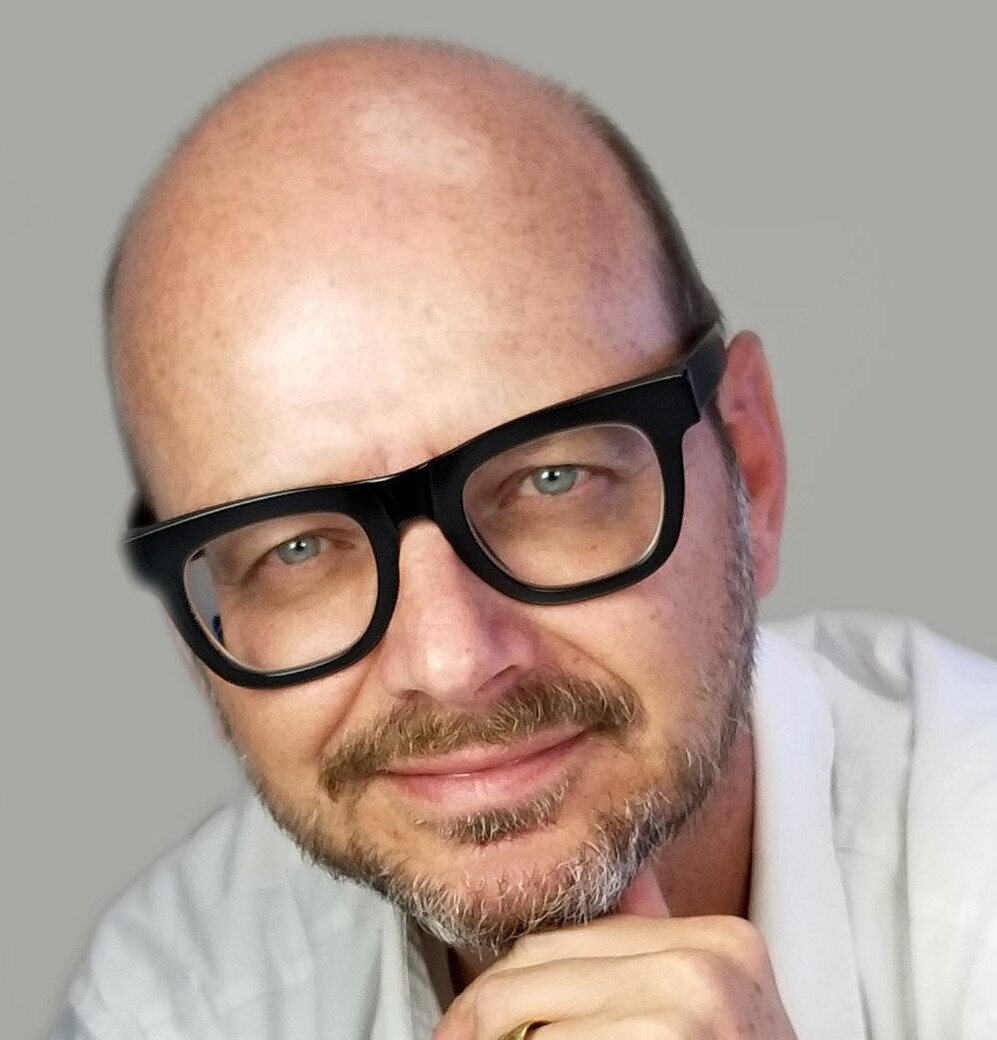
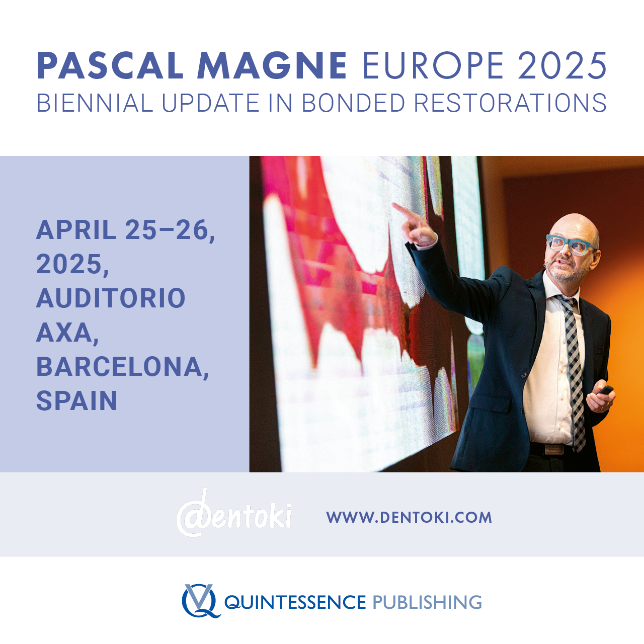
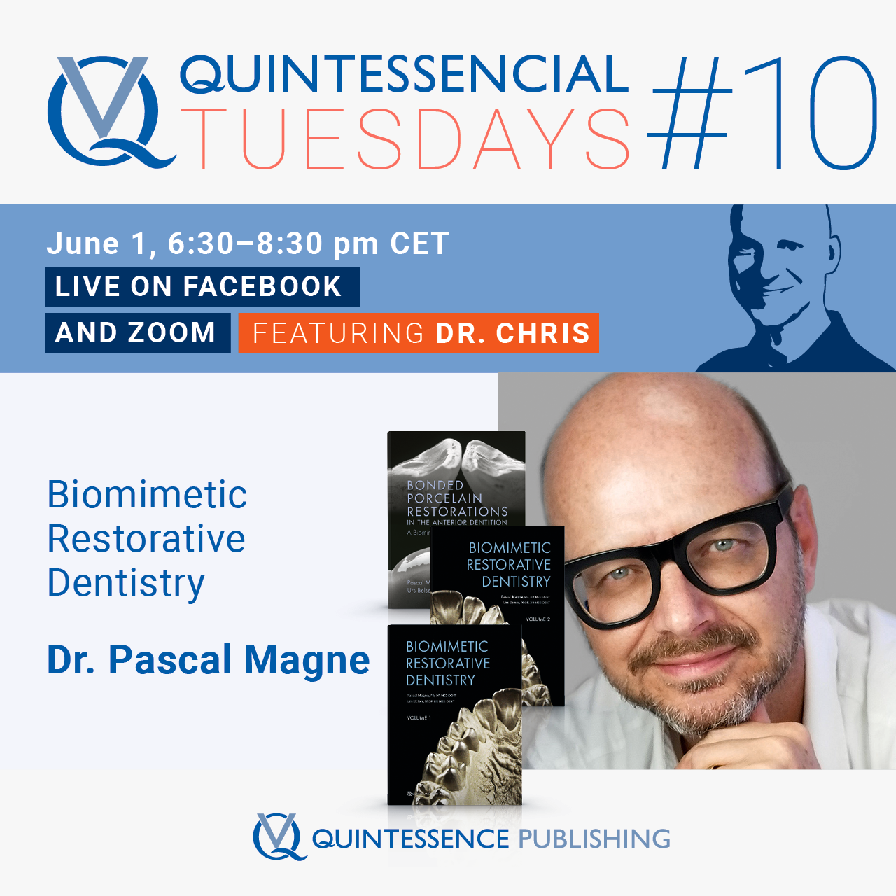
Dr. Pascal Magne ist vor kurzem als Direktor des Center for Education and Research in Biomimetic Restorative Dentistry (CER BRD) zum Beverly Hills Dental Lab gewechselt, nachdem er eine lange und herausragende Karriere in der zahnmedizinischen Lehre und Forschung zurückgelegt hat. Nach Abschluss seines Studiums der Zahnmedizin an der Universität Genf im Jahr 1989 wurde ihm eine Stelle als Dozent an der zahnmedizinischen Fakultät angeboten, während er gleichzeitig praktizierte und sich in festsitzender Prothetik und anderen technischen Fertigkeiten weiterbildete. Parallel dazu promovierte er 1992 (DMD) und 2002 (Ph.D.), letzteres in Verbindung mit der Mitarbeit an dem Buch Bonded Porcelain Restorations in the Anterior Dentition: A Biomimetic Approach mit seinem Mentor, Professor Urs Belser, welches in 12 Sprachen übersetzt wurde. „Das biomimetische Prinzip bezieht sich auf den Einsatz von Technologie und Dentalmaterialien, um die Struktur und Funktion der Zähne zu erhalten und nachzubilden“, erklärt er.
Von 1997 bis 1999 forschte er an der University of Minnesota School of Dentistry an zahnmedizinischen Innovationen, bevor er nach Genf zurückkehrte. Seine Forschungsergebnisse wurden in zahlreichen Fachzeitschriften veröffentlicht und mit Preisen ausgezeichnet.
Bereits vor dem Abschluss seines Zahnmedizinstudiums war Dr. Magne von den beeindruckenden Fotos der naturgetreuen Keramikrestaurationen seines älteren Bruders Michel fasziniert, der als Zahntechniker in einer Zahnarztpraxis arbeitete. 1991 begann Pascal mit Claude Sieber zusammenzuarbeiten, der ein neues, von Vita entwickeltes Keramikmaterial verwendete. Das war der Wendepunkt. „Zum ersten Mal konnte ich die Keramik eines Meisters im Mund meiner eigenen Patienten sehen“, sagt er.
Im Jahr 2004 wurde Pascal als Don und Sybil Harrington Professor für ästhetische Zahnmedizin an die Herman Ostrow School of Dentistry, University of Southern California berufen. Während seiner Amtszeit an der Ostrow School of Dentistry hat Dr. Magne eine Vielzahl von Lehrplaninnovationen in den Bereichen Biomimetik, minimalinvasive Zahnmedizin und Klebetechniken vorangetrieben. Seine wissenschaftliche Tätigkeit brachte über 100 von Experten begutachtete wissenschaftliche Beiträge und über 200 Fortbildungsprogramme im In- und Ausland hervor. Als vorklinischer Leiter (2012-2020) des Moduls Zahnmedizinische Morphologie, Funktion und Ästhetik in Ostrow hat Dr. Magne außerdem einen innovativen Ansatz für die Lehre der zahnmedizinischen Anatomie und Morphologie entwickelt.
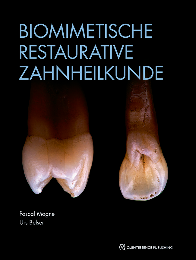
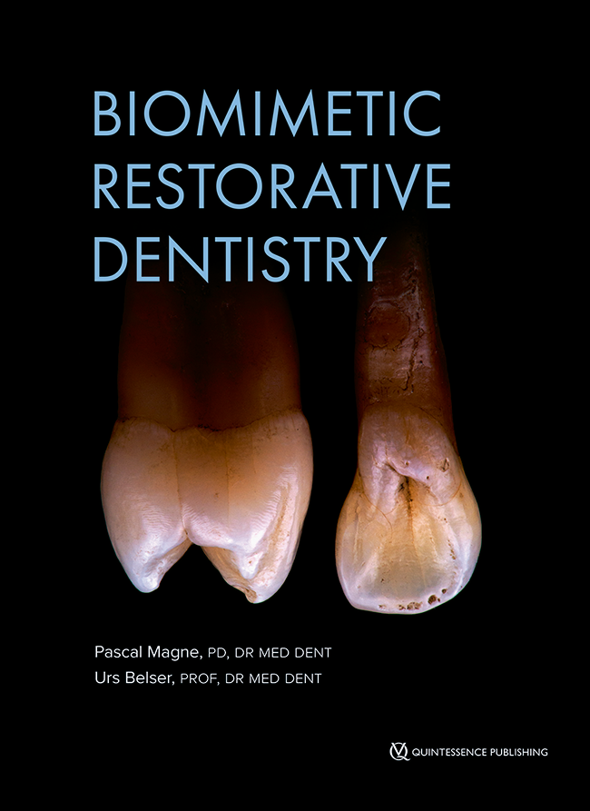
Im Jahr 2021 veröffentlichte Dr. Magne die zweite Auflage seines Bestsellers mit dem Titel Biomimetic Restorative Dentistry. Es ist bereits in mehrere Sprachen übersetzt worden. Nach 33 Jahren in der akademischen Welt hat Dr. Magne im Jahr 2022 die spannende Möglichkeit der CER BRD-Fortbildungsprogramme, angeboten vom Beverly Hills Dental Laboratory, begeistert angenommen.
2. Auflage 2022 Buch Hardcover; Two-volumes book with slipcase; 21 x 28 cm, 888 Seiten, 2500 Abbildungen Sprache: Englisch Kategorien: Restaurative Zahnheilkunde, Ästhetische Zahnheilkunde Artikelnr.: 23491 ISBN 978-0-86715-572-3 QP USA
10. oct 2024 — 12. oct 2024Palacio de congresos de Sevilla, Sevilla, España
Ponentes: Sam Alawie, Markus Blatz, Marco Ferrari, Henriette Terezia Lerner, Antonio Liñares, Arturo Llobell, Ignazio Loi, Michel Magne, Pascal Magne, Francesco Mangano, Eitan Mijiritsky, Nazariy Mykhaylyuk, Mutlu Özcan, George Papavasiliou, Alessandro Pozzi, Marta Revilla-León, Vygandas Rutkūnas, Teresa Sierpińska, Jacobo Somoza, Miguel Stanley, Francesca Vailati
SEPES - Sociedad Española de Prótesis Estomatologías y Estética
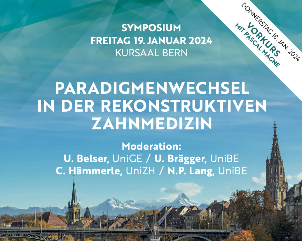
Paradigmenwechsel in der Rekonstruktiven Zahnmedizin
Auswirkungen der Fortschritte in Dentinadhäsion, Implantologie, Digitalisierung & Materialentwicklung für die tägliche Praxis18. ene 2024 — 19. ene 2024Congress Center, Kursaal Bern, Bern, Suiza
Ponentes: Urs C. Belser, Urs Brägger, Daniel Buser, Didier Dietschi, Vincent Fehmer, Ueli Grunder, Christoph Hämmerle, Ronald Jung, Niklaus P. Lang, Pascal Magne, Konrad H. Meyenberg, Frauke Müller, Irena Sailer, Martin Schimmel, Anton Sculean, Nicola Zitzmann
Bern Congress Organiser
Pascal Magne - Europe 2023
One pass. Two game-changing days1. jul 2023 — 2. jul 2023, 9:00 — 18:00 (MEZ)Hotel Villa Pamphili Roma, Rome, Italia
Ponentes: Pascal Magne
dentoki.com
Spear Summit
Growing Together27. abr 2023 — 29. abr 2023, 13:00 — 16:45 JW Marriott Phoenix Desert Ridge, Phoenix, Estados Unidos de América
Ponentes: Edward P. Allen, Álvaro Blasi, Gonzalo Blasi, Ignacio Blasi, Rebecca Bockow, Gary DeWood, Darin Dichter, Didier Dietschi, Michael Gunson, Gregg Kinzer, Pascal Magne, George Mandelaris, Ricardo Mitrani, Amy Morgan, Jason Smithson, Frank Spear, Eric Van Dooren, Robert Winter
Spear Education
The 14th International Symposium on Periodontics and Restorative Dentistry (ISPRD)
9. jun 2022 — 12. jun 2022Boston Marriott Copley Place, Boston, MA, Estados Unidos de América
Ponentes: Tara Aghaloo, Edward P. Allen, Evanthia Anadioti, Wael Att, Vinay Bhide, Markus Blatz, Scotty Bolding, Lorenzo Breschi, Jeff Brucia, Daniel Buser, Luigi Canullo, Daniele Cardaropoli, Stephen J. Chu, Donald Clem, Christian Coachman, Lyndon F. Cooper, Daniel Cullum, Lee Culp, José Carlos Martins da Rosa, Sergio De Paoli, Marco Degidi, Nicholas Dello Russo, Serge Dibart, Joseph P. Fiorellini, Mauro Fradeani, Stuart J. Froum, David Garber, Maria L. Geisinger, William Giannobile, Luca Gobbato, Ueli Grunder, Galip Gürel, Chad Gwaltney, Christoph Hämmerle, Robert A. Horowitz, Marc Hürzeler, David Kim, Gregg Kinzer, Christopher Köttgen, Ina Köttgen, Purnima S. Kumar, Burton Langer, Lydia Legg, Pascal Magne, Kenneth A. Malament, Jay Malmquist, George Mandelaris, Pamela K. McClain, Michael K. McGuire, Mauro Merli, Konrad H. Meyenberg, Craig M. Misch, Julie A. Mitchell, Marc L. Nevins, Myron Nevins, Michael G. Newman, Miguel A. Ortiz, Jacinthe M. Paquette, Stefano Parma-Benfenati, Michael A. Pikos, Giulio Rasperini, Pamela S. Ray, Christopher R. Richardson, Isabella Rocchietta, Marisa Roncati, Marco Ronda, Paul S. Rosen, Maria Emanuel Ryan, Irena Sailer, Maurice Salama, David M. Sarver, Takeshi Sasaki, Todd Scheyer, Massimo Simion, Michael Sonick, Sergio Spinato, Dennis P. Tarnow, Lorenzo Tavelli, Douglas A. Terry, Tiziano Testori, Carlo Tinti, Istvan Urban, Hom-Lay Wang, Robert Winter, Giovanni Zucchelli
Quintessence Publishing Co., Inc. USA
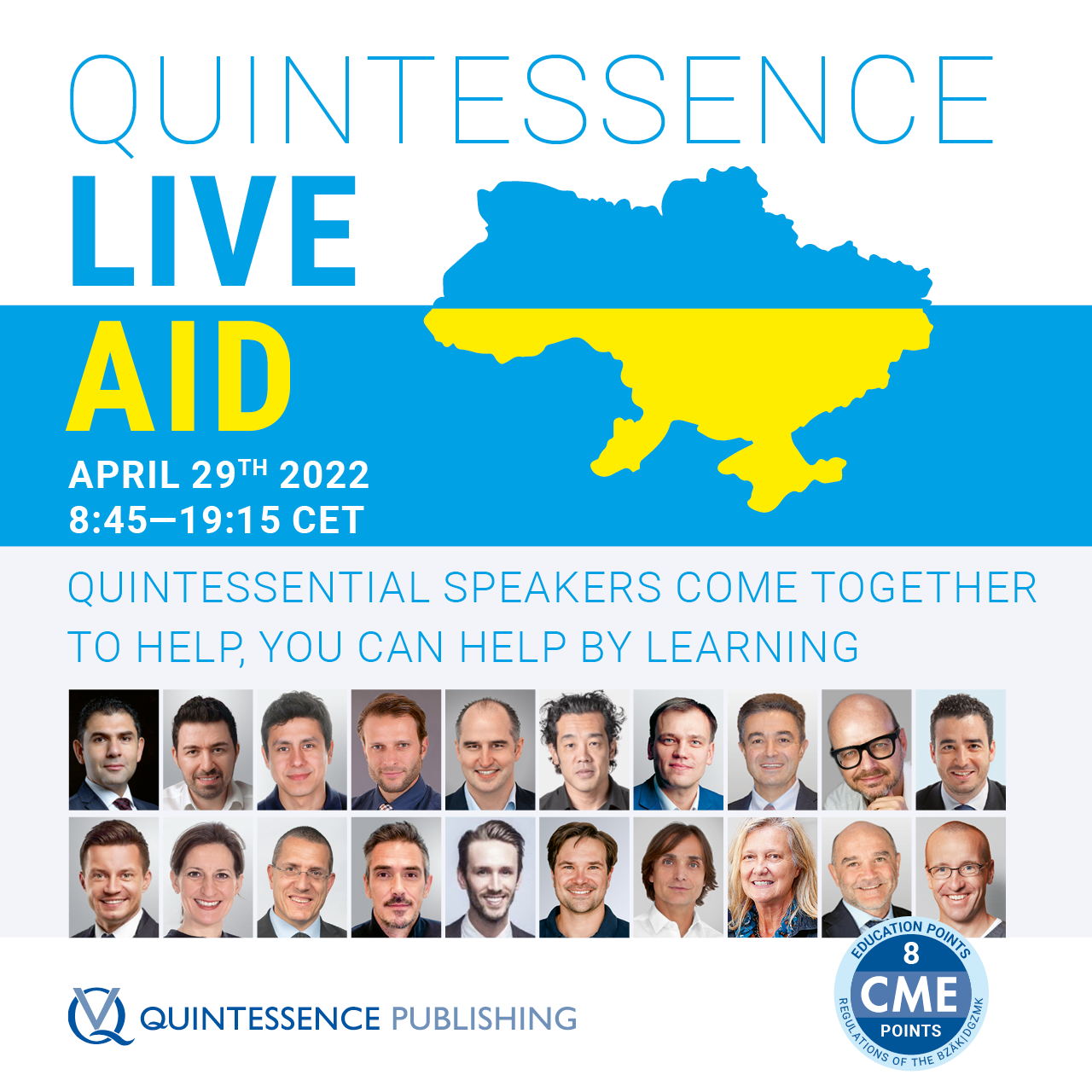
Quintessence Live Aid
Quintessential Speakers from around the world offer a Day of Education that will help those in need29. abr 2022Zoom
Ponentes: Wael Att, Maxim Belograd, Victor Clavijo, Christian Coachman, Stefan Fickl, Ronaldo Hirata, Fouad Khoury, Tomas Linkevičius, Pascal Magne, Nazariy Mykhaylyuk, José M. Navarro, Irena Sailer, Anton Sculean, Kyle Stanley, Miguel Stanley, Markus Tröltzsch, Istvan Urban, Débora R. Vilaboa, Giovanni Zucchelli, Otto Zuhr
Eine retrospektive Studie mit bis zu 27 Jahren Nachbeobachtung
Ziel: In dieser retrospektiven Studie sollte die (technische und biologische) Langzeitbewährung ausgedehnter keramischer Oberkieferfrontzahnveneers mit einer inzisalen Dicke von mehr als 2 mm untersucht werden.
Material und Methoden: Patienten, die zwischen 1990 und 2003 an der Universitätszahnklinik Genf von demselben Zahnarzt mit ausgedehnten Keramikveneers versorgt worden waren, wurden zu einer Nachuntersuchung eingeladen. Von den 37 identifizierten Patienten erklärten sich zehn Patienten mit 50 Veneers zu der Nachuntersuchung bereit und wurden inkludiert. Die Überlebensraten sowie die technischen und biologischen Ergebnisse wurden durch eine klinische Untersuchung bestimmt (anhand der modifizierten United States Public Health Services-Kriterien). Zusätzlich wurden die Patientenakten ausgewertet, um die Patientendaten und alle Komplikationsereignisse zu erfassen. Schließlich wurden Patient-R eported Outcomes (PRO) zu den Aspekten Ästhetik, Funktion und Phonetik, Kaufunktion, Zahnempfindlichkeit und Bereitschaft zu einer Ersatzversorgung im Fall eines Restaurationsversagens mithilfe visueller Analogskalen erhoben. Die Daten wurden deskriptiv ausgewertet. Die Analyse des Restaurationsüberlebens und der Komplikationsereignisse erfolgte mittels Kaplan-Meier-Schätzer.
Ergebnisse: Die Überlebensrate der Veneers betrug 96 % nach im Mittel 20,7 ± 3,7 Jahren in Funktion. Die Rate technischer Komplikationen lag bei 30 %. Diese Komplikationen bestanden in zwei Veneerverlusten, neun reparablen Frakturen, drei Rissbildungen und einer Veneerverlagerung durch Zahnunfall. Es fanden sich weder über Kreideflecken hinausgehende Kariesläsionen noch endodontische Komplikationen dokumentiert. Die PRO bezüglich Zufriedenheit mit der Ästhetik und der Phonetik waren sehr gut.
Schlussfolgerungen: Im Rahmen dieser retrospektiven Studie erwiesen sich ausgedehnte Keramikveneers, gemessen an den klinischen Ergebnissen und der Patientenzufriedenheit, als langfristig erfolgreiche Versorgungsmöglichkeit.
Palabras clave: adhäsive Zahnmedizin, Ästhetik, Keramik, Prothetik, restaurative Zahnmedizin, Veneers
A retrospective study with an up to 27-year follow-up
Aim: The aim of the present retrospective study was to evaluate the long-term results, including technical and biologic outcomes, of maxillary extended porcelain veneers with an incisal edge thickness above 2 mm.
Materials and methods: Patients treated with extended porcelain veneers performed by a single clinician at University of Geneva between 1990 and 2003 were identified and invited to an examination. Of the 37 identified patients, 10 patients with 50 veneers agreed to be examined and were included. A clinical examination was performed to assess survival rates as well as technical and biologic outcomes (modified United States Public Health Services criteria). Patient records were also reviewed to retrieve patient and reconstruction data and every complication event. Patient-reported outcome measures (PROMs) were evaluated using a visual analog scale to measure esthetic satisfaction, functional and phonetic comfort, masticatory improvement, tooth sensitivity, and acceptance of restoration replacement in case of failure. Data were descriptively analyzed, and Kaplan-Meier survival estimators were computed for survival rates and complication events.
Results: The survival rate of the veneers was 96% after a mean follow-up of 20.7 ± 3.7 years in function. The technical complication rate amounted to 30%, including two failures, nine repairable fractures, three cracks, and one displacement due to trauma. No cavitated caries lesions or endodontic complications were registered. PROMs were very high for esthetic satisfaction and phonetic comfort.
Conclusions: Within the limitations of the present retrospective study, extended porcelain veneers appear to be a successful long-term treatment option in terms of clinical outcomes and patient satisfaction.
Palabras clave: adhesive dentistry, esthetics, ceramics, prosthodontics, restorative dentistry, veneers
Acceso libre Sólo en líneaResearchDOI: 10.3290/j.jad.b4051477, ID de PubMed (PMID): 3709705525. abr 2023,Páginas 99-106, Idioma: InglésMagne, Pascal / Milani, Taban
Purpose: To assess the mechanical performance and enamel-crack propensity of large MOD composite-resin restorations on maxillary molars with severely undermined cusps.
Materials and Methods: Thirty-six extracted maxillary third molars (n = 12) received a standardized slot-type MOD preparation (5-mm depth by 5-mm bucco-palatal width) with severe undercuts, leaving unsupported buccal and lingual enamel cusps. A short-fiber reinforced composite resin base (SFRC, everX Flow, GC) was used for both the experimental direct approach and semi-direct CAD/CAM inlays (Cerasmart 270, GC). In the control group using a direct approach, Gradia Direct (GC) composite resin was used alone without SFRC. Optibond FL (Kerr) adhesive was used in all three groups (also for the immediate dentin sealing of inlays). Artificial masticatory forces were simulated under water using closed-loop servo-hydraulics (MTS Acumen 3). Each specimen was mounted at a 30-degree angle and positioned so that a cylindrical antagonistic cusp (actuator) contacted the internal palatal cusp slope of the restoration. Cyclic loading was applied at a frequency of 5 Hz, starting with a load of 200 N, increasing by 100 N every 2000 cycles. Samples were loaded until fracture and the number of endured cycles and failure modes of each specimen was recorded. Each sample was also evaluated for crack propensity during the experiment and for final failure mode (reparable failures above the CEJ [cementoenamel junction] vs irreparable failures below the CEJ).
Results: Shrinkage-induced cracks (>3 mm) were found in most specimens for both direct groups (66% to 83%) but not with inlays. The survival of inlays with a SFRC base was superior to that of the direct SFRC restorations and Gradia Direct (control) restorations (Kaplan-Meier survival analysis and post-hoc log-rank test p < 0.000). The direct control group without SFRC exhibited not only the poorest survival but also 100% catastrophic failure (vs 42% and 17% for SFRC direct and SFRC inlays, respectively).
Conclusion: Large MOD restorations with severely undermined cusps were most favorably restored with an SFRC base and a CAD/CAM inlay, yielding the highest survival rate, more reparable failures and absence of shrinkage-induced cracks. When a low-cost restoration must be chosen, the SFRC base will significantly improve the performance and failure mode of directly layered restorations.
Palabras clave: short-fiber reinforced composite, composite resin, CAD/CAM, fatigue resistance, crack propensity, undermined cusps, shrinkage stress
Purpose: The present study aimed to assess fatigue resistance and color modifications of endodontically treated incisors (ETIs) submitted to internal bleaching and restored using three different techniques.
Materials and methods: Forty-five maxillary central incisors received endodontic treatment and were internally bleached. After the completion of bleaching, the ETIs were divided into three groups (n = 15) according to the different restoration procedures: 1) glass-ionomer cement base covered with composite resin (GI); 2) short fiber-reinforced composite resin base with composite resin (SF); 3) composite resin restoration over a fiberglass post (FP). Specimens were subjected to accelerated fatigue testing: frequency of 5 Hz, beginning with a load of 100 N for 5000 cycles and a 25-N load increase applied every 1700 cycles until a load of 1200 N was reached. Samples were loaded until fracture. The Kaplan-Meier survival analysis with the log-rank post hoc test were performed (α = 0.05). Tooth color was measured 4 weeks after the bleaching treatment and again after the final restoration procedure using a spectrophotometer and the Commission Internationale de l’Eclairage (CIE) L*a*b* system. L* values of the specimens were analyzed using the Shapiro-Wilk and paired sample t tests (α = 0.05).
Results: All groups showed similar survival mean cycles until failure (P = 0.332) and presented a major number of nonrestorable failures. The GI group presented the lowest number of nonrepairable fractures (GI = 68%, SF = 79%, FP = 86%) and showed the most stable L* value (P = 0.987).
Conclusions: The fatigue survival of internally bleached ETIs was not affected by the restorative technique utilized. Retaining the glass-ionomer base and covering the surface with composite resin should provide optimal color stability.
Ziel: In der vorliegenden Studie wurden die Ermüdungsbeständigkeit und Farbstabilität endodontisch behandelter Schneidezähne untersucht, die nach internem Zahnbleaching mit drei verschiedenen Techniken restauriert worden waren.
Material und Methoden: Insgesamt 45 obere zentrale Schneidezähne wurden endodontisch behandelt und erhielten ein internes Bleaching. Nach Abschluss des Bleachings wurden die Zähne drei Gruppen (n = 15) mit jeweils unterschiedlicher Restaurationstechnik zugeordnet: 1. Unterfüllung aus Glasionomerzement bedeckt mit einem Komposit (Gruppe GI), 2. Unterfüllung aus kurzfaserverstärktem Komposit bedeckt mit einem Komposit (Gruppe KF), 3. Kompositrestauration über einem Glasfaserstift (Gruppe FS). Die Proben durchliefen einem beschleunigten Ermüdungstest mit einer Lastfrequenz von 5 Hz, beginnend mit einer Last von 100 N für 5.000 Zyklen und anschließenden Lastinkrementen um 25 N für jeweils 1.700 Zyklen, bis eine Last von 1.200 N erreicht war. Die Proben wurden bis zum Bruch belastet und die Daten mittels Kaplan-Meier-Schätzer und Logrank-Post-hoc-Test ausgewertet (α = 0,05). Die Zahnfarbe wurde vier Wochen nach dem Bleaching und nochmals nach Abschluss der Restaurationstechnik bestimmt, und zwar mithilfe eines Spektralfotometers im CIEL*a*b*-Farbraum. Für die Auswertung wurde der L*-Wert der Proben mit dem Shapiro-Wilk- und dem gepaarten t-Test analysiert (α = 0,05).
Ergebnisse: Die Proben aller drei Gruppen überlebten eine ähnliche mittlere Anzahl an Zyklen bis zum Versagen (p = 0,332) und zeigten eine große Zahl nicht restaurierbarer Frakturen. In der GI-Gruppe fanden sich die geringste Zahl irreparabler Brüche (GI = 68 %, SF = 79 %, FP = 86 %) und der stabilste L*-Wert (p = 0,987).
Schlussfolgerungen: Das Ermüdungsüberleben nach internem Bleaching endodontisch behandelter Schneidezähne wurde nicht vom Restaurationstyp beeinflusst. Die Glasionomerzement-Unterfüllung zu behalten und an der Oberfläche mit einem Komposit zu bedecken, sollte zu einer optimalen Farbstabilität führen.
This study evaluates the fatigue resistance and failure mode of CAD/CAM composite resin and lithium disilicate-bonded screw-retained incisor crowns with long and short titanium bases. Sixty CAD/CAM implant restorations were fabricated using lithium disilicate (IPS e.max CAD, Ivoclar Vivadent) and composite resin (Block HC, Shofu). The central incisor crowns were bonded to a prefabricated titanium base 6 mm tall (groups: Emax6 and Shofu6; n = 15 each) or a modified abutment 4 mm tall (groups: Emax4 and Shofu4; n = 15 each). The intaglio surface of the restorations was conditioned according to the material and bonded to the titanium abutments/bases using dual-cure cement. All assembled crowns were torqued onto implants and subjected to cyclic isometric loading at the incisal edge along the implant axis. Samples were loaded until fracture. Groups were compared using Kaplan-Meier survival analysis (log rank test at P = .05). The number of mean survived cycles differed significantly, with Emax6 and Emax4 at 48,448 and 43,727 cycles, respectively, and Shofu6 and Shofu4 at 44,124 and 37,620 cycles, respectively. Post hoc tests showed similar fatigue resistance for Emax6, Emax4, and Shofu6. Shofu4 was less resistant than all other groups (P < .03). All restorations survived significantly above physiologic load limits. Lithium disilicate screw-retained incisor crowns can be used with long and short titanium bases, while it is recommended to keep a long titanium base for screw-retained composite resin crowns. The composite resin material required the full height of the abutment for optimal strength but may offer enhanced shock absorption and wear-friendliness when considering function and antagonistic wear.
International Journal of Esthetic Dentistry (EN), 4/2021
ID de PubMed (PMID): 34694078Páginas 534-552, Idioma: Inglésde Carvalho, Marco Aurélio / Lazari-Carvalho, Priscilla Cardoso / Del Bel Cury, Altair A. / Magne, Pascal
Purpose: The present study aimed to investigate the resistance and failure mode of broken-down endodontically treated incisors without ferrule restored with CAD/CAM endocrowns.
Materials and methods: Endodontically treated bovine incisors (N = 30) without ferrule were divided into two groups and restored with two types of CAD/CAM endocrowns: lithium disilicate (Eld) or resin nanoceramics (Erc). The preparations included a 4-mm–deep ‘internal ferrule’ and immediate dentin sealing. The samples were subjected to accelerated fatigue testing. Cyclic isometric loading was applied to the incisal edge at a 30-degree angle at a frequency of 5 Hz, beginning with a load of 100 N (5,000 cycles). A 100 N load increase was applied every 15,000 cycles. Specimens were loaded until failure or to a maximum of 140,000 cycles. Previously published data from the same authors regarding lithium disilicate crowns over post-and-core buildups without ferrule (NfPf), core buildups without post without ferrule (NfNpFR), and with a 2-mm ferrule (FNp) using the same experimental setup were included for comparison. Groups were compared using the Kaplan Meier survival analysis for cycles (log rank pairwise post hoc test comparisons at P = 0.05) and Life Table survival analysis for load at failure, followed by the Wilcoxon pairwise comparison at P = 0.05.
Results: All specimens failed before 140,000 load cycles. There was no statistically significant difference between the endocrown materials (Eld: 53,448 mean endured cycles; Erc: 52,397 mean endured cycles; P = 0.844). Endocrowns outperformed the group with lithium disilicate crowns on incisors without ferrule and post-and-core buildup (NfPf with mean endured 35,025 cycles), showed no statistical difference compared with the group with no-post fiber-reinforced composite resin core buildup (NfNpFR with 45,557 mean endured cycles), and had a lower survival rate compared with the group with ferrule (FNp with mean endured 73,244 cycles). Endocrowns generated a majority of non-catastrophic failures (with an advantage for Erc), while 100% of catastrophic failures were found in the group with a post.
Conclusions: CAD/CAM endocrowns of nonvital incisors without ferrule improved the resistance and optimized the failure mode when compared with traditional bonded crowns with adhesive post-and-core and no-post buildups.
Ziel: Ziel der vorliegenden Studie war es, die Ermüdungsbeständigkeit endodontisch behandelter Schneidezähne (EBS) mit CAD/CAM-Endokronen ohne zirkuläre Umfassung (ferrule) sowie die Bruchformen nach Fraktur zu untersuchen.
Material und Methode: Wurzelbehandelte Rinderzähne (n = 30) wurden in zwei Gruppen geteilt und mit CAD/CAM-Endokronen ohne zirkuläre Umfassung aus zwei verschiedenen Materialien restauriert: Lithiumdisilikat (LD) und Nanokomposit (NK). Die Präparation wurde mit 4 mm tiefer „interner Umfassung” und sofortiger Dentinversiegelung durchgeführt. Die Proben durchliefen folgenden Ermüdungstest: Die Schneidekanten wurden zyklischen isometrischen Belastungen im Winkel von 30° und mit einer Frequenz von 5 H z ausgesetzt, die bei einer Kraft von 100 N für 5000 Zyklen begann und anschließend alle 15000 Zyklen um 100 N gesteigert wurde. Der Ermüdungstest wurde bis zum Bruch oder maximal für 140000 Zyklen fortgesetzt. Früher publizierte Daten derselben Autoren zu Lithiumdisilikat-Kronen (1) ohne zirkuläre Umfassung mit Stift (oUmS), (2) ohne zirkuläre Umfassung, ohne Stift mit kurzfaserverstärktem Kompositaufbau (oUoSfv) und (3) mit 2 mm hoher zirkulärer Umfassung ohne Stift (mUoS), die mit der gleichen Versuchsanordnung erhoben worden waren, wurden zu Vergleichszwecken mit eingeschlossen. Die Gruppenvergleiche erfolgten mittels Kaplan-Meier-Schätzer für die Belastungszyklen (mit Post-hoc-Logrank-Test mit p = 0,05) und Sterbetafelanalyse für die Bruchlast, gefolgt von einem Wilcoxon-Paarvergleich (p = 0,05).
Ergebnisse: Alle Proben brachen vor Erreichen von 140000 Lastzyklen. Es gab keine statistisch signifikanten Unterschiede zwischen den Endokronenmaterialien (mittlere durchlaufene Anzahl Lastzyklen: LD: 53448, NK: 52397, p = 0,844). Die LD- und NK-Endokronen bewährten sich besser als die Lithiumdisilikat-Kronen ohne zirkuläre Umfassung mit Stiftaufbau (oUmS) (die durchschnittlich 35025 Lastzyklen erreichten), zeigten keinen statistisch signifikanten Unterschied zur Gruppe mit faserverstärkter Aufbaufüllung ohne Wurzelstift und ohne zirkuläre Umfassung (oUoSfv) (durchschnittlich 45557 erreichte Zyklen) und hatten eine geringere Überlebensrate als die Gruppe mit zirkulärer Umfassung ohne Stift (mUoS) (durchschnittlich 73244 erreichte Zyklen). LD- und NK-Endokronen führten in der Mehrzahl zu nicht fatalen Frakturen (mit einem Vorteil bei der NK-Gruppe), während in der Gruppe mit Wurzelstift zu 100 % fatale Frakturen auftraten.
Schlussfolgerung: Im Vergleich mit adhäsiv befestigten klassischen Kronen mit Stiftaufbau verbessern CAD/CAM-Endokronen ohne zirkuläre Umfassung auf avitalen Schneidezähnen die Frakturbeständigkeit und die Bruchform.
Although it is generally accepted that a prosthetic restoration must take into account the gingiva, smile, and patient’s face, it is often difficult to determine precisely what facial references must be considered. The purpose of this study was to determine the correct vertical and horizontal facial reference planes in esthetic prosthetic treatment. Using photographic analysis of 160 individuals, the different facial reference planes (interpupillary, intermeatic, intercommissural, and incisal edge lines; facial midline; and Camper and Frankfort planes) were compared to the ideal prosthetic reconstruction axis. Additional measurements, including the human eye’s ability to perceive parallelism, were recorded. Most participants (64%) exhibited facial asymmetry. Asymmetry was horizontal (difference between widths of the right and left sides; 52.4%), vertical (difference between heights of the right and left sides; 6.9%), or mixed (4.7%). The interpupillary line is the main horizontal reference in 88.4% of situations, with the intercommissural line the second most important. In the profile view, the horizontal plane was on average 6.5 degrees above the Camper plane and 9 degrees below the Frankfort plane. The human eye’s ability to perceive parallelism between two lines was found to be limited to differences of approximately 1 degree. During anterior tooth reconstruction, it is necessary to take into account the right horizontal and vertical esthetic references. Knowledge of the biometric facial parameters in natural dentition is necessary to define the right reconstruction axes based on the facial symmetry or asymmetry.
Purpose: To evaluate in vitro the vertical seating of computer-assisted design/computer-assisted manufacturing (CAD/CAM) composite resin inlays, onlays, and overlays luted with two different composite resins.
Materials and methods: Sixty plastic typodont molars were prepared for medium-sized MOD inlays, anatomic onlays, and flat overlays (n = 20); 3-mm thick at the central groove with similar morphology (Cerec biogeneric copy). Restorations were milled using Lava Ultimate blocks, and included standardized hemispherical occlusal concavity for seating measurements with an electromechanic system (force = 30 N). Restorations were luted either with preheated composite resin (Filtek Z100) or dual-cure resin cement (RelyX Ultimate). Seating of restorations was first evaluated at try-in (baseline). Seating was reevaluated after airborne-particle abrasion (Step 1), after seating with luting agent (Step 2), and after light polymerization (Step 3). The Friedman test followed by the Wilcoxon post hoc test were used to compare the seating among steps, and the Kruskal-Wallis test followed by the Mann-Whitney post hoc test were used to compare the seating between luting agents at P 0.05.
Results: Seating differences varied significantly from baseline (P 0.0125). All restorations seated 3.85 µm (inlays) to 5.45 µm (onlays) deeper after airborne-particle abrasion (Step 1) (P 0.007). Except for cement-luted inlays, the try-in position (±1 µm) was recovered following unpolymerized luting (Step 2). After polymerization (Step 3), onlays and overlays seated 2.9 to 3.9 µm deeper than during try-in (baseline) using Z100 (P 0.005), and 7.0 to 7.3 µm deeper using RelyX (P = 0.005). Inlays luted with RelyX seated higher than during try-in (baseline), exactly 7.9 µm after Step 2 (P = 0.005), and 7.7 µm after Step 3 (P = 0.008). Luting with Z100 sustained the seating of inlays with no statistical difference when compared to baseline (P = 0.157).
Conclusion: Airborne-particle abrasion significantly deepens the seating of CAD/CAM composite resin restorations, but the presence of unpolymerized restorative composite resin luting agent perfectly compensates for this discrepancy. Following polymerization, onlays and overlays seat deeper compared to inlays, especially when using RelyX. The latter, however, resulted in a slightly higher seating of inlays.
Clinical significance: With the least variation compared to baseline seating (try-in), restorative composite resin used as luting agent resulted in the seating of CAD/CAM inlays, onlays, and overlays closer to baseline when compared to dual-cure resin cement.