Auf unserer Website kommen verschiedene Cookies zum Einsatz: Technisch notwendige Cookies verwenden wir zu dem Zweck, Funktionen wie das Login oder einen Warenkorb zu ermöglichen. Optionale Cookies verwenden wir zu Marketing- und Optimierungszwecken, insbesondere um für Sie relevante und interessante Anzeigen bei den Plattformen von Meta (Facebook, Instagram) zu schalten. Optionale Cookies können Sie ablehnen. Mehr Informationen zur Datenerhebung und -verarbeitung finden Sie in unserer Datenschutzerklärung.
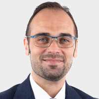
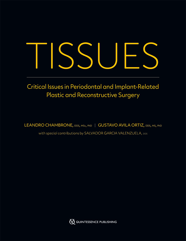
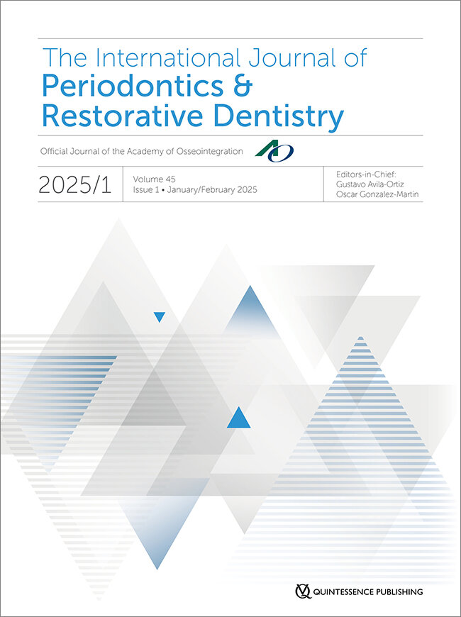
Dr. Gustavo Avila-Ortiz machte seinen Doktor an der Universität von Granada (Spanien), bevor er nach Ann Arbor, Michigan (USA), zog, wo er an der Universität von Michigan einen Master-Abschluss und ein Diplom im Bereich Parodontologie erwarb. Er ist zudem Mitglied des American Board of Periodontology. Dr. Avila-Ortiz verfügt über mehr als 10 Jahre Erfahrung als Ausbilder und hat sich in zahlreichen institutionellen Beiräten und wissenschaftlichen Gesellschaften für die Weiterentwicklung seines Berufsstandes eingesetzt. Er ist ehemaliges Fakultätsmitglied der University of Michigan School of Dentistry und der University of Iowa College of Dentistry, wo er bis 2022 Phillip A. Lainson Professor und Leiter der Abteilung für Parodontologie war. Aktuell ist er als Chefredakteur des International Journal of Periodontics & Restorative Dentistry tätig. Er hat mehr als 100 Beiträge in begutachteten Fachzeitschriften und zahlreiche Buchkapitel verfasst. Er ist Mitautor des Buches TISSUES: Critical Issue in Periodontics and Implant-Related Plastic and Reconstructive Surgery (Quintessence Publishing 2022), das in der Fachwelt große Beachtung gefunden hat. Dr. Avila-Ortiz ist zurzeit Gastprofessor in der Parodontologischen Fachabteilung der Harvard School of Dental Medicine und Mitglied des Atelier Dental Madrid (Spanien), einem Zentrum für zahnmedizinische Fachkompetenz, in dem er eine auf Parodontologie und Implantologie spezialisierte Privatpraxis betreibt und sich regelmäßig an wissenschaftlichen Studien und Fortbildungen beteiligt.
Details make perfection24. Okt. 2024 — 26. Okt. 2024MiCo - Milano Convention Centre, Milano, Italien
Referenten: Bilal Al-Nawas, Gil Alcoforado, Federico Hernández Alfaro, Sofia Aroca, Wael Att, Gustavo Avila-Ortiz, Kathrin Becker, Anne Benhamou, Juan Blanco Carrión, Dieter Bosshardt, Daniel Buser, Francesco Cairo, Paolo Casentini, Raffaele Cavalcanti, Tali Chackartchi, Renato Cocconi, Luca Cordaro, Luca De Stavola, Nuno Sousa Dias, Egon Euwe, Vincent Fehmer, Alberto Fonzar, Helena Francisco, Lukas Fürhauser, German O. Gallucci, Oscar Gonzalez-Martin, Dominik Groß, Robert Haas, Alexis Ioannidis, Simon Storgård Jensen, Ronald Jung, France Lambert, Luca Landi, Georg Mailath-Pokorny jun., Silvia Masiero, Iva Milinkovic, Carlo Monaco, Jose Nart, José M. Navarro, Katja Nelson, Manuel Nienkemper, David Nisand, Michael Payer, Sergio Piano, Bjarni E. Pjetursson, Sven Reich, Isabella Rocchietta, Giuseppe Romeo, Irena Sailer, Mariano Sanz, Ignacio Sanz Martín, Frank Schwarz, Shakeel Shahdad, Massimo Simion, Ralf Smeets, Benedikt Spies, Bogna Stawarczyk, Martina Stefanini, Hendrik Terheyden, Tiziano Testori, Daniel Thoma, Ana Torres Moneu, Piero Venezia, Lukas Waltenberger, Hom-Lay Wang, Stefan Wolfart, Giovanni Zucchelli, Otto Zuhr
European Association for Osseintegration (EAO)
SEPA 24 Bilbao
Clinical evidence based on scientific evidence29. Mai 2024 — 1. Juni 2024Bilbao, Spanien
Referenten: Eduardo Anitua, Sofia Aroca, Serhat Aslan, Gustavo Avila-Ortiz, Juan Blanco Carrión, Gonzalo Blasi, Nagihan Bostanci, Iain L. C. Chapple, Jan Cosyn, Glécio Vaz de Campos, Luca De Stavola, Jan Derks, Vincent Fehmer, Elena Figuero, Sergio García, Alfonso L. Gil, Oscar Gonzalez-Martin, Adrian Guerrero, Sérgio Kahn, Alejandro Lanis, Antonio Liñares, Ferrán Llansana, Francesco Mangano, Dino Calzavara mantovani, Mauro Merli, Juan Mesquida, Alberto Monje, Eduardo Montero, Stefano Parma-Benfenati, Bjarni E. Pjetursson, Pablo Ramírez, Mariano Sanz, Ignacio Sanz Sànchez, Beatriz Solano Mendoza, Jacobo Somoza, Martina Stefanini, Maurizio S. Tonetti, Leonardo Trombelli, Ion Zabalegui
Academy of Osseointegration 2024 Annual Meeting
Going beyond the Boundaries - Optimizing Patient Care Through Scientific and Clinical Innovation7. März 2024 — 9. März 2024Charlotte Convention Center, Charlotte, NC, Vereinigte Staaten von Amerika
Academy of Osseointegration (AO)
The 9th World Dental Meeting in Japan 2023
No Dentistry, No Wellness!29. Sept. 2023 — 1. Okt. 2023Pacifico Yokohama Conference Center, Yokohama, Japan
Referenten: Alessandro Agnini, Andrea Mastrorosa Agnini, Wael Att, Gustavo Avila-Ortiz, Markus B. Blatz, Victor Clavijo, Karim Dada, Glécio Vaz de Campos, Vincent Fehmer, Naoki Hayashi, Mario Imburgia, Guillaume Jouanny, Sérgio Kahn, Bertrand Khayat, Sandra Khong Tai, Christopher Köttgen, Stefen Koubi, Tomas Linkevičius, Nazariy Mykhaylyuk, Ravindra Nanda, Andreas Nolte, Léon Parienté, Jose Manuel Reuss , Domenico Ricucci, Isabella Rocchietta, Irena Sailer, Todd R. Schoenbaum, Werner Schupp, Istvan Urban, Eric Van Dooren, Débora R. Vilaboa, Otto Zuhr
Quintessence Publishing Co. Ltd. Japan
Zeitschriftenbeiträge dieses Autors
International Journal of Periodontics & Restorative Dentistry, Pre-Print
DOI: 10.11607/prd.6935, PubMed-ID: 3781985011. Okt. 2023,Sprache: EnglischGaraicoa-Pazmino, Carlos / Couso-Queiruga, Emilio / Monje, Alberto / Avila-Ortiz, Gustavo / Castilho, Rogerio M. / Amo, Fernando Suárez López del
The aim of this PRISMA-compliant systematic review was to analyze the evidence pertaining to disease resolution after treatment of peri-implant diseases (PROSPERO: CRD42022306999) with the following PICO question: what is the rate of disease resolution following non-surgical and surgical therapy for peri-implant diseases in adult human subjects? A literature search to identify studies that fulfilled a pre-established eligibility criteria was conducted. Data on primary therapeutic outcomes, including treatment success, rate of disease resolution and/or recurrence, as well as a variety of secondary outcomes was extracted and categorized. Fifty-five articles were included. Few studies investigated the efficacy of different non-surgical and surgical therapies to treat peri-implant diseases using a set of pre-defined criteria and with follow-up periods of at least one year. The definition of treatment success and outcomes of disease resolution differed considerably among the included studies. Treatment of peri-implant mucositis was most commonly reported to be successful in arresting disease progression for ≤60% of the cases, whereas most studies on peri-implantitis treatment reported disease resolution occurring in <50% of the fixtures. In conclusion, disease resolution is generally unpredictable and infrequently achieved after the treatment of peri-implant diseases. A great variety of definitions have been used to define treatment success. Notably, percentages of treatment success and disease resolution were generally underreported. The use of standardized parameters to evaluate disease resolution should be considered an integral component in future clinical studies.
Schlagwörter: Peri-implantitis, dental implant, diagnosis, peri-implant endosseous healing, tooth loss, outcome assessment.
This study aimed at characterizing the gingival thickness (GT) and determining correlations with other local phenotypical features. Cone-beam computed tomography scans from adult subjects involving the maxillary anterior teeth were obtained to assess buccal GT at different apico-coronal levels, periodontal supracrestal tissue height (STH), the distance from the cementoenamel junction to the alveolar bone crest (CEJ-BC), and bucco-lingual tooth dimensions in mm. A total of 100 subjects and 600 maxillary anterior teeth constituted the study sample. Variations in mean values of GT were observed as a function of apico-coronal level, tooth type, and gender. GT progressively increased apically. Maxillary central incisors and males generally exhibited thicker GT. Contrarily, females exhibited thinner GT and shorter STH. Tooth dimensions were negatively correlated with GT, as the narrower the tooth crown/root in the bucco-lingual dimension, the thicker the gingiva. GT at the level of the CEJ was dichotomized to differentiate between thin (<1mm) and thick (≥1mm) gingival phenotypes (GP). Teeth with a thin GP displayed greater CEJ-BC and buccolingual tooth width dimensions. Conversely, teeth with a thick GP generally exhibited taller STH and narrower tooth dimensions.
This PRISMA-compliant systematic review aimed to investigate the effect of supportive peri- implant care (SPIC) on peri-implant tissue health and disease recurrence following the non surgical and surgical treatment of peri-implant diseases. The protocol of this review was registered in PROSPERO (CRD42023468656). A literature search was conducted to identify investigations that fulfilled a set of pre-defined eligibility criteria based on the PICO question: what is the effect of SPIC upon peri-implant tissue stability following non-surgical and surgical interventions for the treatment of peri-implant diseases in adult human subjects? Data on SPIC (protocol, frequency, and compliance), clinical and radiographic outcomes, and other variables of interest were extracted and subsequently categorized and analyzed. A total of 8 studies, with 288 patients and 512 implants previously diagnosed with peri-implantitis were included. No studies including peri-implant mucositis fit the eligibility criteria. Clinical and radiographic outcomes were similar independently of specific SPIC features. Nevertheless, a 3-month recall interval was generally associated with a slightly lower percentage of disease recurrence. The absence of disease recurrence at the final follow-up period (mean of 58.7±25.7 months) ranged between 23.3% and 90.3%. However, when the most favorable definition of disease recurrence reported in the selected studies was used, mean disease recurrence was 28.5% at baseline, considered 1 year after treatment for this investigation, and increased to 47.2% after 2 years of follow-up. In conclusion, regardless of the SPIC interval and protocol, disease recurrence tends to increase over time after the treatment of peri-implantitis, occasionally requiring additional interventions.
Schlagwörter: dental implants; peri-implantitis; peri-implant mucositis; disease progression; risk factors
This study aimed to determine the correlation between gingival stippling (GS) and other phenotypic characteristics. Adult subjects in need of CBCT scans and comprehensive dental treatment in the anterior maxilla were recruited. Facial gingival thickness (GT) and buccal bone thickness (BT) were assessed utilizing CBCT. Standardized intraoral photographs were obtained to determine keratinized tissue width (KTW), presence of GS in all facial and interproximal areas between the maxillary canines, and other variables of interest, such as gingival architecture (GA), tooth shape, and location. Statistical analyses were conducted to assess different correlations among recorded variables. A total of 100 participants and 600 maxillary anterior teeth constituted the study population and sample, respectively. Facial GS was observed in 56% of men and 44% of women, and it was more frequently associated with flat GA, triangular and square/tapered teeth, central incisors, and men. Greater mean GT, BT, and KTW values were observed in facial areas that exhibited GS. Interdental GS was present in 73% of the sites, and it was more frequently observed in men, in the central incisor region, and when facial GS was present. Multilevel logistic regression revealed a statistically significant association between the presence of GS and KTW, BT measured at 3 mm apical to the bone crest, and tooth type. This information can be used to recognize common periodontal phenotypical patterns associated with specific features of great clinical significance.
Schlagwörter: dentition, gingiva, oral mucosa, periodontium, permanent, phenotype
The primary aim of this study was to evaluate the efficacy of alveolar ridge preservation (ARP) ther- apy compared with unassisted socket healing (USH) in attenuating interproximal soft tissue atrophy. Adult patients who underwent maxillary single-tooth extraction with or without ARP therapy were included. Surface scans were obtained and CBCT was performed to digitally assess interproximal soft tissue height changes and measure facial bone thickness (FBT), respectively. Logistic regres- sion models were conducted to investigate the individual effect of demographic and clinical vari- ables. Ninety-six subjects (USH = 49; ARP = 47) constituted the study population. Linear soft tissue assessments revealed a significant reduction of the interproximal soft tissue over time within and between groups (P < .0001). ARP therapy significantly attenuated interproximal soft tissue height re- duction compared to USH: –2.0 ± 0.9 mm mesially for USH vs –1.0 ± 0.5 mm mesially for ARP; –1.9 ± 0.7 mm distally for USH vs –1.1 ± 0.5 mm distally for ARP (P < .0001). Thin (≤ 1 mm) facial bone thick- ness (FBT) upon extraction was associated with greater interproximal soft tissue atrophy compared to thick FBT (> 1 mm), independent of the treatment received (P < .0001). Nevertheless, ARP therapy resulted in better preservation of interproximal soft tissue height, especially in thin bone phenotype, by a factor of 2 for the mesial site (+1.3 mm) and by a factor of 1.6 for the distal site (+0.9 mm). This study demonstrated that ARP therapy largely attenuates interproximal soft tissue dimensional re- duction after maxillary single-tooth extraction compared to USH.
Schlagwörter: alveolar ridge preservation, bone resorption, dental implants, digital image processing, tooth extraction
Clinicians, researchers, and policymakers often rely on the available scientific evidence to make strategic decisions. Systematic reviews (SRs) occupy an influential position in the hierarchy of scientific evidence. The findings of wellconducted SRs may provide valuable information to answer specific research questions1,2 and identify existing gaps for future research.3 Therefore, it is of supreme importance that SRs are published promptly, reducing as much as possible the time elapsed between the last date of the search for primary studies and the actual publication date. A study published in 2014 assessed the publication delay of SRs in orthodontics, revealing that the median time interval from the last search to publication was more than 1 year (13.2 months).4 Delays in the publication of SRs or original research articles may depend on author-related factors (eg, timing of resubmission after receiving feedback from reviewers) or journal-related factors (eg, time taken to process a submission).5–7 Regardless of the reasons, clinical recommendations and translation of SR findings may be affected by publication delay. We assessed the extent of publication delay of systematic reviews in dentistry with the purpose of addressing its implications and presenting potential solutions.