Auf unserer Website kommen verschiedene Cookies zum Einsatz: Technisch notwendige Cookies verwenden wir zu dem Zweck, Funktionen wie das Login oder einen Warenkorb zu ermöglichen. Optionale Cookies verwenden wir zu Marketing- und Optimierungszwecken, insbesondere um für Sie relevante und interessante Anzeigen bei den Plattformen von Meta (Facebook, Instagram) zu schalten. Optionale Cookies können Sie ablehnen. Mehr Informationen zur Datenerhebung und -verarbeitung finden Sie in unserer Datenschutzerklärung.
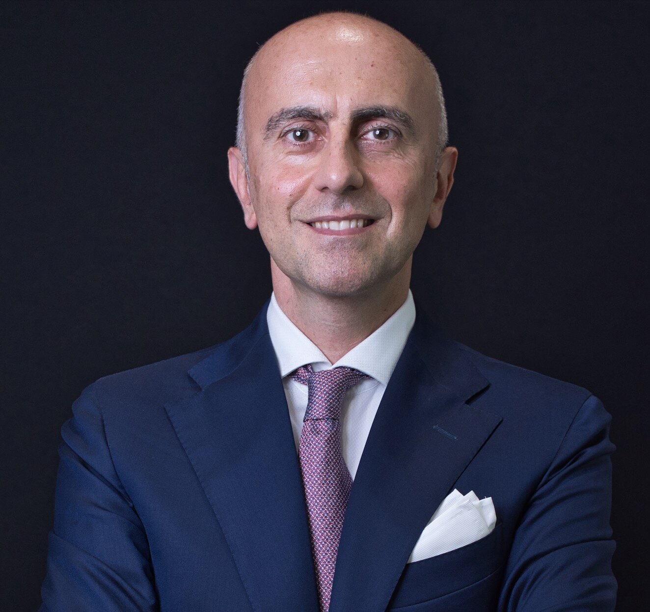
Im Jahr 1992 absolvierte Prof. Carlo Monaco sein Studium in Zahnmedizin und Zahntechnik an der Universität Bologna. Von 1992 bis 1997 arbeitete er in der Abteilung für konservative Zahnheilkunde der Zahnklinik Bologna unter der Leitung von Prof. G. Dondi dall'Orologio. Zwischen 1995 und 1997 erhielt er ein Stipendium für Forschungen zu Komposit-Resinen und deren Korrelation mit Adhäsivsystemen. Seit 1998 ist er als Fellow in der Abteilung für orale und maxillofaziale Prothetik der Abteilung für Odontostomatologische Wissenschaften der Universität Bologna tätig, geleitet von Prof. Scotti, wo er sich hauptsächlich mit indirekten ästhetischen Restaurationen und festsitzenden Prothesen beschäftigt. In den Jahren 2002-04 war er Lehrbeauftragter für Zahnprothetik an der Universität Bologna. Im Jahr 2002 besuchte er die Abteilung für Karyologie an der Universität Genf unter der Leitung von Prof. I. Krejci, mit dem er Forschungen über adhäsive Brücken durchführte. Seit 2003 ist er als visiting Researcher in der Abteilung für Karyologie der Universität Genf tätig. Im Jahr 2003 erlangte er den Master of Science und im Jahr 2005 den PhD in Zahnmaterialien an der Universität Siena bei Prof. M. Ferrari. In den Jahren 2005 und 2008 gewann er den ersten Preis für die beste prothetische Forschung, verliehen von der Italienischen Akademie für Prothetik (AIOP). Im Jahr 2006 erhielt er zwei erste Preise auf der internationalen Konferenz CONSEURO für die beste Forschung im Bereich der Zahnmaterialien und für das beste Poster aller Sitzungen. Seit 2006 ist er Forscher an der Universität Bologna. Als Referent auf nationalen und internationalen Konferenzen hat er wissenschaftliche Artikel über die Anwendungen von Komposit- und Keramikmaterialien im zahnmedizinischen Bereich veröffentlicht. Im Jahr 2017 erlangte er die nationale Qualifikation. Prof. Carlo Monaco leitet den Masterstudiengang in Prothetik und Implantatprothetik mit fortgeschrittenen Technologien.
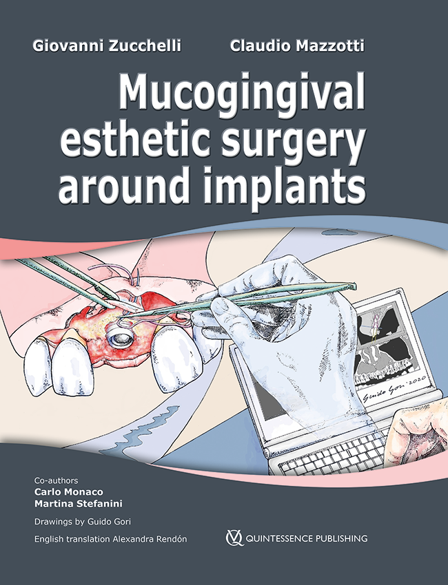
1. Auflage 2022 Buch Hardcover; Two-volume book with slipcase; 21 x 28 cm, 1100 Seiten, 6557 Abbildungen Sprache: Englisch Kategorien: Oralchirurgie, Implantologie Artikelnr.: 24311 ISBN 978-88-7492-091-4 QP Italy
Details make perfection24. Okt. 2024 — 26. Okt. 2024MiCo - Milano Convention Centre, Milano, Italien
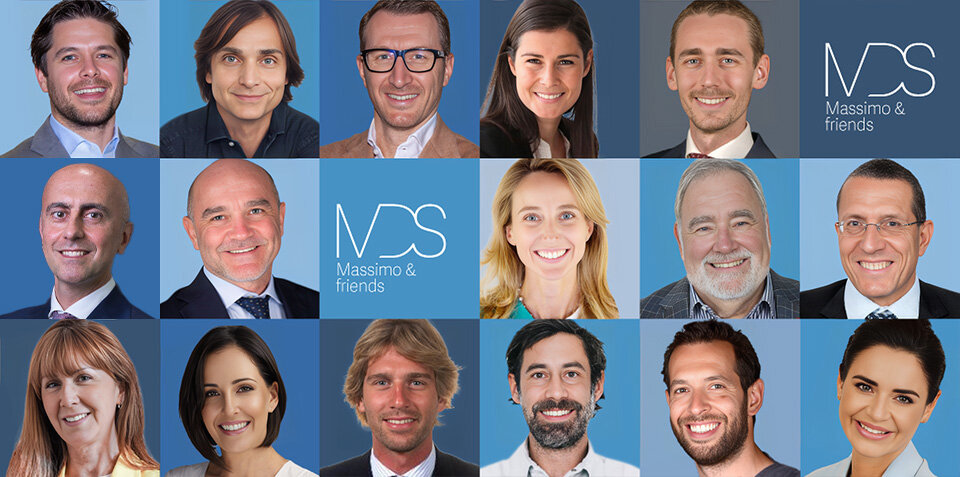
Referenten: Bilal Al-Nawas, Gil Alcoforado, Federico Hernández Alfaro, Sofia Aroca, Wael Att, Gustavo Avila-Ortiz, Kathrin Becker, Anne Benhamou, Juan Blanco Carrión, Dieter Bosshardt, Daniel Buser, Francesco Cairo, Paolo Casentini, Raffaele Cavalcanti, Tali Chackartchi, Renato Cocconi, Luca Cordaro, Luca De Stavola, Nuno Sousa Dias, Egon Euwe, Vincent Fehmer, Alberto Fonzar, Helena Francisco, Lukas Fürhauser, German O. Gallucci, Oscar Gonzalez-Martin, Dominik Groß, Robert Haas, Alexis Ioannidis, Simon Storgård Jensen, Ronald Jung, France Lambert, Luca Landi, Georg Mailath-Pokorny jun., Silvia Masiero, Iva Milinkovic, Carlo Monaco, Jose Nart, José M. Navarro, Katja Nelson, Manuel Nienkemper, David Nisand, Michael Payer, Sergio Piano, Bjarni E. Pjetursson, Sven Reich, Isabella Rocchietta, Giuseppe Romeo, Irena Sailer, Mariano Sanz, Ignacio Sanz Martín, Frank Schwarz, Shakeel Shahdad, Massimo Simion, Ralf Smeets, Benedikt Spies, Bogna Stawarczyk, Martina Stefanini, Hendrik Terheyden, Tiziano Testori, Daniel Thoma, Ana Torres Moneu, Piero Venezia, Lukas Waltenberger, Hom-Lay Wang, Stefan Wolfart, Giovanni Zucchelli, Otto Zuhr
European Association for Osseintegration (EAO)
Zeitschriftenbeiträge dieses Autors
The International Journal of Prosthodontics, Pre-Print
DOI: 10.11607/ijp.9191, PubMed-ID: 3946661428. Okt. 2024,Seiten: 1-24, Sprache: EnglischBonvicini, Martina / Monaco, Carlo / Arena, Antonio / Consolo, Ugo / Silvestri, Davide / Zicari, Francesca
Purpose. The purpose of this in vitro study was to evaluate the effect of a staining solution on color stability of resins for complete dentures produced with conventional and digital technologies. Materials and Methods. 60 resin specimens were prepared and divided in 6 groups of 10 specimens each: Probase Hot (PH, Ivoclar AG), IvoBase CAD (IBC, Ivoclar AG), Ivotion Dent (ID, Ivoclar AG), Ivotion Dent Multi (IDM, Ivoclar AG), Dima Print Denture Base (DPDB, Kulzer), and Dima Print Denture Teeth (DPDT, Kulzer). One surface of each specimen was polished following the manufacturer’s instructions. The CIELab color differences were evaluated before and after immersion for 28 days in a coffee solution and measured as DE using two dental spectrophotometers. One specimen for each group was selected for SEM analysis at baseline and at 28-day. Results. Data analysis was performed by Kruskal-Wallis test and post-hoc Dunn test for comparison. Regarding specimens analyzed with VITA Easyshade, IDM white zone (polished and unpolished) scored higher (∆E= 0.87 and ∆E = 1.15 respectively). All milled samples (IBC, ID, IDM pink zone), scored significantly higher as compared to PH and DPDT. Regarding Spectroshade, IDM pink zone performed better, regarding both polished and unpolished specimens (∆E=1.57 and ∆E=2.07 respectively). ID demonstrated a statistic higher color stability compared to PH and DPDT. Regarding the SEM analysis any differences in surface morphology was observed between baseline and 28-days immersion analyses. Conclusions. Color stability is significantly higher in resin specimens obtained with digital technologies as compared to conventional resin specimens after immersion in a staining solution.
Purpose. Evaluate the influence of different Co-Cr framework manufacturing techniques on the bond strength between metal and two types of veneering ceramic with and without a metal primer. Materials and Methods. 72 metal specimens were fabricated using the same Co-Cr alloy (Keramit NP; Nobil Metal) with three different techniques: ingots for casting (Keramit NP); disks for CAD/CAM milling (Synergy Drive Keramit NP); and powder for SLM (Keramit NP-S). Each of the 24-specimen groups, was divided into two subgroups, according to the application, or not, of Nobil Metal Bonding (NMB) (Nobil Metal). Two different veneering ceramic systems were used: Ceramco iC (Dentsply) and Noritake super porcelain EX-3 (Noritake). The bond strength of the metal-ceramic system was evaluated using the three-point bending test and the modality of failure with SEM analysis. Three-way Anova with interactions was used to evaluate the influence of the metal production process, the presence or absence of the bonding agent and the type of veneering ceramic on ceramic-metal adhesion. Results. All specimens exceeded the minimum bond strength of 25 MPa required by ISO Standard 9693 – 1:2012. The highest values of adhesion were registered for specimens manufactured with casted metal/Noritake veneering ceramic (40.59 ± 7.30 MPa). The lowest values were registered for specimens manufactured with CAD/CAM metal/NMB/Ceramco iC veneering ceramic (29.47 ± 3.38 MPa). Conclusions. The fabrication method and the type of veneering ceramic had a significant influence on the mean bond strength. The application of the NMB did not show an influence on bond strength.
Ziel: Diese In-vitro-Studie untersuchte die Lesetiefe von Intraoralscannern (IOS) im Sulcus gingivalis.
Material und Methode: Am Zahn 16 eines Frasaco-Modells wurde eine Tangentialpräparation für eine Vollkrone durchgeführt. Das Modell wurde gescannt und der Sulkus anschließend mit einer dedizierten Software (Model Creator, Version DentalCAD 2.4 Plovdiv) durch Setzen der Parameter apikale Breite, koronale Breite und Sulkustiefe virtuell modifiziert. Anschließend wurden zwei Modelle mit unterschiedlichen Sulkustiefen (1 mm und 2 mm) im Digital-Light-Processing-Verfahren (DLP) 3-D-gedruckt. Getestet wurden sieben IOS: Emerald, Trios 3, Carestream 3600, Dental Wings DWIO, CondorScan, True Definition Scanner (TDS) und Cerec Omnicam. Jedes der beiden Modelle wurde mit jedem Scanner 10-mal gescannt. Anschließend wurde mit einer Viewer-Software (3Shape 3D) die Sulkustiefe D (Distanz zwischen dem koronalsten Punkt und dem Boden des Sulkus) gemessen. Die Prüfung auf Normalverteilung der Daten erfolgte mit dem Shapiro-Wilk-Test (p < 0,05). Auf Varianzhomogenität wurde mit dem Levene-Test geprüft. Für die statistische Analyse kam der Kruskal-Wallis-Test mit Nemenyi-post-hoc-Test zum Einsatz (α = 0,05).
Ergebnisse: Alle IOS waren in der Lage, in dem 1 mm tiefen Sulkus zu lesen, wenn auch mit einigen statistisch signifikanten Unterschieden (p < 0,001). In dem 2 mm tiefen Sulkus allerdings konnten nur die Trios 3 und das TDS lesen (p < 0,001).
Schlussfolgerungen: Die Lesetiefe verschiedener IOS kann sich signifikant unterscheiden. An dem Modell mit 2 mm tiefem Sulkus war die Sulkuserfassung trotz Abwesenheit von Sulkusflüssigkeit unvollständig, was bedeutet, dass tief subgingival reichende Präparationen mit IOS nur schwer abzuformen sind.
Schlagwörter: Intraoralscanner, Sulcus gingivalis, Lesetiefe, Tangentialpräparation, Federrand, Digital Light Processing
The International Journal of Prosthodontics, 2/2023
DOI: 10.11607/ijp.7591, PubMed-ID: 36445219Seiten: 228-232, Sprache: EnglischMonaco, Carlo / Scheda, Lorenzo / Arena, Antonio / di Fiore, Adolfo / Zucchelli, Giovanni
Purpose: To describe the treatment of malpositioned implants in the esthetic area using the angulated welded abutment (AWA) approach together with peri-implant soft tissue surgery.
Materials and Methods: A clinical case with extreme buccal angulation of two implants in the anterior maxilla was used to illustrate the AWA technique. After implant impression-taking, digital analysis was used to determine the ideal prosthetic angulation of the abutment and the ideal position of its screw hole in relation to the gingival margin of the adjacent teeth. The AWA was designed in two combinable components that were meant to be welded together. Before the welding process, an angulated screw was included in the abutment. Since the angulated screw was inside the abutment, the screwdriver hole could be designed as narrow as possible and put in an ideal position. After periodontal and peri-implant surgery were carried out, the AWA was applied to the implants.
Results: The AWA allowed correction of the prosthetic axis. Moreover, relocation of the screw hole allowed the gingival tissue to creep over the abutment. In this way, a new esthetic restoration can be placed after the mucogingival surgery.
Conclusion: The excessive misangulation of the implants was efficiently recovered. Further studies are needed to evaluate long-term clinical success, and standardization of this technique is required for routine clinical use.
Purpose: To describe the possible adverse effects of sodium hypochlorite (NaOCl) solutions, highconcentration alcohol solutions, and povidone-iodine products indicated for disinfection of inanimate surfaces against human coronavirus of the severe acute respiratory syndrome (SARS-CoV) on prosthesis materials, including zirconia, lithium disilicate, and acrylic resin.
Materials and Methods: A systematic literature research was conducted in the SCOPUS, PubMed/Medline, Web of Science, EMBASE, and Science Direct databases from January 2010 to February 2020 using a combination of the following MeSH/Emtree terms and keywords: “sodium hypochlorite”; “alcohol”; “ethanol”; “povidone-iodine”; “dental ceramic”; “zirconia”; “lithium disilicate”; and “acrylic resin”.
Results: A total of 538 studies were identified in the search during initial screening, 44 were subject to full-text evaluation, and 24 fulfilled the inclusion criteria. Seven articles on zirconia and lithium disilicate investigated the effect of NaOCl (0.5% and 1%), 96% isopropanol, and 80% ethanol on bond strength after saliva contamination. The remaining articles evaluated color alteration, surface roughness modifications, decrease in flexural strength, and bond strength of all cleaning agents on acrylic resin.
Conclusion: NaOCl solution (1%) for 1 minute is recommended to reduce SARSCoV infectivity and to minimize the risk of cross-contamination through prosthetic materials. An increase in surface roughness and color alteration on acrylic resin were recorded using 1% NaOCl, but without any clinical significance. A decrease in bonding strength was determined after using 1% NaOCl, 96% isopropanol, or 80% ethanol solutions on lithium disilicate. Silanization before the try-in procedure and the application of a second layer of silane after cleaning methods are recommended to improve the bond strength.
Purpose: To evaluate the influence of the crown-to-implant ratio (CI) on marginal bone loss (MBL) around short dental implants placed in the posterior mandible.
Materials and Methods: All patients treated with short implants (7-mm length) in the posterior mandible between 1994 and 2003 at the Dental Clinic of the Department of Neuroscience of the University of Padua (Italy) were retrospectively included in the analysis. MBL and clinical CI (cCI) were measured on the radiographs. Implant characteristics including implant diameter, prosthetic type, retention mode, antagonist type, veneering material, and implant surfaces were retrieved from local medical records. A generalized linear mixed model was estimated to identify the predictors of MBL.
Results: A total of 108 dental implants placed in 51 patients were included in the analysis. Mean follow-up was 16 years (range: 11 to 20 years). Mean cCI was 2.21 (SD = 0.31) with a mean crown height of 10.86 mm (SD = 0.99). Mean MBL was 1.42 mm (SD = 0.38). At multivariable analysis, cCI ≥ 2 was associated with higher MBL (regression coefficient: 0.27; 95% CI: 0.15 to 0.40), while implant characteristics, follow-up, and site were not associated with MBL. The effect of a cCI ≥ 2 was estimated in an increase of 0.28 mm in MBL (95% CI: 0.14 to 0.43 mm).
Conclusion: Higher cCI was associated with greater MBL of implant-supported fixed dental prostheses in short dental implants placed in the posterior mandible, while implant characteristics, follow-up, and site were not associated with MBL. However, the increase of 0.28 mm of MBL in patients with a cCI ≥ 2 may not be clinically relevant.
Schlagwörter: crown-to-implant ratio, implant-supported dental prosthesis, marginal bone loss, prosthetic parameters, short implant
International Journal of Oral Implantology, 2/2018
PubMed-ID: 29806668Seiten: 215-224, Sprache: EnglischZucchelli, Giovanni / Felice, Pietro / Mazzotti, Claudio / Marzadori, Matteo / Mounssif, Ilham / Monaco, Carlo / Stefanini, Martina
Purpose: To report the 5-year clinical and aesthetic outcomes of a novel surgical-prosthetic approach for the treatment of buccal soft tissue dehiscence around single dental implants.
Materials and methods: Twenty patients with buccal soft tissues dehiscence around single implants in the aesthetic area were treated by removing the implant-supported crown, reducing the implant abutment, coronally advanced flap in combination with connective tissue graft and final restoration. After the first year, patients were recalled three times a year until the final clinical re-evaluation performed 5 years after the final prosthetic crown. Complications, bleeding on probing (BoP), peri-implant probing depth (PPD), clinical attachment level (CAL), keratinized tissue height (KTH), soft tissue coverage and thickness (STT), patient satisfaction (VAS) and aesthetic assessment (PES/WES) were evaluated 5 years after the final restoration.
Results: Of the 20 patients enrolled in the study, 19 completed the study at 5 years. A total of 99.2% mean soft tissue dehiscence coverage, with 79% of complete dehiscence coverage, was achieved at 5 years. A statistically significant increase in buccal soft tissue thickness (0.3 mm 0.1-0.4 P 0.001) and keratinized tissue height (0.5 mm 0.0-1.0; P 0.001) at 5 years with respect to 1 year was demonstrated. The patient aesthetic evaluation showed high VAS scores with no statistical difference between 1 year and 5 years (8.75 ± 1.02 and 8.95 ± 0.91 respectively). A statistical significant PES/WES score improvement was observed between baseline and 5 years (9.48 ± 2.68; P 0.001), but not between 1 and 5 years.
Conclusions: Successful aesthetic and soft tissue dehiscence coverage outcomes were well maintained at 5 years. The strict regimen of post-surgical control visits and the emphasis placed on the control of the toothbrushing technique could be critical for the successful long-term maintenance of soft tissue dehiscence coverage results.
Schlagwörter: aesthetics, connective tissue, dental implant, mucogingival surgery, soft tissue dehiscence
Conflict-of-interest statement: The authors declare that they have no conflict of interest.