Auf unserer Website kommen verschiedene Cookies zum Einsatz: Technisch notwendige Cookies verwenden wir zu dem Zweck, Funktionen wie das Login oder einen Warenkorb zu ermöglichen. Optionale Cookies verwenden wir zu Marketing- und Optimierungszwecken, insbesondere um für Sie relevante und interessante Anzeigen bei den Plattformen von Meta (Facebook, Instagram) zu schalten. Optionale Cookies können Sie ablehnen. Mehr Informationen zur Datenerhebung und -verarbeitung finden Sie in unserer Datenschutzerklärung.
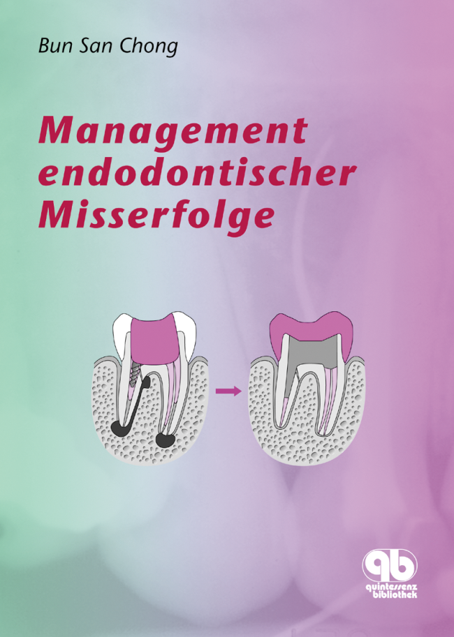
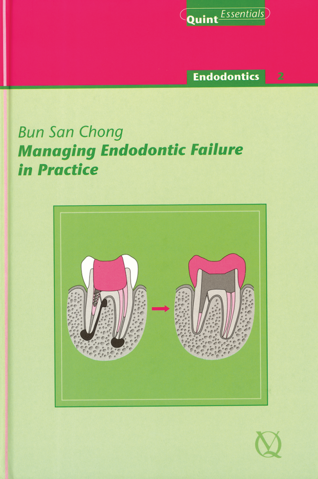
BS Chong is Professor of Restorative Dentistry/Honorary Consultant, Endodontic Lead & Director, Postgraduate Endodontics, Barts & The London School of Medicine and Dentistry, Queen Mary University of London. In addition, he is a General Dental Council (UK) registered Specialist in Endodontics and has been in private practice since 1990. Professor Chong is the Editor and contributing author of two editions (6th and 7th) of Harty's Endodontics in Clinical Practice; author of Managing Endodontic Failure in Practice; contributing author to Self-Assessment Picture Tests in Dentistry: Endodontics and Principles of Endodontics. He is also on the Editorial Board of the International Endodontic Journal, and President, British Endodontic Society (2017-18).
Erscheinungsweise: eingestellt Sprache: Englisch Kategorie: Endodontie QP Deutschland
40,00 €
Veranstaltungen
7 Decades of Quintessence Publishing
10. Jan. 2019 — 12. Jan. 2019Estrel Convention Center
Referenten: Jiro Abe, Michèle Aerden, Wael Att, Stavros Avgerinos, Avijit Banerjee, Vesna Barac Furtinger, Klaus-Dieter Bastendorf, Lars Bergmans, Ashwini Bhalerao, Jaroslav Bláha, Sebastian Bürklein, Daniel Buser, Josette Camilleri, Sevim Canlar, Sandra Chmieleck, Bun San Chong, Victor Clavijo, Carsten Czerny, Bettina Dannewitz, Alessandro Devigus, Didier Dietschi, Irina Dragan, Daniel H.-J. Edelhoff, Peter Eickholz, Karim Elhennawy, Peter Engel, Wolfgang Eßer, Marco Esposito, Susanne Fath, Vincent Fehmer, Federico Ferraris, Stefan Fickl, Mauro Fradeani, Roland Frankenberger, Eiji Funakoshi, Petra Gierthmühlen, Christiane Gleissner, Florian Göttfert, Dennis Grosse, Galip Gürel, Christian Haase, Horst-Wolfgang Haase, Manuela Hackenberg, Jörg Haist, Anke Handrock, Arndt Happe, Karsten Heegewaldt, Rüdiger Henrici, Michael Hülsmann, Hajime Igarashi, Tomohiro Ishikawa, Hideaki Katsuyama, Kathryn Kell, Matthias Kern, Fouad Khoury, Marko Knauf, Ralf J. Kohal, Stefen Koubi, Fabian Langenbach, Henriette Terezia Lerner, Thomas Malik, Siegfried Marquardt, Henrike März, Kathleen Menzel, Helen Möhrke, Kotaro Nakata, Marc L. Nevins, Masayuki Okawa, Rebecca Otto, Mark Stephen Pace, Shanon Patel, Karin Probst, Domenico Ricucci, Katrin Rinke, Irena Sailer, Edgar Schäfer, Ralf Schäfermeier, Jan Schellenberger, Tom Schloss, Gottfried Schmalz, Devorah Schwartz-Arad, Frank Schwarz, Thomas A. Schwenk, Anton Sculean, Bernd Stadlinger, Athanasios Stamos, Ana Stevanovic, Masana Suzuki, Senichi Suzuki, Hiroyuki Takino, Sameh Talaat, Mitsuhiro Tsukiboshi, Hideaki Ueda, Istvan Urban, Luc W. M. van der Sluis, Eric Van Dooren, Bart Van Meerbeek, Paula Vassallo, Juliane von Hoyningen-Huene, Michael Walter, Siegbert Witkowski, Stefan Wolfart, Sylvia Wuttig, Masao Yamazaki, Maciej Zarow, Matthias Zehnder, Raquel Zita Gomes, Giovanni Zucchelli, Otto Zuhr, Bettina Zydatiß
Quintessenz Verlags-GmbH
Zeitschriftenbeiträge dieses Autors
ENDO, 4/2020
Seiten: 247, Sprache: EnglischSchäfer, Edgar / Chong, Bun San
The advent of guided endodontics, combining 3D imaging techniques with digitally designed guides, has led to its utilisation in surgical and nonsurgical root canal treatment. To date, however, guided endodontics has focused mainly on static guidance, with dynamic guidance receiving limited attention. In the present case report, a dynamic navigation implant system was used for guided endodontics. The safe and accurate access cavity preparation, location and negotiation of tahe calcified canals of a maxillary second premolar demonstrate the successful clinical application of dynamic guided endodontics in everyday endodontic practice.
Schlagwörter: access cavity, canal location, dynamic navigation, guided endodontics
Objectives: To investigate the novel use of computer-aided dynamic navigation for guided endodontics.
Method and materials: Dental casts were fabricated from sets of extracted human teeth. A cone beam computed tomography (CBCT) scan of each cast, with a molded thermoplastic stent and a radiographic marker attached, was obtained and imported into the planning software of a dynamic navigation implant surgery system. Simulating implant surgery but for guided endodontics, the drilling entry point, angle, pathway, and depth of virtual implants were planned for 29 selected teeth. The radiographic marker was replaced with a jaw tag and mounted in a phantom head. A drill tag was attached to the drill handpiece. Following calibration, guided by the stereoscopic motion-tracking camera via the tags and images on a computer monitor providing real-time dynamic plus visual intraoperative feedback, the handpiece was aligned accordingly and endodontic access cavity preparation carried out. Successful root canal location was confirmed using periapical radiographs and CBCT.
Results: Conservative access cavities were achieved and all the expected canals were successfully located in 26 teeth (n = 29). Due to tracking difficulties, only one canal was located in two maxillary second molars; in a maxillary first molar, only two canals were located and the access preparation for the third canal was misaligned and off-target.
Conclusions: The results of this study demonstrate the potential of using computer-aided dynamic navigation technology in guided endodontics in clinical practice.
Schlagwörter: access cavity, cone beam computed tomography, dynamic navigation, endodontics, guided endodontics
Objective: To assess the influence of root-end filling (REF) material on apical surgery outcome.
Materials and methods: Cases of apical surgery performed in a private practice limited to endodontics, which had received Intermediate Restorative Material (IRM; Group 1) or mineral trioxide aggregate (MTA; Group 2) as the REF material were collected. Radiographic surgical healing at up to 12 months and 24 months was evaluated using agreed criteria by two trained observers. The results were analysed using the Pearson chi-square and Fisher exact tests, with the level of significance set at P 0.05.
Results: Twenty-two out of the 95 teeth were excluded for various reasons leaving a total of 73 teeth. Group 2 showed a significantly higher healed rate than Group 1 (81% vs 56%; P = 0.0189); however, the difference between 'healed' and 'healing' teeth was not statistically significant for both follow-up periods (≤ 12 months, P = 0.2852; ≤ 24 months, P = 0.1101). As a secondary observation, it was found that 'large' lesions (> 5 mm diameter) required a longer time to heal than 'small' lesions (48% vs 77%; P = 0.0147). Analysis of the REF material in relation to lesion size showed that MTA 'healed' rates were significantly higher for 'small' lesions than 'large' lesions (P = 0.0042), whereas with IRM, the difference was insignificant (P = 0.4217).
Conclusions: MTA showed a higher rate of 'complete healing'. 'Large' lesions took a longer time to heal completely. 'Small' lesions had a higher 'healed' rate when MTA was used as the REF material.
Schlagwörter: apical surgery, Intermediate Restorative Material, mineral trioxide aggregate, outcome, root-end filling