Auf unserer Website kommen verschiedene Cookies zum Einsatz: Technisch notwendige Cookies verwenden wir zu dem Zweck, Funktionen wie das Login oder einen Warenkorb zu ermöglichen. Optionale Cookies verwenden wir zu Marketing- und Optimierungszwecken, insbesondere um für Sie relevante und interessante Anzeigen bei den Plattformen von Meta (Facebook, Instagram) zu schalten. Optionale Cookies können Sie ablehnen. Mehr Informationen zur Datenerhebung und -verarbeitung finden Sie in unserer Datenschutzerklärung.
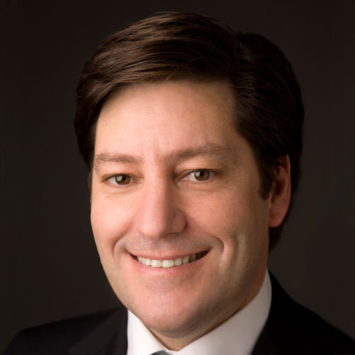
Dr. German Gallucci schloss sein Zahnmedizinstudium (Dr. Med. Dent.) an der Universität Genf, Schweiz, ab. Er absolvierte eine Zusatzausbildung an der Abteilung für festsitzende Prothetik und Okklusion der zahnmedizinischen Fakultät an der Universität Genf, wo er später eine Vollzeittätigkeit in derselben Abteilung aufnahm. Dr. Gallucci erhielt zudem einen Doktortitel von der Freien Universität Amsterdam. Im Jahr 2007 gründete er ein neues Fortbildungsprogramm für orale Implantologie an der Harvard School of Dental Medicine. Zurzeit ist er Vorsitzender der Abteilung für restaurative Zahnmedizin und Biomaterialien und leitet das Harvard Dental Center als Geschäftsführer. Dr. Gallucci nimmt an Vorträgen, Seminaren, Praxiskursen und Diskussionsrunden auf nationalen und internationalen Tagungen und Symposien teil. Er ist Autor wissenschaftlicher Veröffentlichungen auf dem Gebiet der festsitzenden Prothetik und der Implantologie, seine Arbeiten wurden größtenteils in internationalen, begutachteten Fachzeitschriften veröffentlicht. Dr. Gallucci ist Mitglied des Redaktionsausschusses des Journal of Dental Research (JDR), des Clinical Oral Implants Research (COIR) und des International Journal of Oral and Maxillofacial Implants (IJOMI). Er ist Fellow des International Team for Implantology (ITI), Schweiz, und aktives Mitglied der Academy of Osseointegration und der European Academy of Osseointegration.
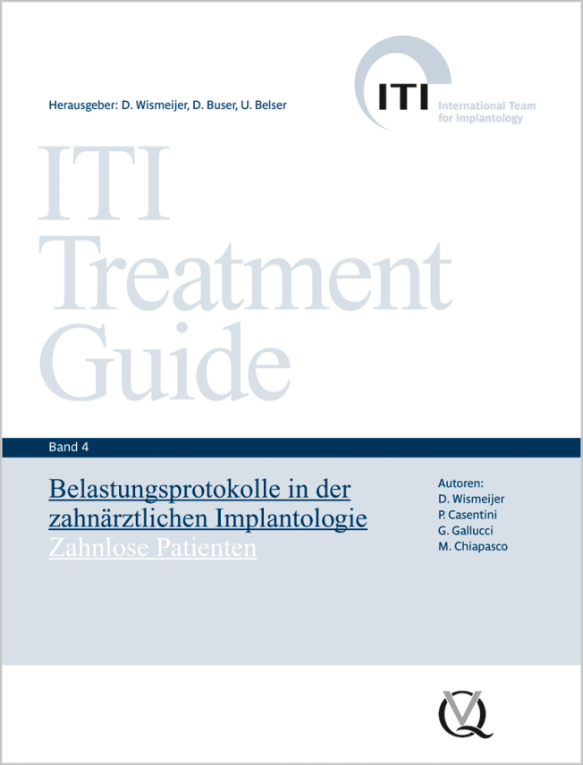
Reihe: ITI Treatment Guide Series, Band 04 1. Auflage 2011 Buch Hardcover, 248 Seiten, 746 Abbildungen Sprache: Deutsch Kategorie: Implantologie Artikelnr.: 17720 ISBN 978-3-938947-17-3 QP Deutschland
86,00 €
Veranstaltungen
31st EAO Annual Scientific Meeting
Details make perfection24. Okt. 2024 — 26. Okt. 2024MiCo - Milano Convention Centre, Milano, Italien
Referenten: Bilal Al-Nawas, Gil Alcoforado, Federico Hernández Alfaro, Sofia Aroca, Wael Att, Gustavo Avila-Ortiz, Kathrin Becker, Anne Benhamou, Juan Blanco Carrión, Dieter Bosshardt, Daniel Buser, Francesco Cairo, Paolo Casentini, Raffaele Cavalcanti, Tali Chackartchi, Renato Cocconi, Luca Cordaro, Luca De Stavola, Nuno Sousa Dias, Egon Euwe, Vincent Fehmer, Alberto Fonzar, Helena Francisco, Lukas Fürhauser, German O. Gallucci, Oscar Gonzalez-Martin, Dominik Groß, Robert Haas, Alexis Ioannidis, Simon Storgård Jensen, Ronald Jung, France Lambert, Luca Landi, Georg Mailath-Pokorny jun., Silvia Masiero, Iva Milinkovic, Carlo Monaco, Jose Nart, José M. Navarro, Katja Nelson, Manuel Nienkemper, David Nisand, Michael Payer, Sergio Piano, Bjarni E. Pjetursson, Sven Reich, Isabella Rocchietta, Giuseppe Romeo, Irena Sailer, Mariano Sanz, Ignacio Sanz Martín, Frank Schwarz, Shakeel Shahdad, Massimo Simion, Ralf Smeets, Benedikt Spies, Bogna Stawarczyk, Martina Stefanini, Hendrik Terheyden, Tiziano Testori, Daniel Thoma, Ana Torres Moneu, Piero Venezia, Lukas Waltenberger, Hom-Lay Wang, Stefan Wolfart, Giovanni Zucchelli, Otto Zuhr
European Association for Osseintegration (EAO)
73rd Annual Scientific Session of the American Academy of Fixed Prosthodontics
Navigating Expectations in Fixed Prosthodontics23. Feb. 2024 — 24. Feb. 2024Chicago Marriott Downtown Magnificent Mile, Chicago, Vereinigte Staaten von Amerika
Referenten: Karen Baker, Lino Calvani, Stephen J. Chu, Arian Deutsch, Terry E. Donovan, Nicholas L. Egbert, German O. Gallucci, Sarit Kaplan, Sonia Leziy, Mariam Malament, Radi Masri, Bradley A. Purcell, Irena Sailer
American Academy of Fixed Prosthodontics
5th ITI Congress Middle East
Innovations and trends in the digital era18. Okt. 2023 — 20. Okt. 2023Hilton Dead Sea Resort & Spa, Sweimeh , Jordanien
Referenten: Motasum Abu-Awwad, Esem Alem, Rola Alhabashneh, Arwa Alsayed, Kai-Hendrik Bormann, Hossam Elsabagh, Vincent Fehmer, German O. Gallucci, Ahmad Hamdan, Najla Kasabreh, Stephanie Khalaf, Lina Khasawneh, Carla Maria Kheirallah, Stephanie Mrad, Dalia Nourah, Mario Roccuzzo, Moustapha Saad, Irena Sailer, Anton Sculean, Mahmoud Shalash, Hani Tohme, Fahad Umer, Ronald Younes
ITI International Team for Implantology
European Prosthodontic Debates 2023
14. Sept. 2023 — 16. Sept. 2023Radisson Blu Hotel Lietuva, Vilnius, Litauen
Referenten: Rolandas Andrijauskas, Adomas Auskalnis, Kazuyoshi Baba, Tord Berglundh, Lawrence E. Brecht, Luigi Canullo, Farronato Davide , Luca De Stavola, Vincent Fehmer, Marco Ferrari, German O. Gallucci, Ieva Gendviliene, Simonas Grybauskas, Sascha Hein, Reinhilde Jacobs, Matthias Kern, Tomas Linkevičius, Diego Lops, Daniele Manfredini, Francesco Mangano, Magda Mensi, Eitan Mijiritsky, Suresh Nayar, Sam Omar, George Papavasiliou, Guillermo Pradíes, Algirdas Puišys, Aušra Ramanauskaitė, Daniele Rondoni, Dmitri Ruzanov, Irena Sailer, Frank Schwarz, Nicola Scotti, Tristan Staas, Eglė Vindašiūtė-Narbutė, Ulrich Wegmann, Lukasz Zadrozny, Ausra Znamenskaite-Levickiene
European Prosthodontic Association
ITI Congress Australasia
It's all about teamwork! Communicating and collaborating within the realm of implant dentistry7. Sept. 2023 — 9. Sept. 2023InterContinental Sydney, Sydney , Australien
Referenten: Merete Aaboe, Catherine Boorer, Kit Chan, Stephen Chen, Kenny Chong, Ivan Darby, Anthony Dickinson, Rebecca Ellis, Tennent Emerson , German O. Gallucci, Nova Gibson, Luiz H. Gonzaga, Adam Hamilton, Janice Kan, Kirthi Kosalram, Emma Lewis , John Lucas, Luan Ngo, Prashant Patel, Ben Sellick , Charlotte Stilwell, Peter Tsakiris, Morgan Wu
ITI International Team for Implantology
ISC 2023 – The 26th International Symposium on Ceramics
Simplicity Meets Esthetics8. Juni 2023 — 11. Juni 2023Town and Country Resort, San Diego, Vereinigte Staaten von Amerika
Referenten: Wael Att, Diego Bechelli, Markus B. Blatz, Domenico Cascione, Gerard J. Chiche, Stephen J. Chu, Victor Clavijo, Florin Cofar, Iván Contreras Molina, Don Cornell, Lee Culp, Vincent Fehmer, Mirela Feraru, German O. Gallucci, Petra Gierthmühlen, Gustavo Giordani, David Guichet, Galip Gürel, Effrat Habsha, Hilal Kuday, Kenneth A. Malament, Adam J. Mieleszko, Ricardo Mitrani, Masayuki Okawa, Jacinthe M. Paquette, Joshua Polansky, Marta Revilla-León, Giuseppe Romeo, Irena Sailer, John Sorensen, Martina Stefanini, Taiseer Sulaiman, Douglas A. Terry, Van Thompson, George Tysowsky, Eric Van Dooren, Yu Zhang
Quintessence Publishing Co., Inc. USA
EAO Digital Days
Implantology: Beyond your expectations12. Okt. 2021 — 14. Okt. 2021online
Referenten: Enrico Agliardi, Alessandro Agnini, Andrea Mastrorosa Agnini, Mauricio Araujo, Goran Benic, Juan Blanco Carrión, Daniel Buser, Raffaele Cavalcanti, Tali Chackartchi, Luca Cordaro, Jan Cosyn, Holger Essig, Vincent Fehmer, Stefan Fickl, Alberto Fonzar, Helena Francisco, German O. Gallucci, Ramin Gomez-Meda, Oscar Gonzalez-Martin, Robert Haas, Arndt Happe, Alexis Ioannidis, Ronald Jung, Niklaus P. Lang, Tomas Linkevičius, Iva Milinkovic, Sven Mühlemann, Katja Nelson, Sergio Piano, Michael A. Pikos, Bjarni E. Pjetursson, Marc Quirynen, Franck Renouard, Isabella Rocchietta, Dennis Rohner, Irena Sailer, Henning Schliephake, Shakeel Shahdad, Massimo Simion, Ali Tahmaseb, Hendrik Terheyden, Jochen Tunkel, Stefan Vandeweghe, Piero Venezia, Stijn Vervaeke, Martin Wanendeya, Georg Watzek, Giovanni Zucchelli
European Association for Osseintegration (EAO)
Zeitschriftenbeiträge dieses Autors
International Journal of Periodontics & Restorative Dentistry, Pre-Print
DOI: 10.11607/prd.7168, PubMed-ID: 392705946. Sept. 2024,Seiten: 1-18, Sprache: EnglischPeña-Cardelles, Juan Francisco / Markovic, Jovana / De Souza, Andrè / Hamilton, Adam / Lanis, Alejandro / Gallucci, German O.
Introduction: The innervation of the hard and soft tissues of the maxillary anterior area depends on the nasopalatine nerve. Due to its anatomy and closeness to implants in the esthetic area, it is essential to fully comprehend its traits and possible effects while performing implant placement procedures. Objective: To assess the prevalence of neurosensorial alteration and the survival and success rate of dental implants in a relationship with the nasopalatine canal. Material and methods: A comprehensive search of the literature was conducted in the following databases: MEDLINE, Web of Science, and Scopus. The articles included had to be case series, studies conducted in patients undergoing dental implant procedures in the incisive canal region or undergone dental implant procedures with incisive canal deflation or neurovascular lateralization. A quantitative synthesis using a meta-analysis software program was performed. Fixed- or random-effect models were applied based on the heterogeneity among studies. Results: Four studies were included. Neurosensorial alterations were presented in three out of four articles included. The range of neurosensorial alterations varied from 30% to 60%. A weighted mean of 29 % ± 13 % of neurosensorial alterations was calculated from the meta-analysis meanwhile a mean of 100 % of implant survival and 100% of implant success were found. In the results, please clarify that "29%±13%" represents the weighted mean calculated from the metaanalysis. Conclusions: Implants in the nasopalatine area are associated with high rates of survival and success being a safe procedure, however, clinicians should be aware that neurosensorial alterations may be present when placing implants in this area.
Computer-assisted implant planning allows for a comprehensive treatment plan by combining radiographic data provided by a Cone Beam Computed Tomography (CBCT) with surface optical scan (IOs) data that includes patient intraoral situation and the intended restorative planning. Integrating a tailored restorative design with the patient’s anatomical conditions through virtual implant planning allows for an ideal bio-restorative treatment planning to maximize biological, functional, and esthetic outcomes. This article discusses dataset registration techniques that combine radiographic CBCT data with restorative information as the main path to create a virtual patient. The described techniques include the use of removable radiographic templates with radiopaque markers, dual scan technique, and direct digital file registration of intra-oral scans using anatomical references. Depending on the individual clinical situation, different factors must be considered to appropriately select methods that achieve an optimal registration of diverse datasets. An inherent challenge lies in the presence of scattering artifacts in CBCT scans. Two approaches are proposed for these situations – the use of chairside-fabricated composite resin markers or adhesive spot-markers fabricated for the use with CBCT scans. Both techniques exhibit limitations that need to be taken into consideration. Further approaches should be developed for situations involving scattering in CBCT.
Schlagwörter: computer-assisted surgery, data superimposition, digital imaging processing, digital workflow, dual technique, scattering
The International Journal of Oral & Maxillofacial Implants, Pre-Print
DOI: 10.11607/jomi.11060, PubMed-ID: 390932921. Aug. 2024,Seiten: 1-13, Sprache: EnglischAkhondi, Samuel / Pala, Kevser / Pedrinaci, Ignacio / Gallucci, German O.
This technical note serves as a practical guide for clinicians aiming to provide solutions to optimize the precision of static computer-assisted implant surgery (sCAIS) and the success of implant placement in free-end situations. Five methods are introduced for enhancing surgical guide stabilization and improving the accuracy of implant placements in situations lacking stabilizing adjacent teeth. Techniques include the use of keratinized soft tissues, implant-borne stabilizers, lateral fixation pins, transitional implants, and digital bone segmentation for guide support. Each method is evaluated for its potential to address the specific challenges faced in sCAIS, aiming to contribute to the field through practical solutions for clinicians.
Introduction: Maxillary sinus floor augmentation is a procedure known for its long-term success and predictable outcomes. However, the perforation of the Schneiderian membrane remains the most common complication associated with this procedure. Objective: This systematic review aims to determine the presence of complications during maxillary sinus floor augmentation procedures using CAD-CAM surgical templates. Material and methods: An electronic search was carried out in MEDLINE (via PubMed), Web of Science, and Scopus. A descriptive analysis of the data was performed. Studies that have performed lateral sinus floor augmentation were included in the inclusion criteria. The CAD-CAM surgical template design and the intraoperative complications were registered. Results: A total of 13 studies were included. Seven were case reports, four were case series, and two were randomized clinical trials. A total of 94 lateral SFA procedures were included (84 using CADCAM templates and 10 without using templates). Three of the 84 maxillary sinus floor augmentation procedures using a CAD-CAM template presented intraoperative complications. Conclusions: Maxillary sinus floor augmentation performed by using CAD-CAM surgical templates could be related to low rates of complications, however, due to the heterogeneity of the articles included, more standardized studies are needed to confirm these outcomes.
International Journal of Periodontics & Restorative Dentistry, 6/2024
DOI: 10.11607/prd.6826, PubMed-ID: 38198437Seiten: 709-719, Sprache: EnglischPeña-Cardelles, Juan Francisco / Markovic, Jovana / Alanezi, Ahmad / Hamilton, Adam / Gallucci, German O. / Lanis, Alejandro
The interforaminal region is considered more favorable for implant placement than the posterior mandible in edentulous patients, mainly because the inferior alveolar nerve can interfere with implant placement in the severely resorbed posterior mandible. However, complications in the interforaminal region may occur due to the presence of the mandibular incisive nerve. This scoping review aims to describe the mandibular incisive nerve anatomy related to the potential interference in implant therapy. A comprehensive literature search was conducted in the following databases: MEDLINE (via PubMed), Web of Science, and Scopus. This scoping review was structured according to the Joanna Briggs Institute method. Thirteen studies were included in the review. All of the studies were observational cohort anatomical studies, carried out mainly by CBCT and on cadavers. A total of 1,471 patients/cadavers were studied. The mandibular incisive nerve was present in 87% to 100% of cases, with an average length of 9.97 mm and an average diameter of 1.97 mm. The mandibular incisive nerve may be damaged during drilling and implant placement, especially when using implant lengths > 12 mm. Damage to the mandibular incisive nerve due to implant placement could be present, but it is necessary to conduct more studies focusing on assessing mandibular incisive nerve damage to understand the clinical relevance of this nerve and its associated morbidities, such as neurosensorial alterations. Due to the different anatomical characteristics of this nerve, CBCT analysis is recommended for implant therapy in the anterior mandible to prevent the described complications.
Schlagwörter: dental implant, mandibular incisive canal, mandibular incisive nerve, neuropathy
Seiten: 65-79, Sprache: DeutschHamilton, Adam / Obermaier, Barbara / Doliveux, Simon / Negreiros, William Matthew / Alnasser, Muhsen / Gallucci, German O.
In der vorliegenden Fallserie sollten Einflussfaktoren für die erfolgreiche Eingliederung CAD/CAM-gefertigter implantatgetragener Provisorien untersucht werden, die auf Grundlage einer virtuell geplanten Implantatposition vor der digital navigierten Implantatinsertion hergestellt werden. Die Daten wurden an Patienten gewonnen, bei denen für eine erforderliche Einzelzahn-Implantatversorgung eine digitale Volumentomografie (DVT) und Intraoralscans in eine Implantatplanungssoftware importiert wurden. Ein Synchronisationstool stellte die Verbindung zwischen der Implantatplanungs- und der CAD-Software her, in der eine digitale diagnostische Zahnaufstellung mit passenden Zahndimensionen und adäquater Weichgewebearchitektur erstellt wurde. Anschließend wurden die virtuelle Implantatplanung abgeschlossen und die geplante Implantatposition in die CAD-Software übertragen, wo eine Restauration konstruiert und zur Herstellung geschickt wurde. Nach der navigierten Implantatinsertion wurde das vorgefertigte Provisorium noch am Tag der Implantatsetzung oder, wenn eine verzögerte Belastung oder gedeckte Implantateinheilung indiziert war, nach der Heilungsphase eingesetzt. Die Auswertung erfolgte mittels deskriptiver Statistik und Z-Test für zwei Proportionen. Insgesamt 23 Patienten mit 28 Einzelimplantatstellen erfüllten die Einschlusskriterien und wurden in die Studie inkludiert. Neunzehn individuelle Gingivaformer und 10 provisorische Kronen für insgesamt 29 Restaurationen wurden digital konstruiert und hergestellt. Für die verglichenen Variablen fanden sich keine statistisch signifikanten Unterschiede. Das Fazit: Auf einer virtuell geplanten Implantatposition basierende, individuell vorgefertigte CAD/CAM-Implantatprovisorien lassen sich erfolgreich konstruieren, herstellen und eingliedern, wenn die Implantation navigiert erfolgt.
Originalpublikation: Hamilton et al. „Digitally Fabricated Provisional Implant Restorations Prior to Implant Placement: A Clinical Case Series.” (Int J Prosthodont 2022;35:94–108)1.
Schlagwörter: digitaler Workflow, CAD/CAM, Provisorium, Gingivaformer, Einzelimplantat, Implantatplanung, navigierte Implantation, DVT, Planungssoftware
The International Journal of Oral & Maxillofacial Implants, 1/2020
Online OnlyDOI: 10.11607/jomi.7668, PubMed-ID: 31923302Seiten: e15-e20, Sprache: EnglischDoliveux, Simon / Jamjoom, Faris Z. / Finelle, Gary / Hamilton, Adam / Gallucci, German O.
This case report describes a digital workflow for a computer-aided design/computer-assisted manufacturing (CAD/CAM) healing abutment used in immediate implant placement in the esthetic zone. The design of the healing abutment was based on the existing tooth anatomy in order to provide anatomical support to the gingival tissues and to preserve the gingival contours of the natural tooth. This approach enhances the esthetic outcome of the definitive implant restoration. The surgical procedure including the guided bone regeneration is simplified, postoperative morbidity is reduced, and excessive occlusal loading during healing is limited.
Schlagwörter: CAD/CAM healing abutment, computer-guided surgery, immediate implant placement
In der dargestellten In-vitro-Studie wurde die Genauigkeit von Implantatschablonen untersucht, die mit drei unterschiedlichen 3-D-Druckverfahren hergestellt wurden. Dabei wurden jeweils zehn identische Implantatschablonen mit vier 3-D-Druckern (Hersteller und Modell zweier Drucker identisch) gedruckt. Alle Implantatschablonen wurden mit einem Referenz-Scanner gescannt und anschließend in die Darstellungssoftware importiert. Die Software berechnete über eine Best-fit-Überlagerung automatisch die dreidimensionalen Abweichungen aller Punkte. Es zeigte sich, dass alle getesteten Drucker ein hohes Maß an Genauigkeit erreichten, was den versuchsweisen Einsatz kostengünstiger Drucker bei der klinischen Herstellung von Implantatschablonen rechtfertigt.
Schlagwörter: additive Fertigung, 3-D-Druck, Stereolithografie, Genauigkeit, Implantatschablone
Purpose: This in vitro study aimed to evaluate the accuracy of surgical templates fabricated using three different 3D printing technologies.
Materials and Methods: Ten identical surgical templates were printed using four 3D printers (two of an identical make and model). Each of the surgical templates was scanned by a reference scanner and then imported into the inspection software. Inspection software utilized a best-fit alignment to automatically calculate the 3D variation at all points.
Results: Statistically significant differences were found among the three groups (χ2[2] = 12.880, P = .0016). Mean 3D deviation was also significantly different between the two printers of an identical make and model (χ2[1] = 8.251, P = .0041).
Conclusion: All of the tested printers had a high level of accuracy in the fabrication of surgical templates, which would justify the trial of cost-effective printers for clinical fabrication of surgical implant templates.
The International Journal of Oral & Maxillofacial Implants, 4/2017
DOI: 10.11607/jomi.5546, PubMed-ID: 28708910Seiten: 801-806, Sprache: EnglischGallucci, German O. / Khoynezhad, Shirin / Yansane, Alfa I. / Taylor, Jacob / Buser, Daniel / Friedland, Bernard
Purpose: The purpose of this study was to examine the anatomy of the mandibular posterior region to develop an anatomical categorization for implant-prosthodontic planning.
Materials and Methods: Using cone beam computed tomography scans, 313 cross-sectional views of edentulous posterior mandibular sites were evaluated with respect to the anatomical ridge morphology. Virtual implant planning was performed, and the need for bone grafting was assessed. The level of complexity for planning implants in those positions was assessed. Sites were classified as straightforward, advanced, or complex sites based on the need for bone grafting.
Results: Five well-defined cross-sectional configurations were observed: straight (53.6%), oblique (26.2%), s-shape (7.4%), hourglass shape (1.9%), and basal bone (10.8%). There was a statistically significant association between the ridge shape and the feasibility of placing an implant with or without bone grafting; the straight and oblique ridge shapes were more likely to be associated with a favorable anatomy for implant placement.
Conclusion: The ridge shape significantly influenced the ease or difficulty of placing an implant. The s-shape, hourglass, and basal bone posterior mandibular cross-sectional shapes were associated with a higher degree of difficulty.
Schlagwörter: dental implants, mandibular classification, posterior edentulous mandible, ridge morphology, virtual implant planning