Auf unserer Website kommen verschiedene Cookies zum Einsatz: Technisch notwendige Cookies verwenden wir zu dem Zweck, Funktionen wie das Login oder einen Warenkorb zu ermöglichen. Optionale Cookies verwenden wir zu Marketing- und Optimierungszwecken, insbesondere um für Sie relevante und interessante Anzeigen bei den Plattformen von Meta (Facebook, Instagram) zu schalten. Optionale Cookies können Sie ablehnen. Mehr Informationen zur Datenerhebung und -verarbeitung finden Sie in unserer Datenschutzerklärung.
Dr. Tomas Linkevicius promovierte im Jahr 2000 an der Universität Kaunas in Litauen. Im Jahr 2004 schloss er das Masterstudium in zahnärztlicher Prothetik an der Universität Vilnius ab und verteidigte 2009 seine Doktorarbeit mit dem Titel "The influence of mucosal tissue thickness on crestal bone stability around dental implants" an der Riga Stradins University in Lettland. Dr. Linkevicius ist derzeit außerordentlicher Professor am Institute of Odontology der Universität Vilnius. Er führt außerdem eine Privatpraxis und ist Gründer eines privaten Forschungszentrums. Dr. Linkevicius ist als Gutachter für mehrere zahnmedizinische Fachzeitschriften tätig, darunter das International Journal of Oral and Maxillofacial Implants, das Journal of Periodontology, Clinical Implant Dentistry and Related Research und das Journal of Clinical Periodontology. Er ist Autor des Bestsellers Zero Bone Loss Concepts sowie zahlreicher Beiträge in internationalen Fachzeitschriften.
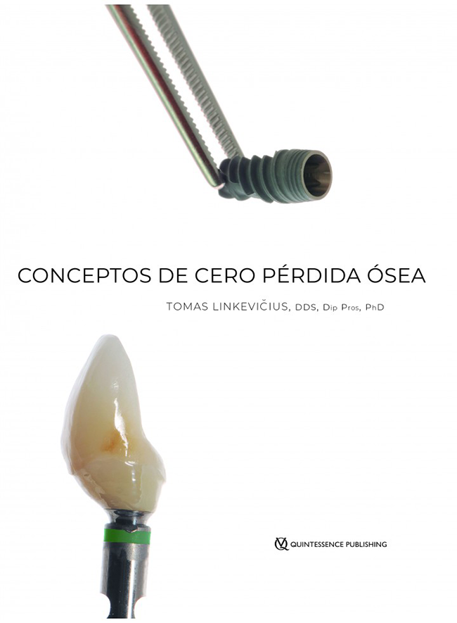
1st Edition 2019 Buch Hardcover, 21,6 x 27,9 cm, 304 Seiten, 1231 Abbildungen Sprache: Spanisch Kategorien: Implantologie, Oralchirurgie Artikelnr.: 21984 ISBN 978-84-89873-87-2 QP USA
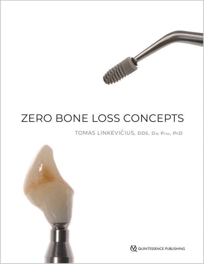
1st Edition 2019 Buch Hardcover, 21,6 x 27,9 cm, 304 Seiten, 1231 Abbildungen Sprache: Englisch Kategorien: Implantologie, Oralchirurgie Artikelnr.: 21981 ISBN 978-0-86715-799-4 QP USA
No Dentistry, No Wellness!29. Sept. 2023 — 1. Okt. 2023Pacifico Yokohama Conference Center, Yokohama, Japan
Referenten: Alessandro Agnini, Andrea Mastrorosa Agnini, Wael Att, Gustavo Avila-Ortiz, Markus B. Blatz, Victor Clavijo, Karim Dada, Glécio Vaz de Campos, Vincent Fehmer, Naoki Hayashi, Mario Imburgia, Guillaume Jouanny, Sérgio Kahn, Bertrand Khayat, Sandra Khong Tai, Christopher Köttgen, Stefen Koubi, Tomas Linkevičius, Nazariy Mykhaylyuk, Ravindra Nanda, Andreas Nolte, Léon Parienté, Jose Manuel Reuss , Domenico Ricucci, Isabella Rocchietta, Irena Sailer, Todd R. Schoenbaum, Werner Schupp, Istvan Urban, Eric Van Dooren, Débora R. Vilaboa, Otto Zuhr
Quintessence Publishing Co. Ltd. Japan
European Prosthodontic Debates 2023
14. Sept. 2023 — 16. Sept. 2023Radisson Blu Hotel Lietuva, Vilnius, Litauen
Referenten: Rolandas Andrijauskas, Adomas Auskalnis, Kazuyoshi Baba, Tord Berglundh, Lawrence E. Brecht, Luigi Canullo, Farronato Davide , Luca De Stavola, Vincent Fehmer, Marco Ferrari, German O. Gallucci, Ieva Gendviliene, Simonas Grybauskas, Sascha Hein, Reinhilde Jacobs, Matthias Kern, Tomas Linkevičius, Diego Lops, Daniele Manfredini, Francesco Mangano, Magda Mensi, Eitan Mijiritsky, Suresh Nayar, Sam Omar, George Papavasiliou, Guillermo Pradíes, Algirdas Puišys, Aušra Ramanauskaitė, Daniele Rondoni, Dmitri Ruzanov, Irena Sailer, Frank Schwarz, Nicola Scotti, Tristan Staas, Eglė Vindašiūtė-Narbutė, Ulrich Wegmann, Lukasz Zadrozny, Ausra Znamenskaite-Levickiene
15. Juni 2022 — 18. Juni 2022Bella Center Copenhagen, Copenhagen, Dänemark
Referenten: Mario Aimetti, Zvi Artzi, Serhat Aslan, Georgios Belibasakis, Florian Beuer, Juan Blanco Carrión, Michael M. Bornstein, Nagihan Bostanci, Philippe Bouchard, Darko Božić, Olivier Carcuac, Maria Clotilde Carra, Nelson Carranza, Iain L. C. Chapple, Pierpaolo Cortellini, Jan Cosyn, Mike Curtis, Francesco D'Aiuto, Bettina Dannewitz, Luca De Stavola, Jan Derks, Nikolaos Donos, Peter Eickholz, Bahar Eren Kuru, Ricardo Faria Almeida, Roberto Farina, Magda Feres, Elena Figuero, Dagmar Fosså Bunæs, Rok Gašperšič, William Giannobile, Cecilie Gjerde Gjengedal, Moshe Goldstein, Marjolaine Gosset, Klaus Gotfredsen, Filippo Graziani, Adrian Guerrero, George Hajishengallis, Hady Haririan, Lisa J. A. Heitz-Mayfield, Palle Holmstrup, Marc Hürzeler, Mark Ide, Søren Jepsen, Ronald Jung, Sérgio Kahn, Anhgela R. Kamer, Alpdogan Kantarci, Moritz Kebschull, Björn Klinge, Thomas Kocher, Odd Carsten Koldsland, Kenneth Kornman, Marja Laine, Markus Laky, Isabelle Laleman, Evanthia Lalla, France Lambert, Luca Landi, Niklaus P. Lang, Antonio Liñares, Tomas Linkevičius, Bruno Loos, Rodrigo Lopez, Eli Machtei, Aslan Mammadov, Mauro Merli, Andrea Mombelli, Eduardo Montero, Niki Moutsopoulos, Jose Nart, Gustavo G. Nascimento, Ian Needleman, Tiernan O'Brien, William Papaioannou, Panos N. Papapanou, Michael A. Pikos, Pawel Plakwicz, Constanza Pontarolo, Philip M. Preshaw, Marc Quirynen, Mia Rakic, Christoph Andreas Ramseier, Hélène Rangé, Papageorgiou Spyridon, Maurizio S. Tonetti, Leonardo Trombelli, Istvan Urban, Fridus van der Weijden, Fabio Vignoletti, Charalambos Vlachopoulos, Nicola West, Asaf Wilensky, Ion Zabalegui, Egija Zaura, Nicola Zitzmann, Giovanni Zucchelli, Otto Zuhr, Fardal Øystein
European Federation of Periodontology (EFP)
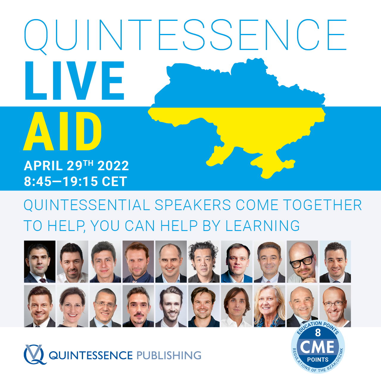
Quintessence Live Aid
Quintessential Speakers from around the world offer a Day of Education that will help those in need29. Apr. 2022Zoom
Referenten: Wael Att, Maxim Belograd, Victor Clavijo, Christian Coachman, Stefan Fickl, Ronaldo Hirata, Fouad Khoury, Tomas Linkevičius, Pascal Magne, Nazariy Mykhaylyuk, José M. Navarro, Irena Sailer, Anton Sculean, Kyle Stanley, Miguel Stanley, Markus Tröltzsch, Istvan Urban, Débora R. Vilaboa, Giovanni Zucchelli, Otto Zuhr
ausgebuchtQuintessenz Verlags-GmbH
40th Congresso Internazionale
La rivoluzione protesica: cosa è cambiato, cosa è ancora attuale18. Nov. 2021 — 20. Nov. 2021Palazzo dei Congressi Bologna, Bologna, Italien
Referenten: Ingo Baresel, Florian Beuer, Cristiano Broseghini, Mauro Broseghini, Gaetano Calesini, Roberto Canalis, Davide Cortellini, Marco Degidi, Stefano Gracis, Stefano Granata, Bernd Kordaß, Tomas Linkevičius, Ignazio Loi, Paolo Miceli, Ricardo Mitrani, Nazariy Mykhaylyuk, Piotr Nagadowsky, José M. Navarro, Jan Schünemann, Clemens Schwerin, Lorenzo Trevisiol, Luca Vailati, Marco Valenti, Naoto Yuasa
Accademia Italiana di Odontoiatria Protesica (AIOP)
EAO Digital Days
Implantology: Beyond your expectations12. Okt. 2021 — 14. Okt. 2021online
Referenten: Enrico Agliardi, Alessandro Agnini, Andrea Mastrorosa Agnini, Mauricio Araujo, Goran Benic, Juan Blanco Carrión, Daniel Buser, Raffaele Cavalcanti, Tali Chackartchi, Luca Cordaro, Jan Cosyn, Holger Essig, Vincent Fehmer, Stefan Fickl, Alberto Fonzar, Helena Francisco, German O. Gallucci, Ramin Gomez-Meda, Oscar Gonzalez-Martin, Robert Haas, Arndt Happe, Alexis Ioannidis, Ronald Jung, Niklaus P. Lang, Tomas Linkevičius, Iva Milinkovic, Sven Mühlemann, Katja Nelson, Sergio Piano, Michael A. Pikos, Bjarni E. Pjetursson, Marc Quirynen, Franck Renouard, Isabella Rocchietta, Dennis Rohner, Irena Sailer, Henning Schliephake, Shakeel Shahdad, Massimo Simion, Ali Tahmaseb, Hendrik Terheyden, Jochen Tunkel, Stefan Vandeweghe, Piero Venezia, Stijn Vervaeke, Martin Wanendeya, Georg Watzek, Giovanni Zucchelli
European Association for Osseintegration (EAO)
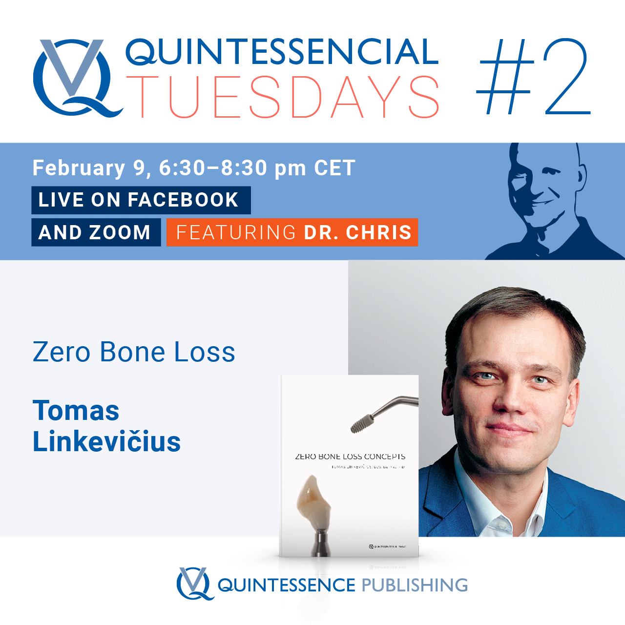
FREE - Quintessencial Tuesday with Dr. Chris #2
Zero Bone Loss Concepts with Dr. Tomas Linkevičius9. Feb. 2021
Referenten: Christopher Köttgen, Tomas Linkevičius
Quintessenz Verlags-GmbH
Zeitschriftenbeiträge dieses Autors
The International Journal of Prosthodontics, 5/2023
DOI: 10.11607/ijp.6896, PubMed-ID: 33651033Seiten: 588-594, Sprache: EnglischLinkevicius, Tomas / Gineviciute, Evelina / Alkimavicius, Jonas / Andrijauskas, Rolandas / Sakalauskas, Danas / Linkeviciene, Laura
Purpose: To evaluate the efficacy of cleaning protocols for the decontamination of organic compounds from polished zirconium oxide samples. Materials and Methods: A total of 24 rectangular plate specimens were sintered from zirconium oxide. All samples were polished with commercially available polishers (coarse, fine, and superfine) and polishing paste. During the first step of the protocol, all specimens were cleaned with steam. Samples were then randomly assigned to one of three groups (n = 8 each): A, B, or C. In group A, no additional cleaning was performed, while specimens in group B underwent ultrasonic cleaning in distilled water. Group C specimens were cleaned in an ultrasonic bath with a special detergent solution. After washing, samples were subjected to energy-dispersive x-ray spectroscopy (EDX) and scanning electron microscopy (SEM) examination. In order to detect organic materials, the level of carbon atoms was measured. Results: EDX analysis revealed that samples in group A had the highest percent of carbon atoms (9.57 ± 3.67) on the surface compared to other cleaning protocols. Following the Group B cleaning protocol resulted in lower carbon levels (4.73 ± 3.56), but this difference was not significant compared to group A (P = .439). None of the specimens in group C had detectable carbon atoms (0), which implies that all wax molecules were removed (P < .05). Conclusion: Total decontamination of organic compounds from a polished zirconium surface can be expected only following the C cleaning protocol; therefore, it is advised to employ an ultrasonic bath with detergent solution for cleaning procedures of zirconium abutments before delivery. Int J Prosthodont 2023;36:588–594.
Purpose: To assess excess cement removal after cementation of implant-supported cement-retained restorations with different cements.
Material and Methods: A dental model with imitation soft tissue, 20 individual zirconium oxide abutments, and 20 zirconium oxide crowns were fabricated. Half of the restorations were cemented using resin cement (RX) and the other half with resin-modified glass-ionomer cement (GC). After cement cleaning, each abutment-crown unit was removed from the model, photographed, and analyzed on four surfaces, resulting in a final sample size of 80 measurements. Radiographic examination and the computerized planimetric method in Adobe Photoshop were used to determine the amount of cement left and evaluate the ratio between the area of cement residue and all abutment-crown surfaces. Significance was set to .05.
Results: GC resulted in 7.4% more cement residue on all surfaces (P < .05) than RX. The P value on three surfaces (all except the mesial) was < .05, indicating that the data were statistically significantly different between groups and surfaces. Complete removal of the cement was impossible in all cases (100%), but in 95% of cases, cement remnants could not be detected radiographically.
Conclusions: More undetected cement remains when using GC. It was impossible to remove excess of both types of cement completely. Most of the cement remnants were located on the distal surface. Radiographic examination could not be considered as a reliable method to identify excess cement.
The International Journal of Oral & Maxillofacial Implants, 2/2022
DOI: 10.11607/jomi.9406Seiten: 320-327, Sprache: EnglischLinkevicius, Tomas / Alkimavicius, Jonas / Linkevicius, Rokas / Gineviciute, Evelina / Linkeviciene, Laura
Purpose: To determine the effect of 0.7- and 2.4-mm transmucosal abutment height titanium bases on the crestal bone stability and peri-implant soft tissue condition of bone-level implants with platform switching in patients with vertically thick soft tissues.
Materials and methods: Sixty bone-level platform-switched implants were placed in the molar and premolar regions of both arches in 60 patients. All epicrestally inserted nonsubmerged implants had a 4.1-mm diameter and, after osteointegration, were randomly allocated into two groups: (1) the short group, with a titanium base of 0.7-mm transmucosal abutment height, and (2) the high group with a 2.4-mm height. Monolithic zirconia restorations were fabricated for all implants. Parallel intraoral radiographs were obtained after the delivery of restorations (T1) and after 1 year (T2). Crestal bone levels and peri-implant soft tissue conditions were calculated for each implant. The significance level was set at α = .05.
Results: After 1 year, 55 patients were evaluated, with a mean bone loss of 0.6 ± 0.51 mm (median: 0.71, range: 0 to 2.09 mm) in the short group (23 patients) and 0.45 ± 0.59 mm (median: 0.65, range: 0 to 2.12 mm) in the high group (22 patients), showing no significant difference between groups (P = .168). A significant increase in marginal bone height was noted between the T1 and T2 time points in the short and high (P = .029 and .001, respectively) groups. The peri-implant soft tissue health parameters did not show statistically significant differences.
Conclusion: Crestal bone stability after 1 year of follow-up around epicrestally placed platform-switched implants is not influenced by transmucosal abutment height, if the vertical soft tissue thickness is ≥ 3 mm.
Schlagwörter: prosthodontics, randomized controlled clinical trial, single implant
International Journal of Periodontics & Restorative Dentistry, 3/2021
Seiten: 347-355, Sprache: EnglischZukauskas, Saulius / Puisys, Algirdas / Andrijauskas, Paulius / Zaleckas, Linas / Vindasiute-Narbute, Egle / Linkevičius, Tomas
This case control study measured early crestal bone changes around subcrestally placed platform-switched implants surrounded by thin soft tissue and compared them with regular, matching-platform implants placed in a supracrestal position and surrounded by thick soft tissue. Sixty-six patients received two-piece internal hex dental implants. Control group patients (n = 33) received implants that had a horizontally matching implant-abutment connection and were placed approximately 0.5 to 1 mm supracrestally. Test group patients (n = 33) received platform-switched implants that were placed about 1.5 mm subcrestally. Clinical examinations were conducted, intraoral radiographs were taken, and statistical analysis was performed. After 2 months, the mean bone loss was 0.2 mm (SD: 0.22 mm; range: 0.1 to 1.2 mm) in the control group and –0.69 mm (SD: 0.65 mm; range: 0 to 2.6 mm) in the test group; this difference was found to be statistically significant (P < .05). After 1 year, mean bone loss was 0.28 mm (SD: 0.36 mm; range: 0.1 to 1.63 mm) in the control group and –0.6 mm (SD: 0.55 mm; range: 0.05 to 1.8 mm) in the test group. Platform-switched implants placed in a subcrestal position in vertically thin soft tissues showed statistically significantly more bone loss than non–platformswitched implants placed supracrestally with vertically thick tissues.
Purpose: The purpose of this study was to survey practicing clinicians and determine if differences existed concerning their use of torque-limiting devices (TLDs) and screw-tightening protocols, comparing this with existing universal industry standards.
Materials and methods: A nine-question survey was administered with 428 dentists providing data for three specific areas: (1) demographic information-TLD ownership, device age, frequency of use, and observations of screw loosening; (2) recognition information-calibration, reading measurements of the TLD, and the meaning of preload; (3) usage information-screw-tightening protocols and effect of speed during actioning of the TLD. Data collection was compared with industry standards for use of hand torque tools including ISO-6789 1,2:2017 and related texts pertaining to screw fastener protocols.
Results: The beam-type TLD was the most popular; however, 33% surveyed used it incorrectly. Most TLDs being used were older than 1 year, with only 6% calibrated. Forty-eight percent observed screw loosening less than once per year, while 44% reported three or more occurrences per year. A similar number used the TLD for implant placement and abutment screw tightening. Screw-tightening protocols varied. Preload was not understood by the majority of those surveyed.
Conclusion: Dentistry does not appear to adhere to the protocols and standards recommended by other industries that also rely on screw-fastening mechanisms and TLDs. Further education and training appears to be warranted in this area of implant dentistry to reduce the risks of screw-associated complications.
Schlagwörter: abutment screw loosening, calibration, implant screws, preload, screw-tightening, torque-limiting device
Purpose: To evaluate the surface roughness values of zirconium oxide samples that were gradually polished using a commercially available polishing system and polishing paste.
Materials and Methods: A total of 50 rectangular specimens of predetermined size (10 × 10 × 3 mm) were sintered from zirconium oxide. Samples were randomly assigned to one of five groups (n = 10 each): control, coarse (Co), fine (F), super fine (SF), or polishing paste (PP). In the control group, no polishing was done; in the Co group, a coarse polisher was used; and the specimens in the remaining three groups underwent additional processing with a fine rubber abrasive. For SF and PP samples, subsequent treatment with a super fine polisher was applied. Finally, for the PP group, a goat-hair brush with diamond polishing paste was used. An optical profilometer was used to evaluate roughness average (Ra) in micrometers (μm). ANOVA and Games-Howell post hoc tests were utilized to detect differences between groups. The significance level was set to α = .05.
Results: Surface roughness gradually decreased with further polishing throughout the groups: control Ra = 0.525 ± 0.099 μm; Co Ra = 0.252 ± 0.038 μm; F Ra = 0.196 ± 0.035 μm; SF Ra = 0.114 ± 0.031 μm; and PP Ra = 0.054 ± 0.020 μm. Statistically significant differences were detected among all groups (P .05).
Conclusion: A surface roughness of 0.054 μm can be achieved if a full zirconia polishing protocol is used. Zirconium oxide can be polished to various surface roughnesses using commercially available polishing products.
This case series aimed to clinically and histologically evaluate porcine-derived membrane used for vertical thickening of thin soft tissues. Twenty porcinederived collagen membranes and bone-level implants were placed in 20 patients. After 2 months, thickened soft tissues were measured and biopsy samples were harvested. All xenografts healed successfully. The average thickness of thin soft tissue before vertical thickening was 1.65 ± 0.36 mm, while tissue thickness increased to 3.45 ± 0.52 mm after the procedure (P .001); the mean thickness increase was 1.8 ± 0.13 mm. Histologic analysis showed complete integration of the graft and no differences (P = .4578) in vascularization between the host (39.74 ± 17.15 vessels/mm2) and graft (30.43 ± 11.26 vessels/mm2). It can be concluded that porcine-derived membrane can be used for vertical soft tissue thickening with substantial gain in tissue height.
Current use of zirconium oxide (ZrO₂)-based screw-retained restorations does not guarantee maximum contact of soft peri-implant tissues with ZrO₂, because veneering porcelain usually covers the major subgingival part of the restoration. Ceramics preclude direct interaction between zirconia and soft tissue cells, thus reducing biocompatibility and benefit to the patient. The four case reports discussed in this article describe the new design modality of the ZrO₂ screwretained restorations, in which zirconia is exposed to the tissues and no veneering porcelain is located below the gingival margin. The article also shows the impact of this treatment on soft peri-implant tissues after 3 years of follow-up. Soft tissue recession, vestibular contour, bleeding on probing, and probing depth were evaluated.
Purpose: The aim of this clinical trial was to evaluate the influence of gingival tissue thickness on crestal bone loss around dental implants after a 1-year follow-up.
Materials and Methods: Forty-six implants (23 test and 23 control) were placed in 19 patients. The test implants were placed about 2 mm supracrestally, whereas the control implants were positioned at the bone level. Before implant placement, the tissue thickness at implant sites was measured with a periodontal probe. After healing, metal-ceramic cement-retained prostheses were constructed. According to tissue thickness, the test implants were divided into A (thin) and B (thick) groups. Intraoral radiographs were performed and crestal bone changes were measured at implant placement and after 1 year.
Results: Mean bone loss around the test implants in group A (thin mucosa) was 1.61 ± 0.24 mm (SE; range, 0.9 to 3.3 mm) on the mesial and 1.28 ± 0.167 mm (range, 0.8 to 2.1 mm) on the distal. Mean bone loss in test group B (thick mucosa) implants was 0.26 ± 0.08 mm (range, 0.2 to 0.9 mm) on the mesial aspect and 0.09 ± 0.05 mm (range, 0.2 to 0.6 mm) on the distal aspect. Mean bone loss around control implants was 1.8 ± 0.164 mm (range, 0.6 to 4.0 mm) and 1.87 ± 0.166 mm (range, 0.0 to 4.1 mm) on the mesial and distal aspects, respectively. Analysis of variance revealed a significant difference in terms of bone loss between test A (thin) and B (thick) groups on both the mesial and the distal.
Conclusion: Initial gingival tissue thickness at the crest may be considered as a significant influence on marginal bone stability around implants. If the tissue thickness is 2.0 mm or less, crestal bone loss up to 1.45 mm may occur, despite a supracrestal position of the implant-abutment interface.
Schlagwörter: biologic width, crestal bone loss, dental implants, microgap, mucosal thickness
Purpose: The aim of this systematic review was to evaluate available evidence for a difference in the stability of peri-implant tissues between titanium abutments versus gold alloy, zirconium oxide, or aluminum oxide abutments.
Materials and Methods: Studies were identified by examining several electronic databases and major dental implant, prosthetic, and periodontal journals. To be selected for the preliminary article pool, the article must have been written in the English language and published from 1980 to March 2007. Articles were sorted based on the nature of the study. In vitro studies and literature reviews were excluded. The included articles were clinical, human histology, and animal studies. Case reports, case series, uncontrolled clinical trials, and clinical studies with teeth treated as a control were excluded from the final review.
Results: The initial article pool included 40 articles of which 9 met the inclusion criteria: 3 animal studies, 2 human histological studies, and 4 randomized clinical trials. Soft tissue recession was not accurately measured in the included clinical studies. Assessment of peri-implant tissues around zirconium oxide and titanium abutments was described only in animal and human histologic studies. Due to differences in study types, timing of follow-ups, and outcome variables, meta-analysis could not be performed.
Conclusions: Included studies revealed that titanium abutments did not maintain a higher bone level in comparison to gold alloy, aluminum oxide, or zirconium oxide abutments. However, there is a lack of information about the clinical performance of zirconium oxide and gold alloy abutments as compared to titanium abutments.
Schlagwörter: abutment, aluminum, biologic width, crestal bone loss, gold alloy, implant, peri-implant soft tissues, zirconium