Various cookies are used on our website: We use technically necessary cookies for the purpose of enabling functions such as login or a shopping cart. We use optional cookies for marketing and optimization purposes, in particular to place relevant and interesting ads for you on Meta's platforms (Facebook, Instagram). You can refuse optional cookies. More information on data collection and processing can be found in our privacy policy.
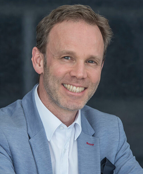
Ronald Jung is trained in oral surgery, prosthodontics and implant therapy. He is currently Head Division of Implantology at the Clinic of Reconstructive Dentistry, Center of Dental Medicine, University of Zürich. In 2006 he worked as Visiting Associate Professor at the Department of Periodontics at the University of Texas Heath Science Center at San Antonio, USA (Chairman: Prof. Dr. D. Cochran). 2008 he finalized his "Habilitation" (venia legendi) in dental medicine and was appointed at the University of Zürich. In 2011 he became his PhD doctorate degree of the University of Amsterdam, ACTA dental school, The Netherlands. In 2013 he worked as Visiting Associate Professor at the Department of Restorative Dentistry and Biomaterials Sciences at Harvard School of Dental Medicine in Boston, USA. In 2015 he has been promoted to full Professorship for Implantology at the University of Zurich. He is an accomplished and internationally renowned lecturer and researcher, best known for his work in the field of hard and soft tissue management and his research on new technologies in implant dentistry. He is currently Treasurer of the EAO, Past President of the Swiss Society of Reconstructive Dentistry, member of the Board of Directors of the Osteology Foundation and active Member of the EAED.
Details make perfection24. Oct 2024 — 26. Oct 2024MiCo - Milano Convention Centre, Milano, Italy
Speakers: Bilal Al-Nawas, Gil Alcoforado, Federico Hernández Alfaro, Sofia Aroca, Wael Att, Gustavo Avila-Ortiz, Kathrin Becker, Anne Benhamou, Juan Blanco Carrión, Dieter Bosshardt, Daniel Buser, Francesco Cairo, Paolo Casentini, Raffaele Cavalcanti, Tali Chackartchi, Renato Cocconi, Luca Cordaro, Luca De Stavola, Nuno Sousa Dias, Egon Euwe, Vincent Fehmer, Alberto Fonzar, Helena Francisco, Lukas Fürhauser, German O. Gallucci, Oscar Gonzalez-Martin, Dominik Groß, Robert Haas, Alexis Ioannidis, Simon Storgård Jensen, Ronald Jung, France Lambert, Luca Landi, Georg Mailath-Pokorny jun., Silvia Masiero, Iva Milinkovic, Carlo Monaco, Jose Nart, José M. Navarro, Katja Nelson, Manuel Nienkemper, David Nisand, Michael Payer, Sergio Piano, Bjarni E. Pjetursson, Sven Reich, Isabella Rocchietta, Giuseppe Romeo, Irena Sailer, Mariano Sanz, Ignacio Sanz Martín, Frank Schwarz, Shakeel Shahdad, Massimo Simion, Ralf Smeets, Benedikt Spies, Bogna Stawarczyk, Martina Stefanini, Hendrik Terheyden, Tiziano Testori, Daniel Thoma, Ana Torres Moneu, Piero Venezia, Lukas Waltenberger, Hom-Lay Wang, Stefan Wolfart, Giovanni Zucchelli, Otto Zuhr
Speakers: Bilal Al-Nawas, Tomas Albrektsson, Michael M. Bornstein, Daniel Buser, Norbert Cionca, Thomas Gander, Ueli Grunder, Christine Jacobsen, Ronald Jung, Sebastian Kühl, Sabrina Maniewicz, Richard J. Miron, Sven Mühlemann, Frauke Müller, Michael Payer, João Pitta, Christoph Andreas Ramseier, Philipp Sahrmann, Giovanni E. Salvi, Martin Schimmel, Patrick R. Schmidlin, David Schneider, Anton Sculean, Dagmar Simon, Alexandra Stähli, Karl Ulrich Volz, Hannes Wachtel, Nicola Zitzmann
Implantat Stiftung Schweiz
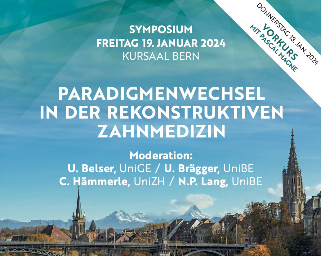
Paradigmenwechsel in der Rekonstruktiven Zahnmedizin
Auswirkungen der Fortschritte in Dentinadhäsion, Implantologie, Digitalisierung & Materialentwicklung für die tägliche Praxis18. Jan 2024 — 19. Jan 2024Congress Center, Kursaal Bern, Bern, Switzerland
Speakers: Urs C. Belser, Urs Brägger, Daniel Buser, Didier Dietschi, Vincent Fehmer, Ueli Grunder, Christoph Hämmerle, Ronald Jung, Niklaus P. Lang, Pascal Magne, Konrad H. Meyenberg, Frauke Müller, Irena Sailer, Martin Schimmel, Anton Sculean, Nicola Zitzmann
Bern Congress Organiser
The Buser & Belser Master Course on Esthetic Implant Dentistry
3 Live-Surgeries, 16 Lectures, 3 Hands-on Workshops (on-site only), 1 Social Dinner (on-site only)11. Oct 2023 — 13. Oct 2023SITEM Insel, Bern, Switzerland
Speakers: Samir Abou-Ayash, Urs C. Belser, Dieter Bosshardt, Daniel Buser, Jordi Caballé-Serrano, Stephen Chen, Vincent Fehmer, Manrique Fonseca, Ronald Jung, Irena Sailer, Anton Sculean
Buser & Sculean Academy
4th Buser & Sculean International Symposium
Regeneration and Esthetics in Periodontology and Implant Dentistry2. Dec 2022 — 3. Dec 2022Congress Center, Kursaal Bern, Bern, Switzerland
Speakers: Sofia Aroca, Daniel Buser, Stephen Chen, Nikolaos Donos, Marc Hürzeler, Sascha Jovanovic, Ronald Jung, Giulio Rasperini, Isabella Rocchietta, Frank Schwarz, Anton Sculean, Shakeel Shahdad, Andreas Stavropoulos, Martina Stefanini, Leonardo Trombelli, Istvan Urban, Giovanni Zucchelli
Buser & Sculean Academy
2nd European Congress Ceramic Implant Dentistry
European Society for Ceramic Implantology20. Oct 2022 — 22. Oct 2022Seminarhotel Bocken, Horgen, Switzerland
Speakers: Owen Addison, Bilal Al-Nawas, Rodrigo Beltrão, Michael M. Bornstein, André Chen, Jérôme Chevalier, Jospeh Choukroun, Alessandro Devigus, Michael Gahlert, Shahram Ghanaati, Elisabeth Jacobi-Gresser, Ronald Jung, Ralf J. Kohal, Sebastian Kühl, Frank-Michael Maier, Jochen Mellinghoff, Michael Payer, Mattias Pettersson, Marc Quirynen, Stefan Röhling, Jens Tartsch
European Society for Ceramic Implantology - ESCI
SEPES – 50+ 1 Annual Congress
13. Oct 2022 — 15. Oct 2022Plaza de las Convenciones, Maspalomas, Las Palmas, Spain
Speakers: Nitzan Bichacho, Markus Blatz, Christian Coachman, Luca De Stavola, Mirela Feraru, Gustavo Giordani, Galip Gürel, Ronald Jung, Nazariy Mykhaylyuk, Nelson R. F. A. Silva, Dennis P. Tarnow
SEPES - Sociedad Española de Prótesis Estomatologías y Estética
EuroPerio10
15. Jun 2022 — 18. Jun 2022Bella Center Copenhagen, Copenhagen, Denmark
Speakers: Mario Aimetti, Zvi Artzi, Serhat Aslan, Georgios Belibasakis, Florian Beuer, Juan Blanco Carrión, Michael M. Bornstein, Nagihan Bostanci, Philippe Bouchard, Darko Božić, Olivier Carcuac, Maria Clotilde Carra, Nelson Carranza, Iain L. C. Chapple, Pierpaolo Cortellini, Jan Cosyn, Mike Curtis, Francesco D'Aiuto, Bettina Dannewitz, Luca De Stavola, Jan Derks, Nikolaos Donos, Peter Eickholz, Bahar Eren Kuru, Ricardo Faria Almeida, Roberto Farina, Magda Feres, Elena Figuero, Dagmar Fosså Bunæs, Rok Gašperšič, William Giannobile, Cecilie Gjerde Gjengedal, Moshe Goldstein, Marjolaine Gosset, Klaus Gotfredsen, Filippo Graziani, Adrian Guerrero, George Hajishengallis, Hady Haririan, Lisa J. A. Heitz-Mayfield, Palle Holmstrup, Marc Hürzeler, Mark Ide, Søren Jepsen, Ronald Jung, Sérgio Kahn, Anhgela R. Kamer, Alpdogan Kantarci, Moritz Kebschull, Björn Klinge, Thomas Kocher, Odd Carsten Koldsland, Kenneth Kornman, Marja Laine, Markus Laky, Isabelle Laleman, Evanthia Lalla, France Lambert, Luca Landi, Niklaus P. Lang, Antonio Liñares, Tomas Linkevičius, Bruno Loos, Rodrigo Lopez, Eli Machtei, Aslan Mammadov, Mauro Merli, Andrea Mombelli, Eduardo Montero, Niki Moutsopoulos, Jose Nart, Gustavo G. Nascimento, Ian Needleman, Tiernan O'Brien, William Papaioannou, Panos N. Papapanou, Michael A. Pikos, Pawel Plakwicz, Constanza Pontarolo, Philip M. Preshaw, Marc Quirynen, Mia Rakic, Christoph Andreas Ramseier, Hélène Rangé, Papageorgiou Spyridon, Maurizio S. Tonetti, Leonardo Trombelli, Istvan Urban, Fridus van der Weijden, Fabio Vignoletti, Charalambos Vlachopoulos, Nicola West, Asaf Wilensky, Ion Zabalegui, Egija Zaura, Nicola Zitzmann, Giovanni Zucchelli, Otto Zuhr, Fardal Øystein
European Federation of Periodontology (EFP)
Farewell Symposium Christoph Hämmerle
Contemporary prosthodontics: Where do we stand, where are we heading?22. Jan 2022, 9:00 — 17:30 (CET)University of Zurich Irchel Campus, Zürich, Switzerland
Speakers: Thomas Attin, Beatrice Beck Schimmer, Tord Berglundh, Rino Burkhardt, Lyndon F. Cooper, Christer Dahlin, Stefano Gracis, Christoph Hämmerle, Lisa J. A. Heitz-Mayfield, Marc Hürzeler, Ronald Jung, Nadja Nänni, Mutlu Özcan, Marc Quirynen, Irena Sailer, Christian S. Stohler, Daniel Thoma
Disconnection and reconnection of abutments multiple times have known to affect the mucosal barrier around implants leading to marginal bone loss. This clinical report describes a novel technique that amalgamates the benefits of digital technologies encompassing the fabrication of surgical guides for implant placement, customized hybrid zirconia abutments and all ceramic lithium disilicate crowns prior to implant placement. A correct 3-dimensional implant positioning along with immediate placement of the definitive hybrid customized abutment and a lithium disilicate crown has the potential to reduce treatment time, visits and costs while delivering optimal esthetic outcomes.
Background: Maxillary premolars have a unique anatomical location. This is an CBCT based study where the suitability of maxillary premolars for immediate implant placement (IIP) is evaluated. Based on prosthetically driven treatment treatment planning a simple classification system is put forth. Materials and Methods: 150 CBCTs of maxillary first premolars were analysed in BlueskyBio software. The topographic position of the tooth was determined by analysing the dimensions of the buccal and lingual cortical plates, the distance between the bucco-lingual plates and the residual bone height from the root apex to the floor of the sinus. Virtual placement of an implant was carried out such that the implant would be positioned 1 mm apical to the buccal bone crest, would engage 3 mm of bone apical to the root apex, and would have a trajectory so that the abutment access was from the central fossa. Four categories were identified and the classification was proposed. Results: It was observed that 74% of cases had buccal bone<1mm,26% had buccal bone >1mm. 79% cases had an average distance >3mm between root apex and maxillary sinus, 21% had an average distance of root apex and maxillary sinus <3mm. The categorizations of implant placement were as follows -Type 1- 24%, Type 2- 56.6%, Type 3-43.3%, Type 4- 0%. Conclusions: In majority of maxillary 1st premolars an IIP is possible with the implants to be placed in the palatal sockets or the furcation area. In cases were the buccal plate thickness is inadequate, simultaneous grafting should be considered between the implant position and buccal plate.
International Journal of Computerized Dentistry, Pre-Print
ScienceDOI: 10.3290/j.ijcd.b5394865, PubMed ID (PMID): 3880119327. May 2024,Pages 1-35, Language: EnglishSadilina, Sofya / Strauss, Franz J. / Jung, Ronald E. / Joda, Tim / Balmer, Marc
Aim: The aim of this scoping review was to identify the scientific evidence related to the utilization of Optical See- Through Head-Mounted Display (OST-HMD) in dentistry, and to determine future research needs.
Methods: The research question was formulated using the "Population" (P), "Concept" (Cpt), and "Context" (Cxt) framework for scoping reviews. Existing literature was designated as P, OST-HMD as Cpt, and Dentistry as Cxt. An electronic search was conducted in PubMed, Embase, Web of Science, and CENTRAL. Two authors independently screened titles and abstracts and performed the full-text analysis.
Results: The search identified 286 titles after removing duplicates. Nine studies, involving 138 participants and 1760 performed tests were included in this scoping review. Seven of the articles were preclinical studies, one was a survey, and one was a clinical trial. The included manuscripts covered various dental fields: three studies in orthodontics, two in oral surgery, two in conservative dentistry, one in general dentistry, and the remaining one in prosthodontics. Five articles focused on educational purposes. Two brands of OST-HMD were used: in eight studies HoloLens Microsoft was used, while Google Glass was utilized in one article.
Conclusions: The overall number of included studies was low; therefore, the available data from this review cannot yet support an evidence-based recommendation for the clinical use of OST-HMDs. However, the existing preclinical data indicate a significant capacity for clinical and educational implementation. Further adoption of these devices will facilitate more reliable and objective quality and performance assessments, as well as more direct comparisons with conventional workflows. More clinical studies must be conducted to substantiate the potential benefits and reliability for patients and clinicians.
Keywords: augmented reality, dental education, digital dentistry, mixed reality, scoping review, virtual reality
International Journal of Computerized Dentistry, 3/2023
ScienceDOI: 10.3290/j.ijcd.b3781703, PubMed ID (PMID): 36632986Pages 237-245, Language: English, GermanGil, Alfonso / Eliades, George / Özcan, Mutlu / Jung, Ronald E. / Hämmerle, Christoph H. F. / Ioannidis, Alexis
Aim: To evaluate the fracture load and type of failure of two different monolithic restorative materials bonded to standardized titanium bases and fabricated by two different procedures regarding the bonding interface.
Materials and methods: All screw-retained implant crown specimens (n = 40), subjected to fatigue by thermomechanical loading, differed in the restorative material (lithium disilicate [LDS] or polymer-infiltrated ceramic network [PICN], referred to as ‘hybrid ceramic’ [HYC]) and the interface type between the restorative material and the titanium base abutment (prefabricated ex-factory or produced during a CAM-milling procedure). This resulted in the following groups (n = 10/group): 1) LDS-M: lithium disilicate crown with a CAM-milled interface; 2) LDS-P: lithium disilicate crown with a prefabricated interface; 3) HYC-M: PICN crown with a CAM-milled interface; and 4) HYC-P: PICN crown with a prefabricated interface. Aged specimens underwent static fracture load testing. The load (N) at which the initial crack occurred was denoted as Finitial, and the maximal load (N) at which the restorations fractured as Fmax. All specimens were examined under a stereomicroscope to determine the failure mode.
Results: The median Finitial values were 180 N for LDS-M, 343 N for LDS-P, 340 N for HYC-M, and 190 N for HYC-P. The median Fmax values were 1822 N for LDS-M, 2039 N for LDS-P, 1454 N for HYC-M, and 1581 N for HYC-P. The intergroup differences were significant for Finitial (KW: P = 0.0042) and for Fmax (KW: P = 0.0010). The failure types also showed differences between the restorative groups.
Conclusions: The choice of restorative material had a stronger influence on the fracture load than the abutment interface workflow. Lithium disilicate showed the highest load for initial crack appearance (Finitial) and for complete fracture of the restoration (Fmax).
Keywords: lithium disilicate, dental materials, polymer-infiltrated ceramic network, thermomechanical aging, fracture load, failure mode, prosthetic dentistry, restorative material, abutment interface
Die wissenschaftliche Evidenz für Zirkonoxidimplantate ist in den letzten Jahren erheblich gestiegen. Mittlerweile liegen Daten aus klinischen Studien vor, die eine Beobachtungszeit von 5−7 Jahren umfassen. Dennoch fehlen weiterhin randomisierte klinische Studien, die den Einsatz von Zirkonoxidimplantaten in verschiedenen klinischen Indikationen vergleichen. Es gibt bislang keine Studie, die den direkten Vergleich zwischen kurzen und längeren Zirkonoxidimplantaten untersucht. Ebenso fehlen klinische Daten über den Einsatz von kurzen Zirkonoxidimplantaten. Als kurze Implantate sind Implantate mit einer Länge von 6 mm oder weniger definiert. 8 mm „kurze“ Zirkonoxidimplantate können jedoch erfolgreich verwendet werden. Zudem zeigen Ergebnisse aus Kohortenstudien, dass die Implantatlänge keinen Einfluss auf das marginale Knochenniveau hat. Kurze Zirkonoxidimplantate könnten im Kieferkamm mit einer reduzierten vertikalen Höhe eine kostengünstige Behandlungsoption mit geringerer Morbidität darstellen, wenn dadurch eine aufwendige Knochenaugmentation verhindert werden kann. In Situationen, in denen ausreichend vertikale Höhe vorhanden ist, scheint die Verwendung von Implantaten mit Standardlänge die sicherere Option zu sein.
Manuskripteingang: 08.07.2023, Annahme: 18.08.2023
Keywords: Zirkonoxidimplantate, vollkeramische Restaurationen, metallfreie Restaurationen, kurze Implantate, wissenschaftliche Evidenz
Ziel: Ziel der vorliegenden retrospektiven Fallserie war die Einführung eines minimalinvasiven zweistufigen lappenfreien Verfahrens für die Kronenverlängerung, das die Gingivektomie vermeidet oder minimiert, indem die Reduktion der Knochenhöhe mithilfe einer Tunnelpräparation vorgenommen wird.
Material und Methode: In diese Studie wurden neun Patientinnen und ein Patient (Altersmedian: 46,6 Jahre, Bereich: 26,9 bis 71,6 Jahre) eingeschlossen. Die Indikationen für die Durchführung der Kronenverlängerung waren: ästhetisch zu kurze Kronen (n = 5), Asymmetrie der Gingivahöhen (n = 2) und die Kombination aus diesen Indikationen (n = 3). Alle Behandelten wiesen einen dicken gingivalen Phänotyp auf, und die mittlere Anzahl der in den Eingriff einbezogenen Zähne betrug 3,7 (Bereich: 2 bis 8 Zähne). Drei Teilnehmende erhielten sechs Wochen nach der Kronenverlängerung eine zusätzliche Gingivektomie.
Ergebnisse: Die Ergebnisse erfüllten in ästhetischer und funktioneller Hinsicht die Erwartungen der Behandelten und des Zahnarztes.
Schlussfolgerung: Das minimalinvasive zweistufige Kronenverlängerungsverfahren unter Anwendung einer Tunneltechnik kann auch ohne offenen Lappen und zusätzliche Gingivektomie vorhersagbare ästhetische Ergebnisse liefern. Prospektive klinische Studien sind nötig, um die Technik zu validieren.
International Journal of Esthetic Dentistry (EN), 4/2022
Clinical ResearchPubMed ID (PMID): 36426612Pages 394-406, Language: EnglishJung, Ronald E. / Bigler, Prisca / Balmer, Marc / Bienz, Stefan P. / Hjerppe, Jenni
Aim: The purpose of the present retrospective case series was to introduce a minimally invasive two-stage flapless crown lengthening procedure where a gingivectomy was avoided or minimized by reducing the bone height through a tunneling technique.
Materials and method: Ten patients (median age 46.6 years, range 26.9 to 71.6 years) were included in the study. The indications for performing a crown lengthening procedure were esthetically short clinical crowns (n = 5), asymmetry of the gingival margin level (n = 2), and both esthetically short clinical crowns and asymmetry of the gingival margin level (n = 3). All the patients presented a thick gingival phenotype, and the mean number of operated teeth per patient was 3.7 (range 2 to 8 teeth). Three patients underwent an additional gingivectomy 6 weeks after the crown lengthening procedure.
Results: The esthetic and functional outcome of the treatments fulfilled the patients’ and dentist’s expectations.
Conclusion: A minimally invasive two-stage crown lengthening procedure applying a tunneling technique has the potential to achieve predictable esthetic outcomes without an open-flap approach and an additional gingivectomy. However, prospective clinical studies are needed to validate this technique.
Aktueller Stand und Wünsche an die neue Technologie
Wenn bei Patientinnen und Patienten restaurativer Bedarf besteht, so macht es aus biologischer Sicht Sinn, möglichst substanzschonend zu arbeiten. Nebst konventionellen Herstellungsverfahren stehen heute auch subtraktive und additive Verfahren für die Herstellung von minimal-invasiven Restaurationen zur Verfügung. Besonders im Zusammenhang mit minimal-invasiven dünnen Restaurationen zeigen subtraktive Fertigungstechniken ihre Limitationen. Die additive Fertigung von Keramiken für minimal-invasive Restaurationen birgt deshalb großes Potenzial. Erste In-vitro-Versuche liefern hoffnungsvolle Ergebnisse. Diese müssen jedoch noch weiter in vitro und später klinisch bestätigt werden.
Keywords: additive Fertigung, minimal-invasive Restaurationen, Lithiumdisilikat, Passung, Belastbarkeit
Festsitzender Zahnersatz, der von oralen Zahnimplantaten getragen wird, ist in der täglichen Praxis zu einer zuverlässigen Behandlungsoption geworden. Dennoch stellt der Ersatz von zwei benachbarten fehlenden Zähnen insbesondere bei ungünstigen anatomischen Verhältnissen eine klinische Herausforderung dar. Um diese Herausforderung zu meistern, hat die Verwendung von implantatgetragenem festsitzendem Zahnersatz mit einem Anhänger an Popularität gewonnen. Dies ist vor allem darauf zurückzuführen, dass keine oder weniger chirurgische Primäreingriffe erforderlich sind, die Kosten geringer ausfallen und somit die Morbidität des Patienten reduziert wird. Ziel der vorliegenden Übersichtsarbeit ist es, den aktuellen Stand der Erkenntnisse über die Verwendung von implantatgetragenem festsitzendem Teilzahnersatz mit Freiendverlängerung zusammenzufassen. Der Artikel befasst sich sowohl mit dem Seitenzahn- als auch mit dem Frontzahnbereich, wobei die Vor- und Nachteile der einzelnen Behandlungen hervorgehoben und die verfügbaren mittel- bis langfristigen Ergebnisse dargestellt werden.
Manuskripteingang: 13.05.2022, Manuskriptannahme: 27.07.2022
Keywords: Zahnimplantate, Freiendverlängerung, klinische Studie