Various cookies are used on our website: We use technically necessary cookies for the purpose of enabling functions such as login or a shopping cart. We use optional cookies for marketing and optimization purposes, in particular to place relevant and interesting ads for you on Meta's platforms (Facebook, Instagram). You can refuse optional cookies. More information on data collection and processing can be found in our privacy policy.
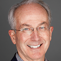
Edward P. Allen, DDS, PhD has served as president of the American Academy of Esthetic Dentistry, the American Academy of Restorative Dentistry, and the American Academy of Periodontology Foundation. He is the recipient of the Master Clinician Award from the American Academy of Periodontology, the President’s Award for Excellence in Dental Education from the American Academy of Esthetic Dentistry, and the Saul Schluger Award for Excellence in Diagnosis and Treatment Planning. In 2019, he was honored with the AAP Gold Medal Award, the highest award bestowed by the Academy. Currently, he serves on the editorial boards of the Journal of Esthetic and Restorative Dentistry, the Journal of Periodontology, and the International Journal of Periodontics and Restorative Dentistry. Dr Allen is founder of the Center for Advanced Dental Education in Dallas, an educational facility where he teaches surgical technique courses. He has over 100 publications and has presented numerous lectures and surgical demonstrations worldwide.
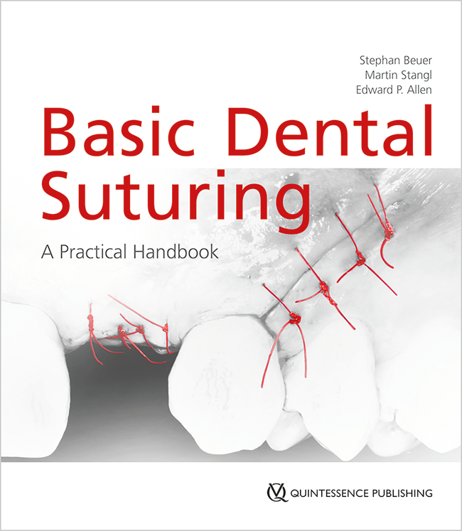
1st Edition 2024 Book Softcover; 21 x 24 cm; incl 8 Videos, 80 pages, 244 illus Language: English Categories: General Dentistry, Oral Surgery Stock No.: 24331 ISBN 978-1-78698-123-3 QP Deutschland
Timelessness in Regeneration18. Oct 2024 — 19. Oct 2024Vigadó Concert Hall (Pesti Vigadó), Budapest, Hungary
Speakers: Edward P. Allen, Matteo Chiapasco, Lisa J. A. Heitz-Mayfield, Giulio Rasperini, Massimo Simion, Istvan Urban, Hom-Lay Wang, Giovanni Zucchelli
Urban Regeneration Institute
Spear Summit
Growing Together27. Apr 2023 — 29. Apr 2023, 13:00 — 16:45 JW Marriott Phoenix Desert Ridge, Phoenix, United States of America
Speakers: Edward P. Allen, Álvaro Blasi, Gonzalo Blasi, Ignacio Blasi, Rebecca Bockow, Gary DeWood, Darin Dichter, Didier Dietschi, Michael Gunson, Gregg Kinzer, Pascal Magne, George Mandelaris, Ricardo Mitrani, Amy Morgan, Jason Smithson, Frank Spear, Eric Van Dooren, Robert Winter
Spear Education
The 14th International Symposium on Periodontics and Restorative Dentistry (ISPRD)
9. Jun 2022 — 12. Jun 2022Boston Marriott Copley Place, Boston, MA, United States of America
Speakers: Tara Aghaloo, Edward P. Allen, Evanthia Anadioti, Wael Att, Vinay Bhide, Markus B. Blatz, Scotty Bolding, Lorenzo Breschi, Jeff Brucia, Daniel Buser, Luigi Canullo, Daniele Cardaropoli, Stephen J. Chu, Donald Clem, Christian Coachman, Lyndon F. Cooper, Daniel Cullum, Lee Culp, José Carlos Martins da Rosa, Sergio De Paoli, Marco Degidi, Nicholas Dello Russo, Serge Dibart, Joseph P. Fiorellini, Mauro Fradeani, Stuart J. Froum, David Garber, Maria L. Geisinger, William Giannobile, Luca Gobbato, Ueli Grunder, Galip Gürel, Chad Gwaltney, Christoph Hämmerle, Robert A. Horowitz, Marc Hürzeler, David Kim, Gregg Kinzer, Christopher Köttgen, Ina Köttgen, Purnima S. Kumar, Burton Langer, Lydia Legg, Pascal Magne, Kenneth A. Malament, Jay Malmquist, George Mandelaris, Pamela K. McClain, Michael K. McGuire, Mauro Merli, Konrad H. Meyenberg, Craig M. Misch, Julie A. Mitchell, Marc L. Nevins, Myron Nevins, Michael G. Newman, Miguel A. Ortiz, Jacinthe M. Paquette, Stefano Parma-Benfenati, Michael A. Pikos, Giulio Rasperini, Pamela S. Ray, Christopher R. Richardson, Isabella Rocchietta, Marisa Roncati, Marco Ronda, Paul S. Rosen, Maria Emanuel Ryan, Irena Sailer, Maurice Salama, David M. Sarver, Takeshi Sasaki, Todd Scheyer, Massimo Simion, Michael Sonick, Sergio Spinato, Dennis P. Tarnow, Lorenzo Tavelli, Douglas A. Terry, Tiziano Testori, Carlo Tinti, Istvan Urban, Hom-Lay Wang, Robert Winter, Giovanni Zucchelli
Quintessence Publishing Co., Inc. USA
This author's journal articles
Quintessence International, 7/2021
DOI: 10.3290/j.qi.b1098307, PubMed ID (PMID): 33749221Pages 576-582, Language: EnglishSculean, Anton / Allen, Edward P. / Katsaros, Christos / Stähli, Alexandra / Miron, Richard J. / Deppe, Herbert / Cosgarea, Raluca
Objectives: To describe the step-by-step procedure of a novel surgical technique consisting of a combination of the laterally closed tunnel (LCT) and the modified coronally advanced tunnel (MCAT) (ie, LCT/MCAT), designed to treat multiple mandibular adjacent gingival recessions (MAGR) and to present the clinical outcomes obtained in 11 consecutively treated patients.
Method and materials: Eleven systemically and periodontally healthy patients (7 females, mean ± SD 33.62 ± 14.6 years, min. 19 years max. 67 years) with a total of 40 adjacent mandibular RT1 (ie, Miller Class 1 and 2) gingival recessions with a minimum depth ≥ 3 mm, were consecutively treated with LCT/MCAT, in conjunction with an enamel matrix derivative (EMD) and subepithelial palatal connective tissue graft (SCTG). Treatment outcomes were assessed at baseline and at 12 months postoperatively. Prior to surgery and at 12 months postoperatively, recession depth (RD) and recession width (RW) were evaluated. The primary outcome variable was complete root coverage (CRC, ie 100% root coverage), the secondary outcome was mean root coverage (MRC).
Results: Postoperative pain and discomfort were low and the healing was uneventful in all cases without any complications. At 12 months, statistically significant (P < .05) root coverage (RC) was obtained in all patients. CRC was obtained in five patients with a total of 21 recessions, while MRC measured 92.9% (ie, 3.75 mm). In seven patients (ie, 63.6%), RC amounted to > 93% while the minimum RC per patient measured 83.76%.
Conclusion: The results of the present case series suggest that the LCT/MCAT is a valuable technique for the treatment of mandibular RT1 MAGR.
Keywords: laterally closed tunnel, modified coronally advanced tunnel, multiple adjacent mandibular gingival recessions, recession coverage, subepithelial palatal connective tissue graft, surgical technique
It is often difficult to perform the tunnel technique for root coverage and soft tissue augmentation in the mandibular anterior region where there is shallow recession and thin soft tissue, particularly when accompanied by prominent roots and alveolar undercuts. The aim of this report is to present a papilla access technique that facilitates tunnel site preparation and graft placement in such sites. This surgical access method may also be applied to sites where there is moderate to severe recession of the mandibular central incisors, and extension of the tunnel to include both the lateral incisors and canines is desired for augmentation. The papillary access provides improved surgical access for both site preparation and graft placement with reduced risk of perforation or injury to thin tissue.
Predictable coverage of deep isolated mandibular gingival recessions is one of the most challenging endeavors in plastic-esthetic periodontal surgery, and limited data is available in the literature. The aim of this paper is to present the rationale, the step-by-step procedure, and the results obtained in a series of 24 patients treated by means of a novel surgical technique (the laterally closed tunnel [LCT]) specifically designed for deep isolated mandibular recessions. A total of 24 healthy patients (21 women and 3 men, mean age 25.75 ± 7.12 years) exhibiting one single deep mandibular Miller Class I (n = 4), II (n = 10), or III (n = 10) gingival recession ≥ 4 mm were consecutively treated with LCT in conjunction with an enamel matrix derivative (EMD) and palatal subepithelial connective tissue graft (SCTG). The following clinical parameters were assessed at baseline and 12 months postoperatively: probing depth (PD), clinical attachment level (CAL), complete root coverage (CRC), mean root coverage (MRC), recession depth (RD), and keratinized tissue width (KTW). The primary outcome variable was CRC. The postoperative morbidity was low, and no complications, such as bleeding, infections/abscesses, or loss of SCTG, occurred. At 12 months, CRC was obtained in 17 of the 24 defects (70.83%), while in the remaining 7 defects RC amounted to 80% to 90% (in 6 cases) and 79% (in 1 case). Of the 17 defects exhibiting CRC, 12 were central incisors and 5 were canines. With respect to defect type, CRC was found in 3 of the 4 Miller Class I, 8 of the 10 Class II, and in 6 of the 10 Class III defects. Mean RD changed from 5.14 ± 1.26 mm at baseline to 0.2 ± 0.37 mm at 12 months, while MRC amounted to 4.94 ± 1.19 mm, representing 96.11% (P .0001). Mean KTW increased from 1.41 ± 1.00 mm at baseline to 4.14 ± 1.67 mm (P .0001) at 12 months, yielding a KTW gain of 2.75 ± 1.52 (P .0001). No statistically significant changes in mean PD occurred following root coverage surgery (1.8 ± 0.2 mm at baseline and 2.1 ± 0.3 mm at 12 months). The present results suggest that the LCT is a valuable approach for the treatment of deep isolated mandibular Miller Class I, II, and III gingival recessions.
International Journal of Periodontics & Restorative Dentistry, 2/2011
PubMed ID (PMID): 21491016Pages 165-173, Language: EnglishStimmelmayr, Michael / Allen, Edward P. / Gernet, Wolfgang / Edelhoff, Daniel / Beuer, Florian / Schlee, Markus / Iglhaut, Gerhard
Covering exposed roots becomes more and more difficult as the gingiva becomes thinner and the vestibule becomes more shallow. Also, the outcome becomes less predictable. In addition, where there is high frenal attachment or muscle pull, such as the mentalis muscle in the mandibular anterior region, secondary retraction of a coronally advanced flap will likely occur. Therefore, a transplanted connective tissue graft may not completely cover the recession. This case series presents a technique where the roots are covered with a combination epithelializedsubepithelial connective tissue graft. The epithelialized portions of the graft are positioned directly over the exposed roots to aid in resistance to the environment of the mouth, and there is no displacement of the mucogingival junction or flattening of the vestibule.
This paper describes a new suturing method, the subpapillary continuous sling suture, for use with soft tissue grafts in tunnel procedures to treat gingival recession. This method combines the graft suture and the sutures used to advance the pouch margins over the graft into a single continuous sling suture. It is indicated particularly for sites with shallow recessions and those treated for augmentation rather than root coverage because of a lack of graft access for standard suture placement. The single-suture method may also be used for sites with moderate to severe recession. The advantages of this method include elimination of the need to place additional sutures for coronal advancement of the pouch, resulting in reduced suturing time and reduced opportunity to inadvertently cut the continuous suture with the needle when suturing the pouch.
An esthetic implant-supported rehabilitation continues to be a major challenge in patients with a thin periodontium. Ridge preservation and immediate implant placement are intended to preserve the hard tissue volume and prevent preimplant bone loss following tooth extraction. Since these techniques are almost always combined with bone grafting, primary wound closure is indispensable. Therefore, a technique for reliable wound closure was developed. This technique employs a combined epithelized-subepithelial connective tissue graft, leaves the mucogingival line in its place, and has the added advantage of thickening the buccal soft tissue with the resultant local conversion of a thin marginal gingiva to a thick marginal gingiva.
The use of connective tissue grafts for root coverage and ridge augmentation is a proven, effective treatment modality. Complications associated with the palatal donor site can arise because of incomplete primary closure of the palatal wound or sloughing of the overlying tissue. This article presents a new technique for the atraumatic harvesting of connective tissue grafts from palatal donor sites. The main advantage of this single-incision technique is the primary closure of the palatal flap, resulting in less pain and sensitivity and fewer postoperative complications. A review of the technique and its indications, rationale, and limitations is presented.