Qdent, 2/2022
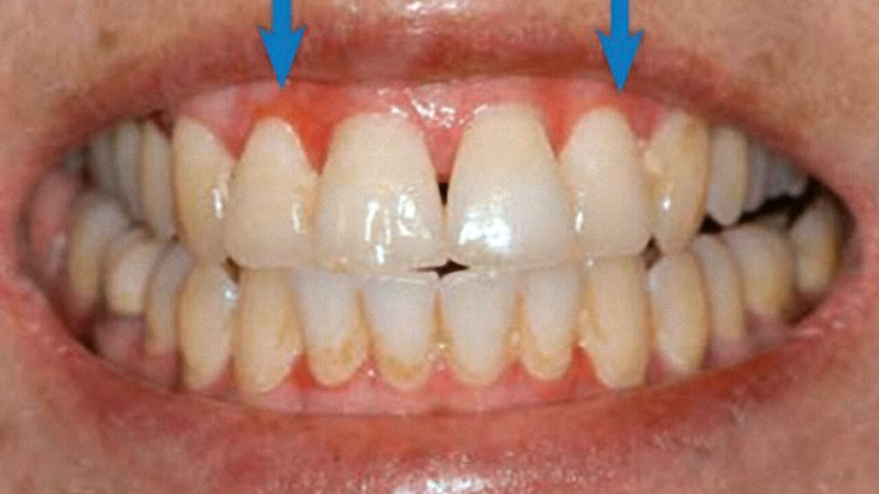
RatgeberPages 40-45, Language: GermanGeibel, Margrit-AnnOrale Inspektion Die orale Inspektion der Mundhöhle durch das geschulte Auge der Zahnärztin und des Zahnarztes gewinnt für die Patienten/-innen zunehmend an Bedeutung. Aus der Sicht derer ist die „Präventive Medizin“ das große Thema der Zukunft. Warum ist in diesem wichtigen Feld die Zahnmedizin eine bisher unterbewertete Dienstleisterin?
Dentista, 2/2019
Pages 49-50, Language: GermanMöller, David / Jerg-Bretzke, Lucia / Geibel, Margrit-AnnEin PlädoyerSenioren-Zahnmedizin, 2/2018
Pages 101-106, Language: GermanOtt, Marie-Carolin / Jerg-Bretzke, Lucia / Geibel, Margrit-AnnEine systematische LiteraturrechercheViele Patienten zahnärztlicher Einrichtungen leiden an Zahnbehandlungsangst. Der Angstentwicklung liegen unterschiedliche Entstehungsgründe, wie z. B. Traumata oder schlechte Erfahrungen, zugrunde. Die Intensität reicht dabei vom unguten Gefühl bis hin zu pathologisch starken Angstzuständen.
Unbestritten trägt die orale Gesundheit einen großen Teil zur Lebensqualität bei. Da vor allem das ältere Patientenkollektiv im zunehmenden Alter immer häufiger mit allgemeinen und oralen Erkrankungen belastet ist, sollte dieser Patientengruppe mehr Zuwendung geschenkt werden.
Mit einer systematischen Literaturrecherche in unterschiedlichen Datenbanken wird in diesem Beitrag der derzeitige Forschungsstand zum Thema Zahnbehandlungsangst geriatrischer Patienten analysiert und dargestellt. Mittels der Recherche konnte herausgearbeitet werden, dass sich die wissenschaftliche Forschung derzeit verhältnismäßig weniger mit dem geriatrischen Patientenkollektiv beschäftigt. Zudem sind zum jetzigen Zeitpunkt nur wenig aussagekräftige Publikationen vorhanden, die eine Geschlechterdifferenzierung vornehmen.
International Journal of Oral Implantology, 2/2017
PubMed ID (PMID): 28555209Pages 197-211, Language: EnglishGeibel, Margrit-Ann / Schreiber, Elena / Bracher, Anna-Katinka / Hell, Erich / Ulrici, Johannes / Sailer, Leif-Konradin / Rasche, VolkerPurpose: The aim of this case series was to compare magnetic resonance imaging (MRI) with cone beam computed tomography (CBCT) in the representation of periapical osteolyses. Based on the histological findings, the potential of MRI for further lesion characterisation was investigated. Materials and methods: Thirteen patients (average age: 41 ± 27 years) with a total of 15 periapical lesions (five molars, five premolars, and five front teeth) were examined. Lesion characterisation was based on the homogeneity/heterogeneity of the lesions, the signal intensity within the lesion compared to the surrounding tissue and differences in the signal intensities between different MRI contrast weightings. Results were compared with CBCT and histological findings. Results: Although all patients presented with dental restorations, such as fixed partial dentures and filling materials, all periapical lesions could be diagnosed with either imaging modality. Histologically, 13 cysts and two apical granuloma were confirmed. In CBCT, the similar appearance of all lesions did not allow any further characterisation. In MRI, radicular cysts and granuloma could be characterised by their appearance in the MRI images with different contrast weightings. The MRI-derived characterisations were consistent with the histological findings. Conclusions: The presented study shows that the application of multi-contrast MRI may lead to better characterisation of apical lesions, thus enabling an improved patient-specific selection of the optimal treatment option.
Keywords: apical osteolysis, cone-beam computer tomography, dental MRI
Conflict-of-interest statement: MAG, ESS, and LKS do not report any potential conflict-of-interest; EH and JU are employees of Sirona Dental Systems; VR is receiving a research grant by Sirona Dental Systems.
International Journal of Computerized Dentistry, 3/2013
PubMed ID (PMID): 24364192Pages 201-207, Language: English, GermanSailer, Benjamin F. / Geibel, Margrit-AnnVariations in angulation of the x-ray tube affect the appearance of insufficient approximal crown margins on intraoral radiographs. This study examines the impact of such angular variation on the assessment of digital radiographs using three different X-ray tubes - Heliodent DS (Sirona), Gendex Expert DC (KaVo Dental) and Focus (KaVo Dental) - as well as the Gendex Visualix eHD CCD sensor (KaVo Dental). The test specimens, crowned teeth 46 from two mandibles provided by the Institute of Anatomy and Cell Biology, were examined with each tube.The results indicate great differences in the angles indicative of insufficient crown margins on X-ray images. Because of beam divergence and the crown marginal gap, the length and width of which frequently varies, it is difficult to infer any optimum angle from the data. This leads to the conclusion that at present, it is not possible to establish ideal angles for visualization of insufficient approximal crown margins.
Keywords: X-ray, intraoral, digital, crown margin, angulation, angle
International Journal of Computerized Dentistry, 4/2012
PubMed ID (PMID): 23457899Pages 287-296, Language: English, GermanNeller, Heiner / Geibel, Margrit-AnnPurpose: To test for differences in image quality between 3D volumetric datasets acquired by cone beam computed tomography (CBCT) with and without simulation of head motion by visual analysis of individual image sections and by comparison of scan-based distance and Hounsfield unit (HU) measurements.
Materials and Methods: A total of 200 volumetric datasets were acquired with and without remote-controlled, simulated movement of a human cadaver head using the KaVo 3D eXam CBCT system.
Results: The "Landscape 8 x 8 cm Slide" mode provided a sufficient field of view at a low radiation dose. All datasets showed reproducible results. Our analysis showed that the level of image quality and image detail increased with increasing resolution. Linear distance measurements and HU measurements in the cone beam CT scans acquired without simulation of head motion were absolutely comparable to those obtained with head motion simulation.
Conclusions: There is no practical difference in image quality between CBCT scans acquired with and without head motion if no long-term change in head position occurs during the acquisition process. Slight head movement has no clinically relevant effect on the geometric accuracy or visual image quality of cone beam CT scans.
Keywords: Cone beam computed tomography, motion artifact, head motion, distance measurement, Hounsfield unit, image quality
Quintessenz Zahnmedizin, 1/2012
Bildgebende VerfahrenPages 101-106, Language: GermanGeibel, Margrit-AnnWie hilfreich ist die Umsetzung von Qualitätsmanagement für das Röntgen?In der zahnärztlichen Radiologie wird für die Dokumentation und Qualitätssicherung ein großer Aufwand betrieben. Die Einführung eines internen Qualitätsmanagement-Systems, wie es seit Ende 2010 von den Praxen verbindlich gefordert wird, ist hilfreich bei der Umsetzung von gesetzgeberischen Vorgaben und Verordnungen. Nach der Implementierung des internen Qualitätsmanagements im Bereich der zahnärztlichen Radiologie müsste es für die Zahnarztpraxen wieder möglich sein, die Patientenversorgung in den Vordergrund zu stellen und gleichzeitig allen gesetzgeberischen Anforderungen nach der Röntgenverordnung 2002 gerecht zu werden.
Keywords: Qualitätsmanagement, Röntgenverordnung 2002, rechtfertigende Indikation, Qualitätsziele
International Journal of Computerized Dentistry, 1/2012
PubMed ID (PMID): 22930946Pages 35-44, Language: English, GermanHellstern, Flurina / Geibel, Margrit-AnnObjective: To evaluate the implementation of quality assurance requirements for digital dental radiography in routine clinical practice. The results should be discussed by radiation protection authorities in the context of the relevant legal requirements and current debates on radiation protection.
Materials and methods: Two hundred digital dental radiographs were randomly selected from the digital database of the Department of Dentistry's Dental and Maxillofacial Surgery Clinic, Ulm University, and evaluated for various aspects of image quality and compliance with radiographic documentation requirements. The dental films were prepared by different radiology assistants (RAs) using one of two digital intraoral radiographic systems: Sirona Heliodent DS, 60 kV, focal spot size: 0.7 mm (group A) or KaVo Gendex 765 DC, 65 kV, focal spot size: 0.4 mm (group B).
Results: Radiographic justification was documented in 70.5% of cases, and the radiographic findings in 76.5%. Both variables were documented in the patient records as well as in the software in 14% of cases. Clinical documentation of the required information (name of the responsible dentist and radiology assistant, date, patient name, department, tube voltage, tube current, exposure time, type of radiograph, film size, department and serial number of the dental radiograph) was 100% complete in all cases. Moreover, the department certified according to DIN ISO 9001:2008 specifications demonstrated complete clinical documentation of radiographic justifications and radiographic findings. The entire dentition was visible on 83% of the digital films. The visible area corresponded to the target region on 85.7% of the digital dental radiographs. Seven to 8.5% of the images were classified as "hypometric" or "hypermetric".
Conclusions: This study indicates that improvements in radiology training and continuing education for dentists and dental staff performing x-ray examinations are needed to ensure consistent high quality of digital dental radiography. Implementation of internal radiological quality assurance programs, as required by public law in Germany since 2010 (SGB V), would appear prudent.
Keywords: Quality management, quality assurance, digital radiography, dental radiograph, radiation protection, justification, dose, dose reduction
Quintessenz Zahnmedizin, 9/2007
Röntgenologie und FotografiePages 981-993, Language: GermanGeibel, Margrit-AnnEin diagnostisches QuizDie radiologische Diagnostik bei der Panoramaschichtaufnahme stellt den Zahnarzt vor unterschiedliche Probleme. Auf der einen Seite ist die Aussagefähigkeit durch das technische Verfahren selbst eingeschränkt, da der dreidimensionale Schädel zweidimensional als Schichtaufnahme mit entsprechenden Überlagerungen abgebildet wird. Auf der anderen Seite muss wegen der Größe des bildgebenden Ausschnitts der Blick für Nebendiagnosen geschärft sein. Oft sind es die radiologischen Zufallsbefunde, die den Zahnarzt zu einer weiterführenden Therapie veranlassen. In dem Beitrag werden 20 Patientenfälle beschrieben und jeweils unterschiedliche Verdachtsdiagnosen zur Wahl gestellt. Die Auflösungen sind im Anschluss an die Fallbeschreibungen abgedruckt.
Keywords: Panoramaschichtaufnahme, Verdachtsdiagnose, Differenzialdiagnose


