Various cookies are used on our website: We use technically necessary cookies for the purpose of enabling functions such as login or a shopping cart. We use optional cookies for marketing and optimization purposes, in particular to place relevant and interesting ads for you on Meta's platforms (Facebook, Instagram). You can refuse optional cookies. More information on data collection and processing can be found in our privacy policy.
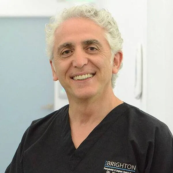
Dr. Homa Zadeh is an internationally renowned periodontist recognized for his clinical and scientific expertise. Dr. Zadeh is a Diplomate of the American Board of Periodontology and Implant Surgery. He has served as tenured associate professor, of the post-doctoral periodontology (2012 to 2019), as well as Chair of Periodontology Section (2003-2006) at USC Ostrow School of Dentistry. Dr. Zadeh graduated from the University of California Los Angeles (UCLA) with a BS degree, followed by Doctor of Dental Surgery (DDS) from USC Ostrow School of Dentistry. He completed post-doctoral clinical training in periodontology, as well as a PhD degree in immunology, at the University of Connecticut Health Center (UCHC) School of Dental Medicine and School of Medicine, respectively. Dr. Zadeh has lectured nationally and internationally in over 30 countries at prestigious conferences on topics ranging from aesthetics to minimally invasive periodontal and implant surgery, as well as tissue engineering.Dr. Zadeh served as president of the Western Society of Periodontology (WSP) in 2017, which is the largest regional periodontology society spanning the 16 western US states and Canadian provinces.Dr. Zadeh is the innovator of the VISTA technique recognized around the world as one of the most effective methods for the treatment of gingival recession. He has also innovated many concepts and devices, which have been patented and developed into products in clinical use in many countries.Dr. Zadeh has an extensive publication track, including over 100 publications in peer-reviewed journals and numerous book chapters. He serves as an editorial reviewer for many scientific journals.He served as a chartered scientific reviewer to the National Institutes of Health (NIH) and chaired a scientific panel at the NIH Center for Scientific Review. Dr. Zadeh has served as the director of USC International Periodontal and Implant Symposium. Dr. Zadeh is the recipient of many honors and awards, including outstanding teaching award and outstanding service award from USC Ostrow School of Dentistry and many research grants.He is an active member of the American Academy of Periodontology, Western Society of Periodontology (WSP), Academy of Osseointegration (AO), American Academy of Implant Dentistry (AAID), American Dental Association (ADA), California Dental Association (CDA), San Fernando Valley Dental Association (SFVDA) and Santa Barbara Ventura Dental Association (SBVDA).
The aim of the present study was to develop a 3D digital image-analysis method to quantitatively assess gingival changes after clear-aligner orthodontic therapy. Using teeth as fixed reference points, 3D image analysis tools have been used to quantify mucosal level changes after specific therapies. This technology has not been applied to orthodontic therapy, primarily because orthodontic tooth movement precludes using teeth as fixed reference points. Rather than superimposing the pre- and posttherapy volumes for the entire dentition, the methodology presented herein superimposed the pre- and post-therapy volumes for individual teeth. The lingual tooth surfaces, which remained unaltered, were used as fixed references. Intraoral scans taken before and after clear-aligner orthodontic therapy were imported for comparison. Volumes were created for each 3D image and were superimposed in a 3D image-analysis software that allowed quantitative measurements. The results demonstrated this technique's ability to measure very small changes in the apicocoronal position of the gingival zenith, as well as alterations of gingival margin thickness, following clear-aligner orthodontic therapy. The present 3D image-analysis method offers a useful tool for investigating the periodontal dimensional and positional changes that accompany orthodontic therapy.
The International Journal of Oral & Maxillofacial Implants, 4/2022
DOI: 10.11607/jomi.9413Pages 722-730, Language: EnglishValentini, Pascal / Zadeh, Homayoun H / Jungo, Sebastien / Mangion, Jean P / Bianca, Giancarlo / Ferrandi, Jean M
Purpose: The aim of the present retrospective clinical study was to evaluate the outcome of a maxillary sinus lateral window augmentation protocol, which sought to shorten the treatment time.
Materials and methods: This protocol entailed sinus augmentation with deproteinized bovine bone minerals (DBBM) and simultaneous implant placement in patients with minimal residual bone height. A total of 89 sinus augmentation procedures were performed in 74 patients, in whom 160 implants were placed between 2005 and 2013. The mean residual bone height was 2.6 ± 0.6 mm. The healing time before loading was 4.18 ± 0.63 months.
Results: In a first evaluation in 2014 the early implant survival rate (EIsR) was 96.8% after a mean period of 5.4 ± 2.2 years. A second evaluation in 2019 after a mean period of 10.4 ± 2.2 years showed a late implant survival rate (LIsR) of 83.1%. The failures after 2014 were all caused by peri-implantitis, which affected 14.6% and 16.8% of patients and implants, respectively. This prevalence of peri-implantitis does not appear to be higher than that usually observed in nonaugmented sites.
Conclusion: This reduction in the duration of treatment compared to the usual duration of 9 to 12 months does not seem to affect the predictability of the technique.
Keywords: atrophic sitres, lateral approach, long-term complications, maxillary sinus augmentation, reduction treatment time
International Journal of Periodontics & Restorative Dentistry, 6/2020
DOI: 10.11607/prd.4571, PubMed ID (PMID): 33151198Pages 907-916, Language: EnglishBotelho, Sabrina V. / Perussolo, Jeniffer / Misawa, Mônica Y. O. / Zadeh, Homayoun H. / Araújo, Mauricio G.
The aim of this study was to describe the basal bone and alveolar process in the maxillary anterior region by assessing patient CBCT scans. Parasagittal reconstructions were made to quantify basal bone and alveolar process dimensions and inclination of teeth in the maxillary anterior region. The CBCT scans of 87 patients and 522 tooth sites were included in this study. The results showed that the surface areas of the basal bone, alveolar process, and palatal triangle varied from 22.1 to 54.1 mm2, 87.8 to 144.0 mm2, and 37.1 to 66.0 mm2, respectively. The basal bone in the canine region had a significantly smaller cross-sectional area than in the incisor region. The alveolar process in the canine region was markedly larger than those of the central and lateral incisor regions. The mean overall thickness of the alveolar facial bone at 3, 5, and 7 mm above the CEJ were 0.6 ± 0.5 mm, 0.9 ± 0.5 mm, and 0.7 ± 0.6 mm, respectively. Additionally, the findings demonstrated that the cross-sectional area of the alveolar process and palatal triangle were greater among men than women. The study identified significant anatomical differences among various tooth regions in the anterior maxilla. The results also demonstrated that the tooth type, but not the tooth inclination or apex location, correlates with the size of the alveolar process.
Immediate implant placement in extraction sockets requires management of postextraction alveolar resorption. This randomized controlled trial evaluated the facial alveolar bone dimension 10 months following immediate implant placement with or without the addition of anorganic xenograft at the time of flapless, one-stage placement of a sloped-platform implant. The primary outcome of facial crestal alveolar bone thickness revealed no difference in the mean dimension (no graft: 1.47 ± 0.85 mm; graft: 1.63 ± 0.71 mm; P = .950). Secondary outcomes, including pink esthetic score, were not different between the two groups. This study suggests that bone formation does occur along the facial surface of implants placed into extraction sockets.
This study sought to evaluate gingival volume changes following root coverage with the vestibular incision subperiosteal tunnel access (VISTA) procedure. Pre- and postoperative surface scans of 21 patients (154 teeth) treated with VISTA using various graft materials were digitally superimposed to quantify volumetric changes. A linear gingival thickness gain of approximately 1 mm and volumetric gain of 5.47 mm3 were achieved. A negative correlation was found between linear thickness gain and root prominence. The thickness achieved was not different with various graft materials. Since gingival thickness has been identified as an important predictor of periodontal root coverage, the methodology described in the present study, along with the identification of predictors of outcome, has important therapeutic implications.
International Journal of Periodontics & Restorative Dentistry, 4/2019
DOI: 10.11607/prd.3865, PubMed ID (PMID): 31226194Pages 545-551, Language: EnglishHameed, Sabina / Bakhshalian, Neema / Alwazan, Essa / Wallace, Stephen S. / Zadeh, Homayoun H.
This retrospective study investigated the changes in the maxillary sinus floor and alveolar crest following extraction of maxillary molars. Pre- and postextraction cone beam computed tomography scans of 23 patients were analyzed. Paired-sample t tests compared pre- and postextraction measurements, and independent-sample t tests were utilized for intergroup comparisons. Pearson correlation was used to assess the association between the measured variables and the outcome measures. The mean alveolar bone height reduction was 3.42 ± 2.40 mm and the alveolar crest loss was 3.07 ± 2.53 mm. The maxillary sinus floor position shifted coronally by a mean of 0.47 ± 0.32 mm. Approximately 88% of postextraction changes in alveolar bone height were due to alveolar crest changes, compared to 12% due to changes in the sinus floor position. The results of this study challenge the commonly held concept of extensive postextraction maxillary sinus floor alterations leading to sinus pneumatization.
The objective of this study was to investigate marginal bone level (MBL) changes as the primary outcome of implants placed following healing of extraction sockets treated with ridge preservation, compared to implants placed following unassisted healing. A total of 268 patients (contributing 572 implants) treated from April 2005 to March 2013 were included in this retrospective study. Following estimation of the overall success rate of implants placed in the practice, 129 patients (contributing 222 implants) met the inclusion and exclusion criteria to be further evaluated. Of these, 78 patients (contributing 105 implants) received ridge preservation procedures, which entailed placement of anorganic bovine bone mineral covered with polytetrafluoroethylene membrane. The other 51 patients (contributing 117 implants), with healed sites without history of bone grafting, served as controls. Digital radiographs were taken at the time of implant placement and during follow-up visits. The mean follow-up period for ridge preservation sites was 33.6 months (range: 7.5 to 61.5 months) and for healed sites was 36.3 months (range: 6 to 91.2 months). The survival rate of implants in sites following healing of ridge preservation was 97.3%, compared to 98.5% in the nongrafted healed sites. The mean MBL changes during the observation period were 0.11 and 0.07 mm on the mesial and 0.06 and 0.06 mm on the distal aspects of the ridge preservation and healed sites, respectively (P > .05). The mean crestal bone remodeling during the observation period was 0.63 and 0.45 mm on the mesial and 0.48 and 0.12 mm on the distal aspects in the ridge preservation and healed sites, respectively (P .05). The results of this study demonstrated stable MBL and survival of implants placed in sites following ridge preservation with xenograft as well as nongrafted healed sites.
The objective of this study was to examine wound healing in extraction sockets following ridge preservation and the outcome of implants placed in those sites. Extraction sockets (N = 31) were grafted with anorganic bovine bone mineral (ABBM) and covered with dense polytetrafluoroethylene membrane. Bone cores obtained during implant placement were examined histologically and histomorphometrically. Percentages of vital bone and residual graft were 37.5% ± 21.3% and 12.5% ± 8.9%, respectively. New vital bone showed a negative correlation with patient age. Percentage of vital bone formation in sockets was correlated with neither postgraft wound healing time nor peri-implant marginal bone level.
A total of 68 extraction sockets were grafted with anorganic bovine bone mineral and covered by dense polytetrafluoroethylene membrane. Quantitative analysis of three-dimensional microcomputed tomography imaging of core samples retrieved after a mean of 21.0 ± 14.2 weeks revealed 40.1% bone volume fraction (bone volume [BV]/total volume [TV]) and 12% residual graft. Evidence of de novo bone formation was observed in the form of discrete islands of newly formed bone in direct apposition to graft particles, separated from parent bone. Anterior sockets exhibited a significantly higher percentage of residual graft compared to premolar sockets (P = .05). The BV/TV and percentage of residual graft correlated well with histomorphometric analysis of the same sites, but not with implant outcomes.
Purpose: Dental implant therapy for posterior partial edentulism may utilize short implants. The advantages of short implants include the ability to avoid the additional surgical procedures that would be required to place longer implants. The aim of this study was to systematically review studies concerning dental implants of = 8.5 mm placed in the posterior maxilla and/or mandible to support fixed restorations.
Materials and Methods: English-language articles published between 1992 and May 2011 were identified electronically and by hand search of the PubMed, Embase, and Cochrane libraries. Data were extracted and compared statistically. Forest plots were generated to compare outcomes of short versus long implants.
Results: An initial screening of 1,354 studies led to direct evaluation of 401 articles. Of these, 33 met the research criteria: 5 randomized clinical studies; 16 prospective, nonrandomized, noncontrolled studies; 12 retrospective, nonrandomized studies; and 1 study with both prospective and retrospective data. These studies indicated that there is no significant difference in the reported survival of short versus long implants. Failure of 59 of 2,573 short implants at 1 year was recorded, with 71% of them failing before loading. Only 101 short implants were followed for 5 years.
Conclusions: The initial survival rate for short implants for posterior partial edentulism is high and not related to implant surface, design, or width. Short implants may constitute a viable alternative to longer implants, which may often require additional augmentation procedures.
Keywords: posterior partial edentulism, short dental implant, systematic review