Various cookies are used on our website: We use technically necessary cookies for the purpose of enabling functions such as login or a shopping cart. We use optional cookies for marketing and optimization purposes, in particular to place relevant and interesting ads for you on Meta's platforms (Facebook, Instagram). You can refuse optional cookies. More information on data collection and processing can be found in our privacy policy.
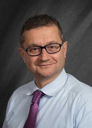
Dr. Ercoli is chairman of the Prosthodontics Postgraduate Training Program at EIOH. A native of Italy where he earned his dental degree, Dr. Ercoli continued his advanced training in ways to restore the comfort, appearance and function of the mouth when patients have missing or damaged teeth (prosthodontics). He is also trained in treatment of the jaw and facial muscles (the areas affected by TMD and TMJ), and of the gums and tissues that support the teeth. Dr. Ercoli is internationally published in prosthodontics and implant dentistry and has lectured locally, nationally and internationally. He is a Diplomate of the American Board of Prosthodontics and past president of the American Prosthodontic Society. He enjoys treating patients, and his double specialization allows him to be your expert for simple and complex needs.
Dr. Carlo Ercoli is Professor of Prosthodontics, Periodontics and Implant Surgery and Chairman of the Prosthodontic Department at the University of Rochester, Eastman Institute for Oral Health where he also serves as the Director for the Center of Excellence for Digital Dentistry. He graduated from the “Enrico Berlinguer” Dental Technology Institute in Rome in 1987 and obtained his DDS from the University of Siena, Italy in 1993. He specialized in Prosthodontics in 1996 at the Eastman Dental Center in Rochester, New York and completed a specialty training in Orofacial Pain and Temporomandibular Joint Disorders in 1997. In 2012, he achieved specialty certification in Periodontology. In 2019, he obtained his MBA from the Simon Business School of the University of Rochester.
Dr. Ercoli is a Past President of the American Prosthodontic Society, Executive Council Member of the Academy of Prosthodontics, Honorary Member of the Italian Academy of Prosthodontics, Founding Member of the Italian Society of Prosthodontics and Oral Rehabilitation, Education and Research Director of the American College of Prosthodontists, Board Member of the American College of Prosthodontists Education Foundation, and Director-Elect of the American Board of Prosthodontics. He served as Executive Council member of The Journal of Prosthetic Dentistry, Scientific Program Chair of the American Prosthodontics Society, American College of Prosthodontists, American Academy of Fixed Prosthodontics, and Academy of Prosthodontics (2024 Program). Dr. Ercoli is a diplomate of the American Boards of Prosthodontics and Periodontology, an ITI Fellow and Chair of the USA ITI East Region, in addition to serving as a Member of the World Workshop on the Classification of Periodontal and Peri-implant Diseases and Conditions. He holds memberships in the American Academy of Periodontology, American College of Prosthodontists, American Dental Education Association, Academy of Osseointegration, American Academy of Fixed Prosthodontics, Greater New York Academy of Prosthodontics, American Prosthodontic Society and American Dental Education Association. He lectures nationally and internationally on dental implantology.
International Journal of Oral Implantology, 4/2022
PubMed ID (PMID): 36377626Pages 353-365, Language: EnglishKotsailidi, Elli Anna / Tatakis, Dimitris N / Chen, Yo-Wei / Caton, Jack G / Ercoli, Carlo / Barmak, Abdul Basir / Tsigarida, Alexandra
Purpose: To compare the outcomes of soft tissue augmentation during one-stage implant placement using grafts harvested from the hard palate or the maxillary tuberosity.
Materials and methods: In this pilot controlled clinical study, non-smoking adults with a single missing tooth in the anterior or premolar region and adequate ridge dimensions for implant placement were enrolled. Each received a single implant and connective tissue graft harvested either from the hard palate (n = 10) or the maxillary tuberosity (n = 10). Digital impressions were taken prior to treatment (T0) and then 2 and 12 months postoperatively (T1 and T2, respectively). The primary study outcome was changes in horizontal ridge dimension. Secondary outcomes included marginal bone level changes over time, pain levels in the first 2 postoperative weeks (W1 and W2) and pink aesthetic score and patient-reported outcome measures at T2. Data analysis included repeated measures analysis of variance for intergroup comparisons.
Results: The horizontal ridge dimension increased significantly in both groups (P ≤ 0.002) at all apico-coronal levels examined, with no significant intergroup differences. There was also no significant intergroup difference in marginal bone level changes (P = 0.376). The hard palate group experienced higher pain levels in the donor site compared to the tuberosity group at W1 (P = 0.023). The pink aesthetic score and patient-reported outcome measures were similar between groups at T2.
Conclusions: Soft tissue augmentation during one-stage implant placement results in significant increases in the horizontal ridge dimension.
Keywords: autogenous tissue graft, connective tissue, dental implants, palate, transplantation, treatment outcome
Conflict-of-interest statement: The authors report no conflicts of interest relating to this study.
An alternative approach was used to increase the buccal vestibular depth of two edentulous patients, using free epithelialized palatal grafts. Two edentulous patients presented with shallow vestibules and inadequate keratinized tissue width in the mandibular anterior region. These sites were treated with vestibuloplasty followed by placement of an epithelialized palatal graft. In order to minimize graft movement and possible mechanical trauma to the area, the graft was covered with the buccal flap during the initial stages of healing. The patients maintained an increase in the vestibular depth as well as the keratinized tissue width at 14 months and 5 years postoperatively. Successful outcomes in terms of increase in vestibular depth can be achieved with the use of epithelialized palatal graft that is covered during the initial stage of healing. The dental practitioner fabricating the complete denture should be aware of the advantages offered by this alternative surgical technique.
Keywords: epithelialized palatal graft, free gingival graft, keratinized tissue augmentation, periodontal surgery, vestibuloplasty
Objective: This study investigated the effect of framework design and modification of porcelain firing cycle on the occurrence of fractures of metal-ceramic and bilayered zirconia crowns after cyclic loading.
Method and materials: Twenty-four zirconia crowns with two different apicocoronal incisal veneering porcelain lengths (ZS = 2.0 mm and ZL = 4.0 mm) were fabricated. Twenty-four metal-ceramic crowns with the same apicocoronal incisal veneering porcelain lengths (MS = 2.0 mm and ML = 4.0 mm) served as the control. All the specimens (n = 48) were thermally and mechanically cycle-loaded (49 N) for 1,200,000 cycles, and then evaluated for cracks and/or bulk fractures with an optical stereomicroscope (×10). In the second part of the study, the influence of firing cycle modification on the success rate of twenty-four bilayered zirconia crowns was also evaluated. Finally, two specimens from each group (n = 12) were sectioned and analyzed using a scanning electron microscope. Load-to-failure of the specimen that did not present bulk fracture was further assessed using a universal testing machine. Statistical analysis was performed with Fisher exact test and Kruskal-Wallis test.
Results: A statistically significant lower occurrence of fractures (P .05) was found after cyclic loading for bilayered zirconia versus metal-ceramic crowns. Modification of the firing cycle did not significantly influence the outcome after cyclic loading for zirconia bilayered crowns (P > .05).
Conclusion: Within the limitations of this study, porcelain-firing cycle with an extended cooling time did not appear to influence the occurrence of cracks and fractures of bilayered zirconia crowns.
Keywords: all-ceramic, cyclic loading, dental technology, failure analysis, firing cycle, metal-ceramic, porcelain, prosthodontics, zirconia
Objective: To compare the thickness of buccal bone around single dental implants placed in the anterior maxilla (premolar to premolar) inserted with different placement protocols.
Data Sources: An electronic search was conducted using MEDLINE (PubMed), Cochrane Central Register of Controlled Trials (CENTRAL), and EMBASE, from January 1980 to July 2015. Mean buccal bone thickness around single dental implants was measured and correlation with implant placement protocols, loading protocols, and augmentation method was assessed. A Q-test was used to access the homogeneity of levels of effect. A univariate meta-regression analysis was used for further investigation of the between-study heterogeneity. Two randomized clinical trials and 12 cohort studies were included for statistical analysis. The difference in buccal bone thickness for implants placed with different implant placement protocols (early vs immediate vs delayed) was not statistically significant (P > .05). Loading protocols (immediate vs delayed) also did not significantly influence the thickness of buccal bone. Descriptive analysis showed different buccal bone thickness for dental implants that received different bone grafting materials at the time of placement.
Conclusion: Different implant placement and loading protocols may not significantly affect the thickness of the buccal bone around single dental implants in the anterior maxilla. Different bone graft materials at the time of implant placement may have an effect on buccal bone thickness.
Keywords: bone graft, buccal bone thickness, CT imaging, dental implants, early placement, facial bone thickness, immediate implant
Objective: The purpose of this systematic review was to assess the difference in the incidence of intraoperative and postoperative complications between the conventional and alternative surgical techniques, during sinus floor augmentation surgery.
Data Sources: Electronic databases were searched for publications related to sinus floor augmentation surgery performed with different techniques (ie, conventional lateral window, piezosurgery, osteotome, trephine) and the incidence of complications. The articles were evaluated by independent reviewers, according to predetermined inclusion/exclusion criteria and processed for meta-analysis, following the PRISMA protocol. Initial search returned 3,940 articles, after inclusion/exclusion criteria, and quality assessment 11 articles were included in the meta-analysis: five randomized clinical trials and six retrospective studies. All the 11 included studies compared the incidence of complications in conventional lateral window sinus augmentation surgery versus alternative techniques, including: osteotome (five articles), piezosurgery (four articles), sonic surgery (one article), and trephine (one article). Meta-analysis of the incidence of intraoperative and postoperative complications during conventional lateral window sinus floor augmentation surgery versus alternative techniques, showed a significantly lower incidence of intraoperative complications for the retrospective studies. However, when data from the clinical trials alone were included in the meta-analysis, a statistically significant difference could not be detected for the incidence of intraoperative and postoperative complications.
Conclusions: The use of alternative techniques for sinus floor augmentation surgery (ie, piezosurgery, sonic surgery, osteotome, and trephine) does not necessarily reduce the incidence of intraoperative and/or postoperative complications compared to the conventional technique.
Keywords: dental implants, intraoperative complications, piezosurgery, postoperative complications, sinus floor augmentation, review
Sjogren's syndrome (SS) is a systemic autoimmune disease that has several oral manifestations, with reduced salivary flow being the most prevalent. As a result of the dry mouth and irritated oral tissues, dental treatment may be challenging. In particular, a patient's satisfaction with removable prosthesis may be limited. This case report and the literature review discuss the feasibility of implant-supported dental prostheses. The clinical and functional advantages provided by implant prostheses might outweigh the slightly lower overall implant success rate in SS patients. The impacts of SS on dental treatment are summarized in this review.
Keywords: dental implant, dry mouth, oral, prosthesis, rehabilitation, Sjogren's syndrome
International Journal of Periodontics & Restorative Dentistry, 1/2014
DOI: 10.11607/prd.1387, PubMed ID (PMID): 24396845Pages 104-112, Language: EnglishWeitz, Daniel S. / Geminiani, Alessandro / Papadimitriou, Dimitrios E. V. / Ercoli, Carlo / Caton, Jack G.
This study reports the incidence of complications and sinus membrane perforations when using sonic instruments to prepare the lateral window osteotomy for maxillary sinus augmentation. The charts of 33 consecutive patients (40 sinuses) were reviewed. Sinus membrane perforations were reported in 7 cases (17.5% of procedures), with 3 perforations occurring during preparation of the osteotomy window (7.5% of procedures). Postoperatively, 2 cases of sinus infection (5%) and 2 cases of flap dehiscence (5%) were recorded. This study demonstrates that elevation of the sinus floor with a combination of hand and sonic instruments is feasible and safe, with a limited incidence of complications. (Int J Periodontics Restorative Dent 2014;34:105-112. doi: 10.11607/prd.1387)
Anteroposterior (AP) deficiencies present a restorative treatment challenge. Complex, multidisciplinary planning is necessary for the success of the treatment. This clinical report describes an approach to managing a complex complete oral rehabilitation of an edentulous patient with skeletal transverse and AP deficiencies with a history of facial trauma to the left zygomaticomaxillary complex. This was further complicated by a hopeless remaining dentition and pneumatization of the maxillary sinuses. Treatment included initial bony augmentation of the vertically and horizontally deficient maxilla, dental implant placement, provisional restoration in a Class III malocclusion with bilateral posterior crossbite, and Le Fort I osteotomy with transverse widening and advancement to correct the skeletal deficiency. Definitive restoration was accomplished with implant-supported fixed prostheses that provided ideal facial balance and occlusion.
Purpose: The purpose of this study was to assess the clinical effectiveness of an experimental technique versus the conventional method for the fabrication of implant-supported fixed complete dentures.
Materials and Methods: Between January 2005 and June 2010, edentulous patients who had received rehabilitation by means of nonsegmented fixed implant-supported complete dentures were identified from the pool of individuals treated at Eastman Institute for Oral Health, University of Rochester. Data collection consisted of a chart review and recording of treatment variables in a customized database. Variables of interest included number of implants per patient, time of implant placement, number of appointments required to complete prosthesis fabrication, type of appointments, manufacturing process used for creation of the framework, and fit of the framework. The number of appointments for conventional and experimental protocols was evaluated for statistical significance using two-way analysis of variance. Presence or absence of clinically acceptable prosthesis fit with the two techniques was evaluated using the Fisher exact test and exact logistic regression analysis.
Results: Forty-two patients (48 arches) were included. When the experimental technique was used, prosthesis fabrication and delivery required an average of 4 appointments, whereas the conventional technique required an average of 7.8 appointments to deliver the definitive prosthesis. The prostheses fabricated with the experimental technique showed clinically passive fit on the implants in 17 of 18 arches. The frameworks fabricated with the conventional technique achieved clinically passive fit in 18 of 30 arches.
Conclusions: The experimental technique significantly reduced the number of appointments required to fabricate a nonsegmented fixed implant-supported prosthesis. Moreover, this experimental technique provided clinically acceptable fit of the prosthesis in a significantly greater number of cases compared to a conventional implant elastomeric impression technique.
Keywords: dental implants, edentulous mandible, edentulous maxilla, immediate loading, implant-supported prosthesis
The International Journal of Oral & Maxillofacial Implants, 6/2007
PubMed ID (PMID): 18271374Pages 928-938, Language: EnglishGarine, Wael N. / Funkenbusch, Paul D. / Ercoli, Carlo / Wodenscheck, Joseph / Murphy, William C.
Purpose: The specific aims of this study were to measure the implant and abutment hexagonal dimensions, to measure the rotational misfit between implant and abutments, and to correlate the dimension of the gap present between the abutment and implant hexagons with the rotational misfit of 5 abutment-implant combinations from 2 manufacturers.
Materials and Methods: Twenty new externally hexed implants (n = 10 for Nobel Biocare; n = 10 for Biomet/3i) and 50 new abutments were used (n = 10; Procera Zirconia; Procera Alumina; Esthetic Ceramic Abutment; ZiReal; and GingiHue post ZR Zero Rotation abutments). The mating surfaces of all implants and abutments were imaged with a scanning electron microscope before and after rotational misfit measurements. The distances between the corners and center of the implant and abutment hexagon were calculated by entering their x and y coordinates, measured on a measuring microscope, into Pythagoras' theorem. The dimensional difference between abutment and implant hexagons was calculated and correlated with the rotational misfit, which was recorded using a precision optical encoder. Each abutment was rotated (3 times/session) clockwise and counterclockwise until binding. Analysis of variance and Student-Newman-Keuls tests were used to compare rotational misfit among groups (a = .05).
Results: With respect to rotational misfit, the abutment groups were significantly different from one another (P .001), with the exception of the Procera Zirconia and Esthetic Ceramic groups (P = .4). The mean rotational misfits in degrees were 4.13 ± 0.68 for the Procera Zirconia group, 3.92 ± 0.62 for the Procera Alumina group, 4.10 ± 0.67 for the Esthetic Ceramic group, 3.48 ± 0.40 for the ZiReal group, and 1.61 ± 0.24 for the GingiHue post ZR group. There was no correlation between the mean implant-abutment gap and rotational misfit.
Conclusions: Within the limits of this study, machining inconsistencies of the hexagons were found for all implants and abutments tested. The GingiHue Post showed the smallest rotational misfit. All-ceramic abutments without a metal collar showed a greater rotational misfit than those with a metal collar.
Keywords: ceramic abutment, metal abutment, misfit, rotational freedom, screw