Various cookies are used on our website: We use technically necessary cookies for the purpose of enabling functions such as login or a shopping cart. We use optional cookies for marketing and optimization purposes, in particular to place relevant and interesting ads for you on Meta's platforms (Facebook, Instagram). You can refuse optional cookies. More information on data collection and processing can be found in our privacy policy.
Studium der Zahnmedizin in Düsseldorf, 1984-1996 Wissenschaftlicher Mitarbeiter und anschließend Oberarzt der Poliklinik für Zahnärztliche Prothetik der Westdeutschen Kieferklinik Düsseldorf, 1994 Habilitation, 1995 Spezialist für "Zahnärztliche Prothetik" der Deutschen Gesellschaft für Zahnärztliche Prothetik und Werkstoffkunde (DGZPW/DGPro), 1997 Professur und Leitung der Abteilung für "Zahnmedizinische Propädeutik/Community Dentistry" an der Universität Greifswald, 1998 stellv. Direktor der Poliklinik für Zahnärztliche Prothetik und Werkstoffkunde in Greifswald, seit 2004 Vize-Präsident der Deutschen Gesellschaft für Computerunterstützte Zahnmedizin DGCZ, 2006 Spezialist für "Zahnärztliche Funktionsdiagnostik und -therapie" der Deutschen Gesellschaft für Zahnärztliche Funktionsdiagnostik und -therapie (DGFDT), seit 2007 Leitung des Arbeitskreises "Kaufunktion und orale Physiologie" in der DGFDT, seit 2009 Leitung der Abteilung für "CAD/CAM- und CMD-Behandlung" in Greifswald (2013 umbenannt in "Digitale Zahnmedizin: Okklusions- und Kaufunktionstherapie").
1. Auflage 2018 Book Hardcover, 21 x 28 cm, 488 pages, 970 illus Language: German Categories: Functional Therapy, Prosthodontics Stock No.: 19390 ISBN 978-3-86867-378-4 QP Deutschland
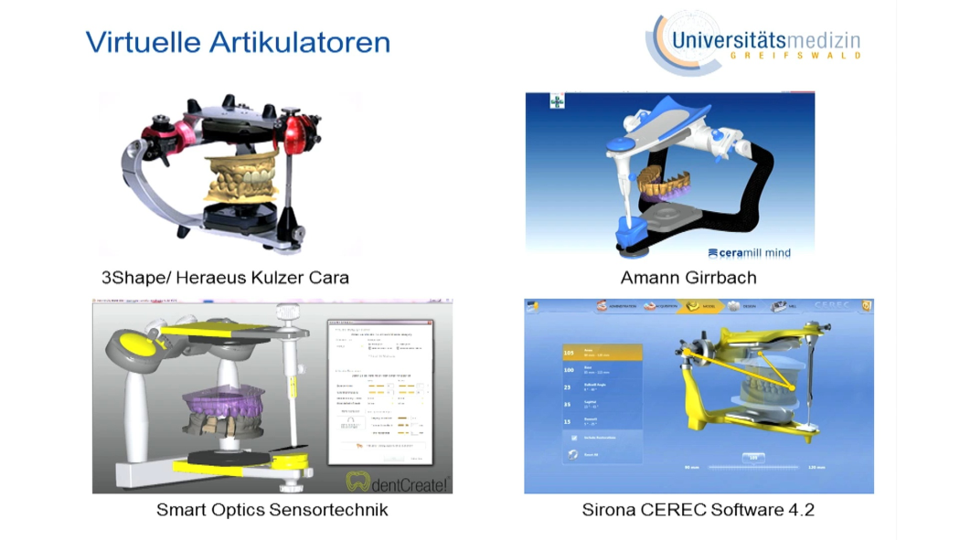
Length: 35 minutes Production year: 2018 Language: English, German Series: Dental Video Journal available since: 11. Jul 2018
Events
40th Congresso Internazionale
La rivoluzione protesica: cosa è cambiato, cosa è ancora attuale18. Nov 2021 — 20. Nov 2021Palazzo dei Congressi Bologna, Bologna, Italy
Speakers: Ingo Baresel, Florian Beuer, Cristiano Broseghini, Mauro Broseghini, Gaetano Calesini, Roberto Canalis, Davide Cortellini, Marco Degidi, Stefano Gracis, Stefano Granata, Bernd Kordaß, Tomas Linkevičius, Ignazio Loi, Paolo Miceli, Ricardo Mitrani, Nazariy Mykhaylyuk, Piotr Nagadowsky, José M. Navarro, Jan Schünemann, Clemens Schwerin, Lorenzo Trevisiol, Luca Vailati, Marco Valenti, Naoto Yuasa
Accademia Italiana di Odontoiatria Protesica (AIOP)
Deutscher Zahnärztetag 2019
8. Nov 2019 — 9. Nov 2019Congress Center Messe Frankfurt
Speakers: Karl-Ludwig Ackermann, Sarah Al-Maawi, Kurt Werner Alt, Jassin Arnold, Thomas Attin, Mustafa Ayna, Anna Greta Barbe, Ingo Baresel, Jens Baresel, Tobias Bauer, Ursula Becker, Wilfried Beckmann, Christoph Benz, Lisa Bitterich, Dirk Bleiel, Uwe Blunck, Martin Boost, Andreas Braun, Anne Bredel-Geissler, Wolfgang Buchalla, Egon Burian, Sebastian Bürklein, Iain L. C. Chapple, Wolfgang Christian, Fabian Cieplik, Bettina Dannewitz, Monika Daubländer, Sybille David-Hebgen, Isabel Deckwer, James Deschner, Annika Döding, Christof Dörfer, Heike Dyrna, Norbert Engel, Peter Engel, Susanne Fath, Michael Frank, Roland Frankenberger, Rene Franzen, Cornelia Frese, Tobias Fretwurst, Michael Gahlert, Roland Garve, Werner Geurtsen, Shahram Ghanaati, Christiane Gleissner, Ulrike Gonder, Werner Götz, Dominik Groß, Knut A. Grötz, Martin Guffart, Norbert Gutknecht, Cornelius Haffner, Thorsten Halling, Frederic Hermann, Carlos Herrera-Vizcaino, Tim Hilgenfeld, Jürgen Hoffmann, Martin Hoffmann, Fabian Huettig, Alfons Hugger, Christine Hutschenreuter, Bruno Imhoff, Silke Jacker-Guhr, Søren Jepsen, A. Rainer Jordan, Alexander Jürchott, Bärbel Kahl-Nieke, Peer W. Kämmerer, Philipp Kanzow, Nele Kettler, Christian Kirschneck, Lydia Kogler, Bernd Kordaß, Franz-Josef Kramer, Norbert Krämer, Felix Krause, Matthis Krischel, Joachim Krois, Christina Kühne, Conrad Kühnöl, Bernd Lapatki, Silke Lehmann-Binder M.Sc., Christian Leonhardt, Ivona Leventic, Daniel Lindel, Jörg Alexander Lisson, Ulrike Lübbert, Elmar Ludwig, Anne-Katrin Lührs, Michael Lüpke, Frank Georg Mathers, Wibke Merten, Georg Meyer, Wolfram Misselwitz, Karin Mölling, Mhd Said Mourad, Dietmar Friedrich Müller, Moritz Mutschler, Katja Nickel, Nicole Nicklisch, Ina Nitschke, Olaf Oberhofer, Karina Obreja, Dietmar Oesterreich, Rebecca Otto, Simon Peroz, Peter Pospiech, Florian Probst, Monika Probst, Michael Rädel, Sven Reich, Katharina Reichenmiller, Katharina Reinecke, Daniel R. Reißmann, Bernd Reiss, Stefan Ries, Christiane Rinnen, Katharina Röher, Jerome Rotgans, Uwe Rudol, Michael Rumpf, Heidrun Schaaf, Claudia Schaller, Karina Schick, Ulrich Schiffner, Maximiliane Amelie Schlenz, Alexander Schmidt, Mathias Schmidt, Andrea-Maria Schmidt-Westhausen, Julian Schmoeckel, Wolfgang Schneider, Sigmar Schnutenhaus, Holger Schön, Andreas Schulte, Nelly Schulz-Weidner, Karola Schulze, Ralf Schulze, Falk Schwendicke, Thomas A. Schwenk, Andreas Simka, Ralf Smeets, Önder Solakoglu, David Sonntag, Hansmartin Spatzier, Benedikt Spies, Norbert Staab, Sabine Steding, Angela Stillhart, Marcus Stoetzer, Hendrik Terheyden, Andrea Thumeyer, Marin Vodanovic, Kai Voß, Maximilian Voß, Wolfgang Wahlster, Michael Walter, Sandra Weber, Almut Johanna Weigel, Paul Weigl, Michael Weiss, Hans-Jürgen Wenz, Johannes-Simon Wenzler, Christian Wesemann, Jens Westemeier, Lotta Westphal, Matthias Widbiller, Annette Wiegand, Horst Willeweit, Karl Frederick Wilms, Sandra Windecker, Michael M. Wolf, Anne Wolowski, Bernd Wöstmann, Sylvia Wuttig
Quintessenz Verlags-GmbH
Deutscher Zahnärztetag 2018
MISSERFOLGE - erkennen, beherrschen, vermeiden9. Nov 2018 — 10. Nov 2018Congress Center Messe Frankfurt
Speakers: Karl-Ludwig Ackermann, Sarah Al-Maawi, Bilal Al-Nawas, Kurt Werner Alt, Anna Greta Barbe, Tobias Bauer, Daniel Bäumer, Marco Baz Bartels, Grietje Beck, Katrin Bekes, Christoph Benz, Dirk Bleiel, Johannes Boesch, Martin Boost, Wolfgang Buchalla, Oskar Bunz, Fabian Cieplik, Monika Daubländer, Sybille David-Hebgen, Andreas Dehler, Renate Deinzer, Sonja H. M. Derman, Konstanze Diekmeyer, Ingmar Dobberstein, Heike Dyrna, Thomas Eger, Guido Elsäßer, Anne Sophie Engel, Peter Engel, Norbert Enkling, Susanne Fath, Stefan Fickl, Michael Frank, Roland Frankenberger, Rene Franzen, Kerstin Galler, Carolina Ganß, Roland Garve, Christian Ralf Gernhardt, Werner Geurtsen, Shahram Ghanaati, Petra Gierthmühlen, Christiane Gleissner, Steffani Görl, Werner Götz, Susanne Grässel, Dominik Groß, Stefan Grümer, Claus Grundmann, Martin Guffart, Heinz-Michael Günther, Norbert Gutknecht, Peter Hahner, Elmar Hellwig, Christian Henrici, Katrin Hertrampf, Fabian Huettig, Michael Hülsmann, Bruno Imhoff, Holger Jentsch, A. Rainer Jordan, Ana Elisa Kauling, Moritz Kebschull, Christian Kirschneck, Joachim Klimek, Andrea Klink, Thomas Klinke, Birte Koch, Thomas Kocher, Eva Köllensperger, Heike Maria Korbmacher-Steiner, Bernd Kordaß, Hannah Kottmann, Pablo Krämer-Fernandez, Gabriel Krastl, Birgit Krause, Till Kreutzer, Conrad Kühnöl, Stefanie Kurzschenkel, Thorsten Kuypers, Günter Lauer, Hans-Christoph Lauer, Elfi Laurisch, Tina Lawall, Karl Martin Lehmann, Silke Lehmann-Binder M.Sc., Dirk Leisenberg, Ulrike Lübbert, Michael Lüpke, Thomas Malik, Jutta Margraf-Stiksrud, Lorenz Meinel, Gudrun Mentel, Wibke Merten, Louisa Mewes, Johanna Isabel Moosmüller, Martin U. Müller, Wolfgang Müller, Nicole Nicklisch, Ina Nitschke, Michael J. Noack, Marina Nörr-Müller, Karina Obreja, Dietmar Oesterreich, Puria Parvini, Ingrid Peroz, Waldemar Petker, Oksana Petruchin, Andree Piwowarczyk, Peter Pospiech, Peter Proff, Sven Reich, Katharina Reichenmiller, Katharina Reinecke, Bernd Reiss, Svenja Rink, Christiane Rinnen, Jerome Rotgans, Didem Sahin, Sonja Sälzer, Petra Santander, Heidrun Schaaf, Jürgen Schäffer, Elisabeth Schiffner, Ulrich Schiffner, Markus Schlee, Maximiliane Amelie Schlenz, Peter Schmidt, Andrea-Maria Schmidt-Westhausen, Claas Ole Schmitt, Sigmar Schnutenhaus, Jörg Schröder, Gerd Schröter, Andreas Schulte, Philipp Schwaab, Frank Schwarz, Falk Schwendicke, Clemens Schwerin, Sinan Sen, Önder Solakoglu, Hansmartin Spatzier, Christian H. Splieth, Norbert Staab, Bernd Stadlinger, Sabine Steding, Marcus Stoetzer, Giorgio Tabanella, Gisela Tascher, Hendrik Terheyden, Valentina A. Tesky, Jan Tetsch, Juliane von Hoyningen-Huene, Maximilian Voß, Michael Walter, Alexander Welk, Dietmar Weng, Hans-Jürgen Wenz, Jens Westemeier, Lotta Westphal, Annette Wiegand, Karl Frederick Wilms, Michael M. Wolf, Diana Wolff, Anne Wolowski, Johann-Dietrich Wörner, Sylvia Wuttig, Mohamed Younis, Stefan Zimmer, Lisa Zumpe
Quintessenz Verlags-GmbH
This author's journal articles
Journal of Craniomandibular Function, 2/2024
SciencePages 117-132, Language: English, GermanKordaß, Bernd / Ruge, Sebastian / Imhoff, Bruno / Güth, Jan-Frederik / Reich, Sven / Schlenz, Maximiliane A. / Hugger, Sybille / Hugger, Alfons
Position Paper of the Study Group for Oral Physiology and Masticatory Function (DGFDT)
Rapid developments in digital dentistry, such as digital workflows and CAD/CAM systems, have led to questions about digital occlusion, including the capabilities of occlusal analysis. There is a need for clear definitions and terminology. What do we mean when we talk about “occlusion” in the context of digitization, especially in the case of digital models? What are the capabilities of digital occlusal analysis? The following article presents our initial thoughts on this important topic, which may be useful in the development of future guideline.
Keywords: digital occlusion, digital occlusion analysis, virtual articulator, digital articulator, digital patient, digital functionally generated path technique (FGP technique)
WissenschaftPages 938-947, Language: German, EnglishKordaß, Bernd / Ruge, Sebastian / Imhoff, Bruno / Güth, Jan-Frederik / Reich, Sven / Schlenz, Maximiliane A. / Hugger, Sybille / Hugger, Alfons
Rapid developments in digital dentistry, such as digital workflows and CAD/CAM systems, have led to questions about digital occlusion, including the capabilities of occlusal analysis. There ist a need for clear definitions and terminology. What do we mean when we talk about “occlusion” in the context of digitization, especially in the case of digital models? What are the capabilities of digital occlusal analysis? The following article presents our initial thoughts on this important topic, which may be useful in the development of future guideline.
Die Digitalisierung in der Zahnmedizin und Zahntechnik schreitet unaufhaltsam voran. Auch die Funktionsdiagnostik und -therapie einschließlich der Kieferrelationsbestimmung profitiert davon. Allerdings ist es wichtig zu wissen, was der digitalen Okklusion zugrunde liegt und welche Gesichtspunkte bei der Interpretation zu berücksichtigen sind. Gegenüber der analogen Welt liegt alles in „Zahlen“ vor, was ohne Zweifel ein eminenter Vorteil ist. Vor allem lassen sich auch die Fehler und Unschärfen besser einschätzen, mit denen wir es beim Scannen und Registrieren, aber letztlich auch beim Umsetzen und Fertigen von Restaurationen zu tun haben. In der praktischen Anwendung kommt es trotz allem auf viel Erfahrungswissen und letztlich intuitives Ausprobieren an, bis wirklich ein zufriedenstellendes Optimum erreicht werden kann.
Manuskripteingang: 14.03.2022, Manuskriptannahme: 17.03.2022
Keywords: Digitale Okklusion, digitale Simulation, CAD/CAM, Okklusionsanalyse
When utilizing CAD/CAM systems to design and manufacture dental prostheses and occlusal splints, one soon wonders: How accurate is virtual occlusion? Conventional methods involving dental impressions, plaster casts, articulators, and manual verification tools such as articulating paper have well-known sources of error and error chains, and tried and tested error-handling strategies for many of them already exist. Digital workflows, on the other hand, are still very new and unfamiliar to some dentists. Besides digital processes such as intraoral scanning and optoelectronic jaw motion tracking, analog processes may also be required. The penetration of maxillary and mandibular dental models is one aspect of digital dentistry that immediately attracts attention. Virtual penetration occurs because digital technology, like every measuring system, is subject to measurement error, and because virtual reality can only approximate the true variable nature of the physiologic masticatory system. The present article aims to identify and discuss variables that affect the accuracy of virtual occlusion. Some errors are first discovered in the digital world. Virtual capabilities open up new perspectives that have yet to be explored and understood.
Keywords: occlusion, virtual articulator, intraoral scan, jaw motion tracking
Evaluations of the regional baseline study (SHIP-1) with the Greifswald Digital Analyzing System (GEDAS)
Sufficient occlusion is a basic prerequisite for the functional efficiency of the occlusal surfaces. Exactly where and in what number the occlusal contacts in the posterior region should be present for this purpose is controversial.
Aim: The present study investigated the number and location of occlusal contacts on posterior teeth without dental findings, ie, without caries or restorative restorations such as fillings, crowns, etc. Such natural posterior teeth were present in 709 subjects (males [m] = 446: 48.9 ± 13.04 years, females [f] = 283: 52.4 ± 14.23 years) of a subject collective of 1223 subjects (m = 648, f = 575) of the regional baseline study ‘Study of Health in Pomerania 1’ (SHIP-1).
Materials and method: Silicone bite registrations in habitual intercuspation (IP) were evaluated, whereby the test persons were asked to fix the bite block with biting force without biting firmly. The registrations were scanned with a document scanner in incident and transmitted light; a calibration strip was used to determine the transparency threshold of a layer thickness of 20 μm, below and equal to which the transparent zone was considered as a contact or contact area. The Greifswald Digital Analyzing System 2 (GEDAS 2) software was used to determine the number and location of occlusal contact areas tooth by tooth. To define the localization of the contacts, a cross with two concentric circles symmetric to the longitudinal fissure was superimposed on the occlusal surface; this resulted in four inner and four outer quadrants. Thus, the number of pixels in occlusal contact areas per inner and outer quadrant could be determined. The image resolution was 300 dpi.
Results: On average (median), the premolars had two occlusal contacts each, the posterior teeth had four to five, and Vieltooth 46 had six contacts. The right and left teeth did not differ in the frequency of occlusal contacts in the Mann-Whitney U test for independent samples. In the maxillary premolars, frequent contact areas were primarily located mesially on the inner and outer slopes of the palatal cusp. In the maxillary molars, the palatal slope of the distopalatal cusp and the inner slopes of the mesiopalatal and distopalatal cusps were frequently addressed. On the mandibular premolars, the inner slopes of the buccal cusps and the buccal slope of the distobuccal cusp were particularly frequently addressed; in teeth 35 and 45, the buccal slope of the mesiobuccal cusp was also somewhat more frequently addressed. Teeth 36 and 46 frequently had contact areas on the buccal slope of the distobuccal cusp as well as on the inner slopes of the distal cusps (distobuccal and distolingual), whereas teeth 37 and 47 tended to behave similarly.
Conclusion: Epidemiologically, the focus of the frequent contact areas on the respective supporting cusps of the maxillary and mandibular posterior teeth and a distribution of contacts stabilizing the tooth in its position in the dental arch through the interlocking were confirmed. It makes sense to take this into account when designing occlusal surfaces in the posterior region.
Keywords: occlusion, occlusal contacts, posterior teeth, epidemiology
Sufficient occlusion is a basic prerequisite for the functional efficiency of the occlusal surfaces. Exactly where and in what number the occlusal contacts in the posterior region should be present for this purpose is controversial. The present study investigated the number and location of occlusal contacts on posterior teeth without dental findings, ie, without caries or restorative restorations such as fillings, crowns, etc. Such natural posterior teeth were present in 709 subjects (males (m) = 446: 48.9 ± 13.04 years, females (f) = 283: 52.4 ± 14.23 years) of a subject collective of 1223 subjects (m = 648, f = 575) of the regional baseline study ‘Study of Health in Pomerania 1’ (SHIP-1).
Silicone bite registrations in habitual intercuspation (IP) were evaluated, whereby the test persons were asked to fix the bite block with biting force without biting firmly. The registrations were scanned with a document scanner in incident and transmitted light; a calibration strip was used to determine the transparency threshold of a layer thickness of 20 μm, below and equal to which the transparent zone was considered as a contact or contact area. The Greifswald Digital Analyzing System 2 (GEDAS 2) software was used to determine the number and location of occlusal contact areas tooth by tooth. To define the localization of the contacts, a cross with two concentric circles symmetric to the longitudinal fissure was superimposed on the occlusal surface; this resulted in four inner and four outer quadrants. Thus, the number of pixels in occlusal contact areas per inner and outer quadrant could be determined. The image resolution was 300 dpi.
On average (median), the premolars had two occlusal contacts each, the posterior teeth had four to five, and tooth 46 had six contacts. The right and left teeth did not differ in the frequency of occlusal contacts in the Mann-Whitney U test for independent samples. In the maxillary premolars, frequent contact areas were primarily located mesially on the inner and outer slopes of the palatal cusp. In the maxillary molars, the palatal slope of the distopalatal cusp and the inner slopes of the mesiopalatal and distopalatal cusps were frequently affected. On the mandibular premolars, the inner slopes of the buccal cusps and the buccal slope of the distobuccal cusp were particularly frequently addressed; in teeth 35 and 45, the buccal slope of the mesiobuccal cusp was also somewhat more frequently addressed. Teeth 36 and 46 frequently had contact areas on the buccal slope of the distobuccal cusp as well as on the inner slopes of the distal cusps (distobuccal and distolingual), whereas teeth 37 and 47 tended to behave similarly.
Epidemiologically, the focus of the frequent contact areas on the respective supporting cusps of the maxillary and mandibular posterior teeth and a distribution of contacts stabilizing the tooth in its position in the dental arch through the interlocking were confirmed. It makes sense to take this into account when designing occlusal surfaces in the posterior region.
Keywords: occlusion, occlusal contacts, posterior teeth, epidemiology
Aim: The present prospective clinical study aimed to validate the Greifswald Digital Analyzing System (GEDAS) as a method for digital assessment of the occlusion in primary and mixed dentition.
Materials and methods: The reproducibility of GEDAS in primary and mixed dentition was assessed using the intraclass correlation coefficient (ICC). In addition, the acceptability of the method to the dentist, the child, and the parent/caregiver was assessed using a modified visual analog scale of faces, the Frankl behavior scale, and the 10-point Likert scale. In total, 20 participants aged between 3 and 9 years (mean age: 6; standard deviation: ± 1.74) with primary (n = 10) and mixed (n = 10) dentition were recruited.
Results: The ICC for the number of contact points in all teeth was 0.94 and for the area of contact points was 0.97, indicating good to excellent reproducibility. The average total number of contacts per bite registration per arch in the primary and mixed dentition was 36.5 (17 to 66) and 37.9 (9 to 74), respectively. The average of the total area of interocclusal contact area in the primary and mixed dentition was 25.55 mm2 (5.39 to 70.20) and 29.59 mm2 (2.80 to 78.53), respectively. During the procedure, the majority of dentists reported the child’s behavior to be positive (85%) and found the procedure easy to perform (80%), short (6.0 min), and tolerable (80%).
Conclusion: GEDAS is an occlusal analysis tool with good acceptability and reproducibility in children and could be considered for the planning and assessment of restorative and orthodontic treatment in the intermediate stages.
Keywords: GEDAS, occlusion, digital, primary dentition, mixed dentition, bite registration