Various cookies are used on our website: We use technically necessary cookies for the purpose of enabling functions such as login or a shopping cart. We use optional cookies for marketing and optimization purposes, in particular to place relevant and interesting ads for you on Meta's platforms (Facebook, Instagram). You can refuse optional cookies. More information on data collection and processing can be found in our privacy policy.
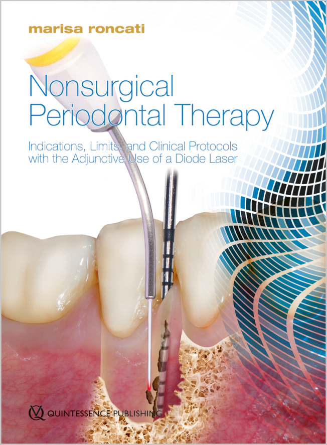
Marisa Roncati, RDH, DDS, is a dental hygiene teacher at the Albanian University dental school. She also teaches oral laser applications at the University of Parma and Rome University and oral surgery and pathology at the University of Parma. Dr Roncati is a registered dental hygienist and the author of two books published in English: Get Sharp: Nonsurgical Periodontal Instrument Sharpening (Quintessence, 2011) and Nonsurgical Periodontal Therapy: Indications, Limits, and Clinical Protocols with the Adjunctive Use of Diode Laser (Quintessence, 2017).
Indications, Limits and Clinical Protocols with the Adjunctive Use of Diode Laser
1st Edition 2017 Book Hardcover, 21 x 28 cm, 416 pages, 1390 illus Language: English Category: Periodontics Stock No.: 20281 ISBN 978-88-7492-045-7 QP Italy
1. Auflage 2011 DVD Amaray-Box, Laufzeit: 40 Min. Language: German Categories: Dental Team, Prophylaxis Stock No.: 6090 ISBN 978-3-86867-044-8 QP Deutschland
The 14th International Symposium on Periodontics and Restorative Dentistry (ISPRD)
9. Jun 2022 — 12. Jun 2022Boston Marriott Copley Place, Boston, MA, United States of America
Speakers: Tara Aghaloo, Edward P. Allen, Evanthia Anadioti, Wael Att, Vinay Bhide, Markus Blatz, Scotty Bolding, Lorenzo Breschi, Jeff Brucia, Daniel Buser, Luigi Canullo, Daniele Cardaropoli, Stephen J. Chu, Donald Clem, Christian Coachman, Lyndon F. Cooper, Daniel Cullum, Lee Culp, José Carlos Martins da Rosa, Sergio De Paoli, Marco Degidi, Nicholas Dello Russo, Serge Dibart, Joseph P. Fiorellini, Mauro Fradeani, Stuart J. Froum, David Garber, Maria L. Geisinger, William Giannobile, Luca Gobbato, Ueli Grunder, Galip Gürel, Chad Gwaltney, Christoph Hämmerle, Robert A. Horowitz, Marc Hürzeler, David Kim, Gregg Kinzer, Christopher Köttgen, Ina Köttgen, Purnima S. Kumar, Burton Langer, Lydia Legg, Pascal Magne, Kenneth A. Malament, Jay Malmquist, George Mandelaris, Pamela K. McClain, Michael K. McGuire, Mauro Merli, Konrad H. Meyenberg, Craig M. Misch, Julie A. Mitchell, Marc L. Nevins, Myron Nevins, Michael G. Newman, Miguel A. Ortiz, Jacinthe M. Paquette, Stefano Parma-Benfenati, Michael A. Pikos, Giulio Rasperini, Pamela S. Ray, Christopher R. Richardson, Isabella Rocchietta, Marisa Roncati, Marco Ronda, Paul S. Rosen, Maria Emanuel Ryan, Irena Sailer, Maurice Salama, David M. Sarver, Takeshi Sasaki, Todd Scheyer, Massimo Simion, Michael Sonick, Sergio Spinato, Dennis P. Tarnow, Lorenzo Tavelli, Douglas A. Terry, Tiziano Testori, Carlo Tinti, Istvan Urban, Hom-Lay Wang, Robert Winter, Giovanni Zucchelli
Quintessence Publishing Co., Inc. USA
This author's journal articles
International Journal of Periodontics & Restorative Dentistry, 3/2023
A 12-month prospective consecutive case series study was conducted to evaluate the effectiveness of a novel porcine collagen matrix to correct moderate to severe buccogingival recession defects. A total of 10 healthy patients (8 women and 2 men; age range: 30 to 68 years) with 26 maxillary and mandibular recession gingival defects > 4 mm deep were included. Healthy maturation of gingival tissues with natural color and texture matching the adjacent soft tissue areas were observed at all reevaluation visits. Complete root coverage was not achieved in all cases, possibly due to severe buccal bone loss in most of the selected cases, which negatively impacted the results. Nevertheless, when a novel porcine collagen matrix was used, a mean root coverage of 63.15% was achieved, and gains in clinical attachment level and keratinized tissue height were seen. Future randomized clinical trials are needed to further explore the potential of porcine collagen matrix for treatment of localized gingival recession defects.
This case report emphasizes how home and professional care can impact clinical outcomes following nonsurgical mechanical therapy with the adjunctive use of a diode laser to treat failing implants. Localized severe peri-implant disease was diagnosed in a patient who was susceptible to periodontal disease, not compliant, and did not accept the surgical approach. A cause-related, diode laser-assisted, nonsurgical treatment was therefore implemented, according to a dedicated protocol, in four closely scheduled appointments. The patient's oral hygiene skills became extremely effective. She then adhered to a bimonthly maintenance protocol in the first follow-up year and quarterly in the subsequent year. Bone remineralization was detected on comparative periapical radiographs 2 years after the diagnosis of peri-implantitis. At the 2-year observation, the patient's high standard of home care and dedicated maintenance arrested the progression of peri-implantitis and yielded clinical and radiographic improvements following nonsurgical mechanical therapy with adjunctive repeated application of a diode laser.
The aim of this classification is to diagnose and grade four different types of soft tissue deficiency around loaded, osseointegrated implants according to increasing severity. The suggested soft tissue augmentation to increase the width of the peri-implant keratinized mucosa will improve the long-term stability of peri-implant tissues.
MAPA-cision, named after those who first introduced the method, is a novel simplified regenerative technique for periodontal-orthodontic cases that can be used in all circumstances where bone thickening is required. It is an innovative, minimally invasive piezoelectric surgical procedure designed to facilitate orthodontic tooth movement while simultaneously increasing bone thickness with guided bone regeneration principles. A new regenerative device consisting of a resorbable collagen membrane with filling materials (a “bone bundle” or “small sausage”) is inserted through a tunneling procedure to increase the bone envelope width by allowing the teeth to move within an enhanced periodontal support.
The aim of this retrospective study was to evaluate long-term clinical and radiologic outcomes of submerged and nonsubmerged guided bone regenerative treatments for peri-implantitis lesions. Strict methods of implant-surface decontamination and detoxification were performed. Data on clinical probing depth, soft tissue measures, and marginal bone level that were documented by comparative radiographs were obtained from 45 patients, for a total of 57 implants prior to treatment and at the latest follow-up. The average followup period was 6.9 years (range: 2 to 21 years). Analysis of implant-based data revealed a success rate of 70.2% for a total of 40 implants. Recurrence of periimplantitis was observed on 9 implants, and 8 implants were removed. The regenerative procedures, under a strict periodontal control, were effective in the treatment of moderate to advanced peri-implantitis lesions.
The clinical effects of treatment with 808- and 980-nm diode lasers as an adjunct to a uniform and consistent nonsurgical periodontal treatment protocol (NSPTP) for initial probing pockets ≥ 6 mm with a 10-year follow-up were evaluated. In total, 24 patients (16 test cases and 8 control cases) were treated with the NSPTP. The control group (CG) was treated only with nonsurgical conventional therapy, while the test group (TG) received treatment with thermal diode lasers (wavelength of 808 or 980 nm). All patients had recall visits every 3 months and were reevaluated at 1 year after NSPTP. Following the 1-year follow-up, patients were monitored at 4-month intervals for the remaining 9 years. In total, 646 teeth (433 in TG [124 multirooted teeth and 309 single-rooted teeth] and 213 in CG [64 multirooted teeth and 149 single-rooted teeth]) were included. The initial probing pocket depth (PPD) in single-rooted teeth from TG patients decreased from an average of 6.7 mm to 3.3 mm after 1 year, and to 2.3 mm after 10 years. The mean increase in clinical attachment level (CAL) was 3.8 mm. The initial PPD of single-rooted teeth in CG patients decreased from an average of 6.4 mm at baseline to 4 mm after 1 year and 3.3 mm after 10 years. The mean increase in CAL was 2.5 mm in CG. In the TG, the PPDs of multirooted teeth decreased from 6.2 mm to 3.3 mm after 1 year, where they remained stable for 10 years. The mean increase in CAL was 2.2 mm in this group. In the CG, the PPDs for multirooted teeth decreased from 7 mm to 5.8 mm after 1 year and were 5.1 mm after 10 years. The mean increase in CAL was 1.9 mm. The TG had significantly better clinical parameters than the CG, including reduced tooth loss. Compared to conventional debridement alone, the use of thermal diode lasers with NSPTP improves PPD and increases CAL in periodontal pockets ≥ 6 mm over the long term.
A total of 25 patients were treated with a nonsurgical periodontal treatment protocol (NSPTP) consisting of four appointments (three within 1 week and one approximately 30 days later). Nonsurgical periodontal instrumentation was implemented, with the adjunctive use of thermal diode lasers (wavelength of 808 or 980 nm). The patients were scheduled for recall visits every 3 months and reevaluated 1 year post-NSPTP. They were subsequently monitored at 4-month intervals for the remaining 2-year follow-up maintenance period. In total, 698 teeth (210 multirooted and 488 single-rooted) were included in the study. The mean bleeding on probing was 43% at baseline and decreased to 12% at 12 months and to 8% at 3 years. The initial ≥ 6-mm probing pocket depth (PPD) in single-rooted teeth decreased from 6.2 mm at baseline to a mean of 1.8 mm at 12 months and remained at 1.8 mm at 3 years, with a mean clinical attachment level (CAL) gain of 4.4 mm. In multirooted teeth, the PPD decreased from 6.7 mm to a mean of 3.9 mm at 12 months and 3.6 mm at 3 years, with a mean CAL gain of 2.9 mm. The mean overall recession was 0.3 mm at baseline, 0.1 mm at 1 year, and 0.3 mm at the 3-year follow-up. In the short-term followup period, uniform and consistent implementation of the NSPTP used here with adjunctive use of thermal diode lasers seemed to convey therapeutic benefits, stable periodontal soft tissue levels, and satisfactory esthetics in patients with moderate to severe chronic periodontitis. The present protocol is relevant as a treatment option for medically compromised patients, those who refuse or delay surgical treatment, or those who present with other limitations.
This case series presents clinical outcomes on reentry using regenerative submerged and nonsubmerged approaches in peri-implant defects; pre- and posttreatment assessments of nine implants in six patients are presented. A mean bone fill value of 91.3% with a 4.88-mm mean bone gain was obtained. Neither approach led to additional bone loss or required additional bone augmentation procedures. Strict methods of implant surface decontamination and detoxification were used on all patients, regardless of implant surface characteristics. The regenerative procedure was effective in the treatment of moderate to advanced peri-implantitis lesions without compromising the previous fixed implant-supported prostheses. These preliminary results are reasonably encouraging in that all cases showed bone gains. Nevertheless, caution must be exercised when determining reosseointegration, because it is not possible to ascertain it in clinical practice.
This case series presents the use of a resorbable "dome device" made of a slow, long-lasting resorbable suturing material to support the barrier creating and maintaining a secluded space to promote bone regeneration. Acellular dermal matrix or cross-linked resorbable collagen membrane, as barriers, combined with mineralized freeze-dried bone allograft, with simultaneous implant placement, were utilized in reconstructing non-space-making defects. Eight implants in six healthy patients were treated with a combination of these resorbable regenerative materials. Only one of seven was treated with a nonsubmerged approach. All sites remained completely covered and no implant exposure occurred during healing. At the 9- to 24-month reentry surgeries, the clinical bone density was equivalent to that of the native bone and the mean number of final exposed threads was 0.5. The mean buccal bone thickness achieved was 3.12 mm, with a mean total coverage of exposed threads in approximately 87.5% of the cases.
Peri-implantitis is a frequently occurring inflammatory disease mediated by bacterial infection that results in the loss of supporting bone. Peri-implantitis should be treated immediately, but there is a lack of evidence regarding the most effective therapeutic interventions. Nonsurgical periodontics may be the treatment of choice in cases of peri-implant mucositis or if the patient has medical contraindications or refuses to consent to more appropriate treatment. Peri-implantitis defects will dictate the therapeutic approach and present a guideline for relative clinical management. The suggested therapeutic solutions are derived from clinical experience and are meant to be a useful guide.